Loading
PDBj
メニューPDBj@FacebookPDBj@TwitterPDBj@YouTubewwPDB FoundationwwPDB
RCSB PDBPDBeBMRBAdv. SearchSearch help
Search by PDB author
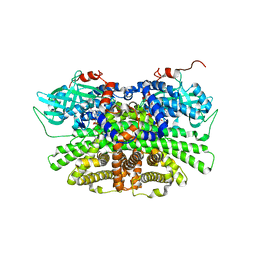
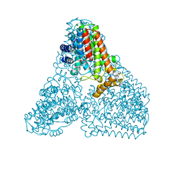
6KD7
DownloadVisualize
BU of 6kd7 by Molmil
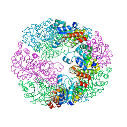
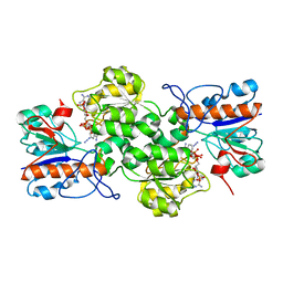
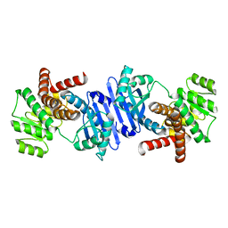
Crystal structure of geranylgeranyl pyrophosphate synthase
分子名称: GLYCEROL, MAGNESIUM ION, PYROPHOSPHATE, ...
著者Kim, S, Kim, K.-J.
登録日2019-07-01
公開日2019-09-11
最終更新日2023-11-22
実験手法X-RAY DIFFRACTION (2 Å)
主引用文献Crystal structure of geranylgeranyl pyrophosphate synthase (crtE) from Nonlabens dokdonensis DSW-6.
Biochem.Biophys.Res.Commun., 518, 2019
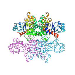
5C6F
DownloadVisualize
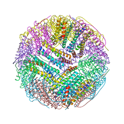
BU of 5c6f by Molmil
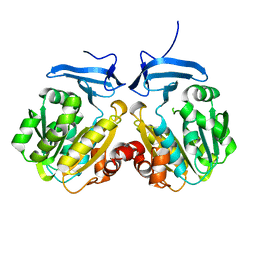
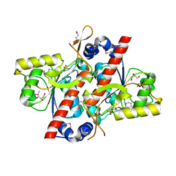
Crystal structures of ferritin mutants reveal side-on binding to diiron and end-on cleavage of oxygen
分子名称: Bacterial non-heme ferritin, FE (III) ION, IMIDAZOLE
著者Kim, S, Kim, K.H, Seok, J.H, Park, Y.H, Jung, S.W, Chung, Y.B, Lee, D.B, Lee, J.H, Han, K.R, Cho, A.E, Lee, C, Chung, M.S.
登録日2015-06-23
公開日2016-07-27
最終更新日2024-03-20
実験手法X-RAY DIFFRACTION (2 Å)
主引用文献Structural Basis of Novel Iron-Uptake Route and Reaction Intermediates in Ferritins from Gram-Negative Bacteria.
J. Mol. Biol., 428, 2016
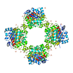
4JDY
DownloadVisualize
BU of 4jdy by Molmil
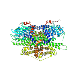
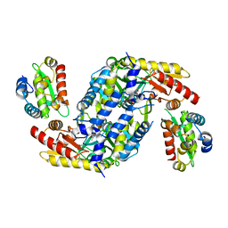
Crystal structure of Rv2606c
分子名称: GLYCEROL, Pyridoxal biosynthesis lyase PdxS
著者Kim, S, Kim, K.-J.
登録日2013-02-25
公開日2013-05-22
最終更新日2023-11-08
実験手法X-RAY DIFFRACTION (1.8 Å)
主引用文献Crystal structure of Mycobacterium tuberculosis Rv2606c: a pyridoxal biosynthesis lyase.
Biochem.Biophys.Res.Commun., 435, 2013
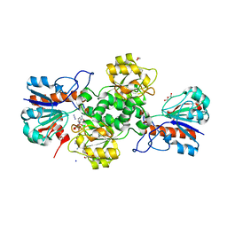
8IYI
DownloadVisualize
BU of 8iyi by Molmil
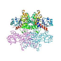
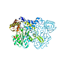
Spermidine synthase from Kluyveromyces lactis
分子名称: KLLA0B09372p
著者Kim, S, Chang, J.H.
登録日2023-04-05
公開日2023-06-28
最終更新日2024-05-29
実験手法X-RAY DIFFRACTION (1.9 Å)
主引用文献Structural Analysis of Spermidine Synthase from Kluyveromyces lactis.
Molecules, 28, 2023
5Y9D
DownloadVisualize
BU of 5y9d by Molmil
Crystal structure of acyl-coA oxidase1 from Yarrowia lipolytica
分子名称: Acyl-coenzyme A oxidase 1, FLAVIN-ADENINE DINUCLEOTIDE
著者Kim, S, Kim, K.-J.
登録日2017-08-24
公開日2018-01-03
最終更新日2023-11-22
実験手法X-RAY DIFFRACTION (2.5 Å)
主引用文献Structural insight into the substrate specificity of acyl-CoA oxidase1 from Yarrowia lipolytica for short-chain dicarboxylyl-CoAs.
Biochem. Biophys. Res. Commun., 495, 2018
4PRL
DownloadVisualize
BU of 4prl by Molmil
Crystal structure of D-lactate dehydrogenase with NAD+ from Lactobacillus jensenii
分子名称: 4-phosphoerythronate dehydrogenase, NICOTINAMIDE-ADENINE-DINUCLEOTIDE
著者Kim, S, Kim, K.J.
登録日2014-03-06
公開日2014-06-18
最終更新日2023-11-08
実験手法X-RAY DIFFRACTION (2.6 Å)
主引用文献Crystal structure and thermodynamic properties of d-lactate dehydrogenase from Lactobacillus jensenii.
Int.J.Biol.Macromol., 68C, 2014
5YS9
DownloadVisualize
BU of 5ys9 by Molmil
Crystal structure of acyl-coA oxidase3 from Yarrowia lipolytica
分子名称: Acyl-coenzyme A oxidase 3, FLAVIN-ADENINE DINUCLEOTIDE
著者Kim, S, Kim, K.-J.
登録日2017-11-13
公開日2018-02-28
最終更新日2023-11-22
実験手法X-RAY DIFFRACTION (2.5 Å)
主引用文献Crystal Structure of Acyl-CoA Oxidase 3 fromYarrowia lipolyticawith Specificity for Short-Chain Acyl-CoA.
J. Microbiol. Biotechnol., 28, 2018
4PRK
DownloadVisualize
BU of 4prk by Molmil
Crystal structure of D-lactate dehydrogenase (D-LDH) from Lactobacillus jensenii
分子名称: 4-phosphoerythronate dehydrogenase
著者Kim, S, Kim, K.J.
登録日2014-03-06
公開日2014-06-18
最終更新日2024-03-20
実験手法X-RAY DIFFRACTION (2.13 Å)
主引用文献Crystal structure and thermodynamic properties of d-lactate dehydrogenase from Lactobacillus jensenii.
Int.J.Biol.Macromol., 68C, 2014
7VG9
DownloadVisualize
BU of 7vg9 by Molmil
Crystal structure of phosphotransbutyrylase from Clostridium acetobutylicum
分子名称: Phosphate butyryltransferase, SULFATE ION
著者Kim, S, Kim, K.-J.
登録日2021-09-15
公開日2021-10-27
最終更新日2023-11-29
実験手法X-RAY DIFFRACTION (2.91 Å)
主引用文献Crystal structure and molecular mechanism of phosphotransbutyrylase from Clostridium acetobutylicum .
J Microbiol Biotechnol., 31, 2021
7YA4
DownloadVisualize
BU of 7ya4 by Molmil
Formate dehydrogenase from Novosphingobium sp. AP12 with NAD and Azide
分子名称: 1,2-ETHANEDIOL, ACETATE ION, AZIDE ION, ...
著者Kim, S, Kim, K.-J.
登録日2022-06-27
公開日2023-07-12
最終更新日2023-11-29
実験手法X-RAY DIFFRACTION (1.8 Å)
主引用文献Dual cofactor specific formate dehydrogenase from Novosphingobium sp. AP12 with high activity.
To Be Published
7YA3
DownloadVisualize
BU of 7ya3 by Molmil
Formate dehydrogenase from Novosphingobium sp. AP12 with NADP and Azide
分子名称: AZIDE ION, Formate dehydrogenase, NADP NICOTINAMIDE-ADENINE-DINUCLEOTIDE PHOSPHATE
著者Kim, S, Kim, K.-J.
登録日2022-06-27
公開日2023-07-12
最終更新日2023-11-29
実験手法X-RAY DIFFRACTION (3 Å)
主引用文献Dual cofactor specific formate dehydrogenase from Novosphingobium sp. AP12 with high activity.
To Be Published
4YED
DownloadVisualize
BU of 4yed by Molmil
TcdA (CsdL)
分子名称: ADENOSINE MONOPHOSPHATE, GLYCEROL, tRNA threonylcarbamoyladenosine dehydratase
著者Kim, S, Park, S.Y.
登録日2015-02-24
公開日2016-01-13
実験手法X-RAY DIFFRACTION (1.9 Å)
主引用文献The Structure of Escherichia coli TcdA (Also Known As CsdL) Reveals a Novel Topology and Provides Insight into the tRNA Binding Surface Required for N(6)-Threonylcarbamoyladenosine Dehydratase Activity
J.Mol.Biol., 427, 2015
4LW4
DownloadVisualize
BU of 4lw4 by Molmil
Structural changes during cysteine desulfurase CsdA and sulfur-acceptor CsdE interactions provide insight into the trans-persulfuration
分子名称: Cysteine desulfuration protein CsdE, Cysteine sulfinate desulfinase, PYRIDOXAL-5'-PHOSPHATE
著者Kim, S, Park, S.Y.
登録日2013-07-26
公開日2013-08-07
最終更新日2023-11-08
実験手法X-RAY DIFFRACTION (2.01 Å)
主引用文献Structural changes during cysteine desulfurase CsdA and sulfur acceptor CsdE interactions provide insight into the trans-persulfuration.
J.Biol.Chem., 288, 2013
4LW2
DownloadVisualize
BU of 4lw2 by Molmil
Structural changes during cysteine desulfurase CsdA and sulfur-acceptor CsdE interactions provide insight into the trans-persulfuration
分子名称: Cysteine sulfinate desulfinase, GLYCEROL, PYRIDOXAL-5'-PHOSPHATE
著者Kim, S, Park, S.Y.
登録日2013-07-26
公開日2013-08-07
最終更新日2023-11-08
実験手法X-RAY DIFFRACTION (1.8 Å)
主引用文献Structural changes during cysteine desulfurase CsdA and sulfur acceptor CsdE interactions provide insight into the trans-persulfuration.
J.Biol.Chem., 288, 2013
1JE4
DownloadVisualize
BU of 1je4 by Molmil
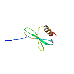
Solution structure of the monomeric variant of the chemokine MIP-1beta
分子名称: macrophage inflammatory protein 1-beta
著者Kim, S, Jao, S, Laurence, J.S, LiWang, P.J.
登録日2001-06-15
公開日2001-10-03
最終更新日2021-10-27
実験手法SOLUTION NMR
主引用文献Structural comparison of monomeric variants of the chemokine MIP-1beta having differing ability to bind the receptor CCR5.
Biochemistry, 40, 2001
7E43
DownloadVisualize
BU of 7e43 by Molmil
Structural insights into a bifunctional peptide methionine sulfoxide reductase MsrA/B fusion protein from Helicobacter pylori
分子名称: ACETATE ION, Peptide methionine sulfoxide reductase MsrA/MsrB
著者Kim, S, Lee, K, Hwang, K.Y.
登録日2021-02-10
公開日2021-04-21
最終更新日2023-11-29
実験手法X-RAY DIFFRACTION (2.2 Å)
主引用文献Structural Insights into a Bifunctional Peptide Methionine Sulfoxide Reductase MsrA/B Fusion Protein from Helicobacter pylori .
Antioxidants (Basel), 10, 2021
1RYU
DownloadVisualize
BU of 1ryu by Molmil
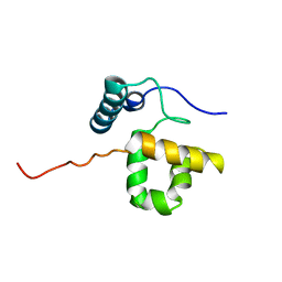
Solution Structure of the SWI1 ARID
分子名称: SWI/SNF-related, matrix-associated, actin-dependent regulator of chromatin subfamily F member 1
著者Kim, S, Zhang, Z, Upchurch, S, Isern, N, Chen, Y.
登録日2003-12-22
公開日2004-05-25
最終更新日2024-05-22
実験手法SOLUTION NMR
主引用文献Structure and DNA-binding sites of the SWI1 AT-rich interaction domain (ARID) suggest determinants for sequence-specific DNA recognition.
J.Biol.Chem., 279, 2004
7VID
DownloadVisualize
BU of 7vid by Molmil
The crystal structure of L-leucine dehydrogenase from Pseudomonas aeruginosa
分子名称: GLYCEROL, Leucine dehydrogenase
著者Kim, S, Kang, W, Yang, J.K.
登録日2021-09-26
公開日2022-06-29
最終更新日2023-11-29
実験手法X-RAY DIFFRACTION (2.5 Å)
主引用文献The Crystal Structure of L-Leucine Dehydrogenase from Pseudomonas aeruginosa.
Mol.Cells, 45, 2022
3VL1
DownloadVisualize
BU of 3vl1 by Molmil
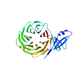
Crystal structure of yeast Rpn14
分子名称: 26S proteasome regulatory subunit RPN14
著者Kim, S, Nishide, A, Saeki, Y, Takagi, K, Tanaka, K, Kato, K, Mizushima, T.
登録日2011-11-28
公開日2012-05-02
最終更新日2023-11-08
実験手法X-RAY DIFFRACTION (1.6 Å)
主引用文献New crystal structure of the proteasome-dedicated chaperone Rpn14 at 1.6 A resolution
Acta Crystallogr.,Sect.F, 68, 2012
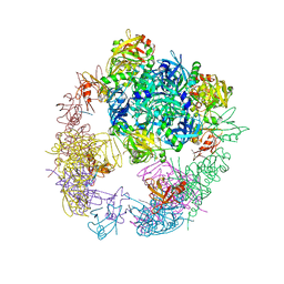
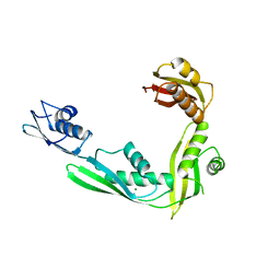
3OTP
DownloadVisualize
BU of 3otp by Molmil
Crystal structure of the DegP dodecamer with a model substrate
分子名称: Lysozyme C, Protease do
著者Kim, S, Grant, R.A, Sauer, R.T.
登録日2010-09-13
公開日2011-01-19
最終更新日2024-02-21
実験手法X-RAY DIFFRACTION (3.76 Å)
主引用文献Covalent Linkage of Distinct Substrate Degrons Controls Assembly and Disassembly of DegP Proteolytic Cages.
Cell(Cambridge,Mass.), 145, 2011
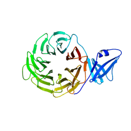
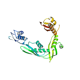
3ACP
DownloadVisualize
BU of 3acp by Molmil
Crystal Structure of Yeast Rpn14, a Chaperone of the 19S Regulatory Particle of the Proteasome
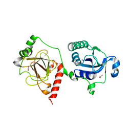
分子名称: WD repeat-containing protein YGL004C
著者Kim, S, Saeki, Y, Suzuki, A, Takagi, K, Fukunaga, K, Yamane, T, Kato, K, Tanaka, K, Mizushima, T.
登録日2010-01-08
公開日2010-03-16
最終更新日2011-07-13
実験手法X-RAY DIFFRACTION (2 Å)
主引用文献Crystal structure of yeast Rpn14, a chaperone of the 19S regulatory particle of the proteasome
J.Biol.Chem., 285, 2010
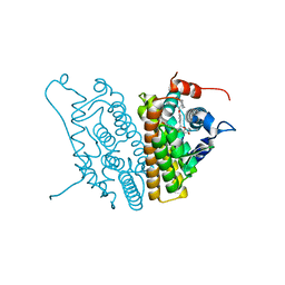
1SJ0
DownloadVisualize
BU of 1sj0 by Molmil
Human Estrogen Receptor Alpha Ligand-binding Domain in Complex with the Antagonist Ligand 4-D
分子名称: (2S,3R)-2-(4-(2-(PIPERIDIN-1-YL)ETHOXY)PHENYL)-2,3-DIHYDRO-3-(4-HYDROXYPHENYL)BENZO[B][1,4]OXATHIIN-6-OL, Estrogen receptor
著者Kim, S, Wu, J.Y, Birzin, E.T, Chan, W, Pai, L.Y, Yang, Y.T, Mosley, R.T, Fitzgerald, P.M, Sharma, N, DiNinno, F, Rohrer, S.P, Schaeffer, J.M, Hammond, M.L.
登録日2004-03-02
公開日2004-04-27
最終更新日2024-02-14
実験手法X-RAY DIFFRACTION (1.9 Å)
主引用文献Estrogen Receptor Ligands. II. Discovery of Benzoxathiins as Potent, Selective Estrogen Receptor alpha Modulators.
J.Med.Chem., 47, 2004
2QDF
DownloadVisualize
BU of 2qdf by Molmil
Structure of N-terminal domain of E. Coli YaeT
分子名称: MAGNESIUM ION, Outer membrane protein assembly factor yaeT
著者Kim, S, Malinverni, J.C, Sliz, P, Silhavy, T.J, Harrison, S.C, Kahne, D.
登録日2007-06-20
公開日2007-09-04
最終更新日2023-08-30
実験手法X-RAY DIFFRACTION (2.2 Å)
主引用文献Structure and function of an essential component of the outer membrane protein assembly machine.
Science, 317, 2007
2QCZ
DownloadVisualize
BU of 2qcz by Molmil
Structure of N-terminal domain of E. Coli YaeT
分子名称: Outer membrane protein assembly factor yaeT
著者Kim, S, Malinverni, J.C, Sliz, P, Silhavy, T.J, Harrison, S.C, Kahne, D.
登録日2007-06-20
公開日2007-08-28
最終更新日2024-02-21
実験手法X-RAY DIFFRACTION (2.7 Å)
主引用文献Structure and function of an essential component of the outer membrane protein assembly machine.
Science, 317, 2007
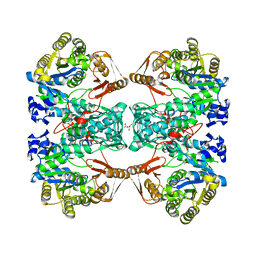
7C11
DownloadVisualize
BU of 7c11 by Molmil
Formate--tetrahydrofolate ligase from Methylobacterium extorquens CM4 strain
分子名称: ACETATE ION, CITRATE ANION, Formate-tetrahydrofolate ligase, ...
著者Kim, K.-J, Kim, S, Seo, H, Lee, S.
登録日2020-05-02
公開日2020-10-28
最終更新日2023-11-29
実験手法X-RAY DIFFRACTION (2.815 Å)
主引用文献Biochemical properties and crystal structure of formate-tetrahydrofolate ligase from Methylobacterium extorquens CM4.
Biochem.Biophys.Res.Commun., 528, 2020

221051

件を2024-06-12に公開中

PDB statisticsPDBj update infoContact PDBjnumon