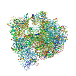
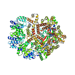
5J88
 
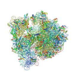 | | Structure of the E coli 70S ribosome with the U1060A mutation in 16S rRNA | | 分子名称: | (4S)-2-METHYL-2,4-PENTANEDIOL, 1,2-ETHANEDIOL, 1,4-DIAMINOBUTANE, ... | | 著者 | Cocozaki, A, Ferguson, A. | | 登録日 | 2016-04-07 | | 公開日 | 2016-07-06 | | 最終更新日 | 2016-12-07 | | 実験手法 | X-RAY DIFFRACTION (3.32 Å) | | 主引用文献 | Resistance mutations generate divergent antibiotic susceptibility profiles against translation inhibitors.
Proc.Natl.Acad.Sci.USA, 113, 2016
|
|
5J5B
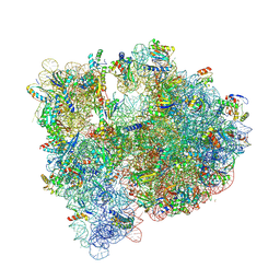 | | Structure of the WT E coli ribosome bound to tetracycline | | 分子名称: | (4S)-2-METHYL-2,4-PENTANEDIOL, 1,2-ETHANEDIOL, 1,4-DIAMINOBUTANE, ... | | 著者 | Cocozaki, A, Ferguson, A. | | 登録日 | 2016-04-01 | | 公開日 | 2016-07-27 | | 最終更新日 | 2018-08-15 | | 実験手法 | X-RAY DIFFRACTION (2.8 Å) | | 主引用文献 | Resistance mutations generate divergent antibiotic susceptibility profiles against translation inhibitors.
Proc.Natl.Acad.Sci.USA, 113, 2016
|
|
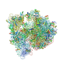
5IT8
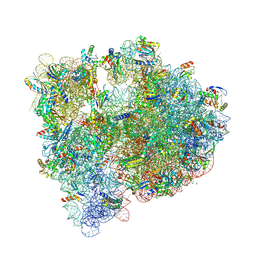 | | High-resolution structure of the Escherichia coli ribosome | | 分子名称: | (4S)-2-METHYL-2,4-PENTANEDIOL, 1,2-ETHANEDIOL, 1,4-DIAMINOBUTANE, ... | | 著者 | Cocozaki, A, Ferguson, A. | | 登録日 | 2016-03-16 | | 公開日 | 2016-07-27 | | 最終更新日 | 2023-11-15 | | 実験手法 | X-RAY DIFFRACTION (3.12 Å) | | 主引用文献 | Resistance mutations generate divergent antibiotic susceptibility profiles against translation inhibitors.
Proc.Natl.Acad.Sci.USA, 113, 2016
|
|
5J8A
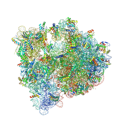 | | Structure of the E coli 70S ribosome with the U1052G mutation in 16S rRNA bound to tigecycline | | 分子名称: | (4S)-2-METHYL-2,4-PENTANEDIOL, 1,2-ETHANEDIOL, 1,4-DIAMINOBUTANE, ... | | 著者 | Cocozaki, A, Ferguson, A. | | 登録日 | 2016-04-07 | | 公開日 | 2016-07-06 | | 最終更新日 | 2016-08-03 | | 実験手法 | X-RAY DIFFRACTION (3.1 Å) | | 主引用文献 | Resistance mutations generate divergent antibiotic susceptibility profiles against translation inhibitors.
Proc.Natl.Acad.Sci.USA, 113, 2016
|
|
5JC9
 | | Structure of the Escherichia coli ribosome with the U1052G mutation in the 16S rRNA | | 分子名称: | (4S)-2-METHYL-2,4-PENTANEDIOL, 1,2-ETHANEDIOL, 1,4-DIAMINOBUTANE, ... | | 著者 | Cocozaki, A, Ferguson, A. | | 登録日 | 2016-04-14 | | 公開日 | 2016-07-06 | | 最終更新日 | 2016-08-03 | | 実験手法 | X-RAY DIFFRACTION (3.03 Å) | | 主引用文献 | Resistance mutations generate divergent antibiotic susceptibility profiles against translation inhibitors.
Proc.Natl.Acad.Sci.USA, 113, 2016
|
|
5J7L
 | | Structure of the 70S E coli ribosome with the U1052G mutation in the 16S rRNA bound to tetracycline | | 分子名称: | (4S)-2-METHYL-2,4-PENTANEDIOL, 1,2-ETHANEDIOL, 1,4-DIAMINOBUTANE, ... | | 著者 | Cocozaki, A, Ferguson, A. | | 登録日 | 2016-04-06 | | 公開日 | 2016-07-27 | | 最終更新日 | 2018-08-15 | | 実験手法 | X-RAY DIFFRACTION (3 Å) | | 主引用文献 | Resistance mutations generate divergent antibiotic susceptibility profiles against translation inhibitors.
Proc.Natl.Acad.Sci.USA, 113, 2016
|
|
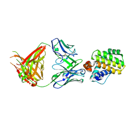
3GLG
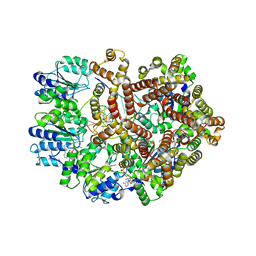 | | Crystal Structure of a Mutant (gammaT157A) E. coli Clamp Loader Bound to Primer-Template DNA | | 分子名称: | ADENOSINE-5'-DIPHOSPHATE, BERYLLIUM TRIFLUORIDE ION, DNA (5'-D(*CP*TP*GP*GP*CP*CP*TP*AP*TP*A)-3'), ... | | 著者 | Simonetta, K.R, Seyedin, S.N, Kuriyan, J. | | 登録日 | 2009-03-12 | | 公開日 | 2009-05-26 | | 最終更新日 | 2024-02-21 | | 実験手法 | X-RAY DIFFRACTION (3.25 Å) | | 主引用文献 | The mechanism of ATP-dependent primer-template recognition by a clamp loader complex.
Cell(Cambridge,Mass.), 137, 2009
|
|
3GLF
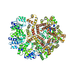 | |
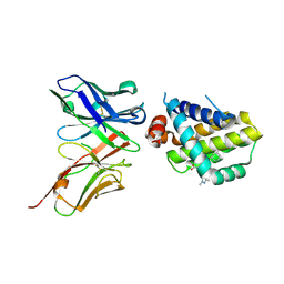
3GLI
 | | Crystal Structure of the E. coli clamp loader bound to Primer-Template DNA and Psi Peptide | | 分子名称: | ADENOSINE-5'-DIPHOSPHATE, BERYLLIUM TRIFLUORIDE ION, DNA (5'-D(*CP*TP*GP*GP*CP*CP*TP*AP*TP*A)-3'), ... | | 著者 | Simonetta, K.R, Cantor, A.J, Kuriyan, J. | | 登録日 | 2009-03-12 | | 公開日 | 2009-05-26 | | 最終更新日 | 2024-02-21 | | 実験手法 | X-RAY DIFFRACTION (3.5 Å) | | 主引用文献 | The mechanism of ATP-dependent primer-template recognition by a clamp loader complex.
Cell(Cambridge,Mass.), 137, 2009
|
|
4Z8D
 | |
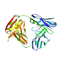
6QB6
 | | Mcl1 in complex with a Fab | | 分子名称: | Fab Heavy Chain, Fab Light Chain, Induced myeloid leukemia cell differentiation protein Mcl-1 | | 著者 | Hargreaves, D. | | 登録日 | 2018-12-20 | | 公開日 | 2019-11-06 | | 最終更新日 | 2019-11-13 | | 実験手法 | X-RAY DIFFRACTION (2.24 Å) | | 主引用文献 | Antibody fragments structurally enable a drug-discovery campaign on the cancer target Mcl-1.
Acta Crystallogr D Struct Biol, 75, 2019
|
|
6QB4
 | | Mcl1-scFv complex with an indole acid inhibitor | | 分子名称: | 3-[3-[[(1~{R})-1,2,3,4-tetrahydronaphthalen-1-yl]oxy]propyl]-7-(1,3,5-trimethylpyrazol-4-yl)-1~{H}-indole-2-carboxylic acid, Induced myeloid leukemia cell differentiation protein Mcl-1, scFv55 | | 著者 | Hargreaves, D. | | 登録日 | 2018-12-20 | | 公開日 | 2019-11-06 | | 最終更新日 | 2019-11-13 | | 実験手法 | X-RAY DIFFRACTION (2.38 Å) | | 主引用文献 | Antibody fragments structurally enable a drug-discovery campaign on the cancer target Mcl-1.
Acta Crystallogr D Struct Biol, 75, 2019
|
|
6QB9
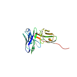 | | Structure of an anti-Mcl1 scFv | | 分子名称: | L(+)-TARTARIC ACID, scFv55 | | 著者 | Hargreaves, D. | | 登録日 | 2018-12-20 | | 公開日 | 2019-11-06 | | 最終更新日 | 2019-11-13 | | 実験手法 | X-RAY DIFFRACTION (1.85 Å) | | 主引用文献 | Antibody fragments structurally enable a drug-discovery campaign on the cancer target Mcl-1.
Acta Crystallogr D Struct Biol, 75, 2019
|
|
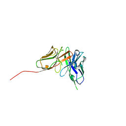
6QBC
 | | structure of anti-Mcl1 Fab | | 分子名称: | Anti-Mcl1 Fab Heavy Chain, Anti-Mcl1 Fab Light Chain | | 著者 | Luptak, J. | | 登録日 | 2018-12-20 | | 公開日 | 2019-11-06 | | 最終更新日 | 2019-11-13 | | 実験手法 | X-RAY DIFFRACTION (1.56 Å) | | 主引用文献 | Antibody fragments structurally enable a drug-discovery campaign on the cancer target Mcl-1.
Acta Crystallogr D Struct Biol, 75, 2019
|
|
6QF9
 | | Structure of an anti-Mcl1 scFv | | 分子名称: | scFv | | 著者 | Luptak, J. | | 登録日 | 2019-01-09 | | 公開日 | 2019-11-06 | | 最終更新日 | 2019-11-13 | | 実験手法 | X-RAY DIFFRACTION (1.43 Å) | | 主引用文献 | Antibody fragments structurally enable a drug-discovery campaign on the cancer target Mcl-1.
Acta Crystallogr D Struct Biol, 75, 2019
|
|
6QFC
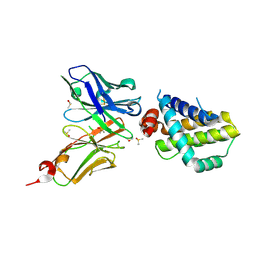 | | Structure of an anti-Mcl1 scFv | | 分子名称: | DIMETHYL SULFOXIDE, Induced myeloid leukemia cell differentiation protein Mcl-1, scFv55 | | 著者 | Luptak, J. | | 登録日 | 2019-01-09 | | 公開日 | 2019-11-06 | | 最終更新日 | 2019-11-13 | | 実験手法 | X-RAY DIFFRACTION (1.96 Å) | | 主引用文献 | Antibody fragments structurally enable a drug-discovery campaign on the cancer target Mcl-1.
Acta Crystallogr D Struct Biol, 75, 2019
|
|
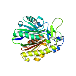
1EM8
 | | Crystal structure of chi and psi subunit heterodimer from DNA POL III | | 分子名称: | DNA POLYMERASE III CHI SUBUNIT, DNA POLYMERASE III PSI SUBUNIT | | 著者 | Gulbis, J.M, Finkelstein, J, O'Donnell, M, Kuriyan, J. | | 登録日 | 2000-03-16 | | 公開日 | 2003-08-26 | | 最終更新日 | 2024-02-07 | | 実験手法 | X-RAY DIFFRACTION (2.1 Å) | | 主引用文献 | Crystal structure of the chi:psi sub-assembly of the Escherichia coli DNA polymerase clamp-loader complex.
Eur.J.Biochem., 271, 2004
|
|


