Loading
PDBj
メニューPDBj@FacebookPDBj@TwitterPDBj@YouTubewwPDB FoundationwwPDB
RCSB PDBPDBeBMRBAdv. SearchSearch help
Search by PDB author
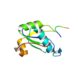
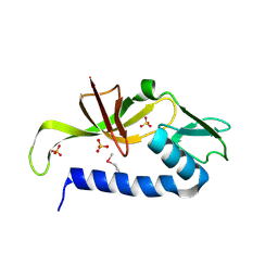
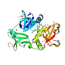
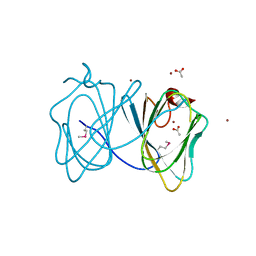
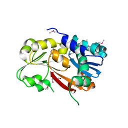
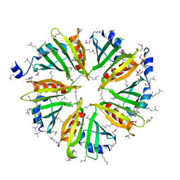
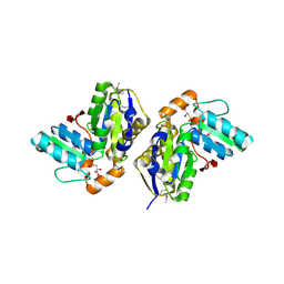
2EVR
DownloadVisualize
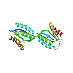
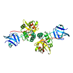
BU of 2evr by Molmil
CRYSTAL STRUCTURE OF A PUTATIVE GAMMA-D-GLUTAMYL-L-DIAMINO ACID ENDOPEPTIDASE (NPUN_R0659) FROM NOSTOC PUNCTIFORME PCC 73102 AT 1.60 A RESOLUTION
分子名称: 1,2-ETHANEDIOL, CHLORIDE ION, COG0791: Cell wall-associated hydrolases (invasion-associated proteins), ...
著者Joint Center for Structural Genomics (JCSG)
登録日2005-10-31
公開日2005-11-22
最終更新日2024-10-16
実験手法X-RAY DIFFRACTION (1.6 Å)
主引用文献Structural Basis of Murein Peptide Specificity of a gamma-D-Glutamyl-L-Diamino Acid Endopeptidase.
Structure, 17, 2009
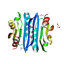
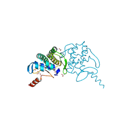
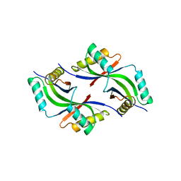
2FG0
DownloadVisualize
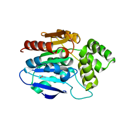
BU of 2fg0 by Molmil
Crystal structure of a putative gamma-d-glutamyl-l-diamino acid endopeptidase (npun_r0659) from nostoc punctiforme pcc 73102 at 1.79 A resolution
分子名称: COG0791: Cell wall-associated hydrolases (invasion-associated proteins), GLYCEROL
著者Joint Center for Structural Genomics (JCSG)
登録日2005-12-20
公開日2006-01-10
最終更新日2023-01-25
実験手法X-RAY DIFFRACTION (1.79 Å)
主引用文献Structural Basis of Murein Peptide Specificity of a gamma-D-Glutamyl-L-Diamino Acid Endopeptidase.
Structure, 17, 2009
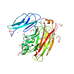
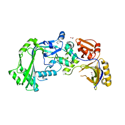
2FEA
DownloadVisualize
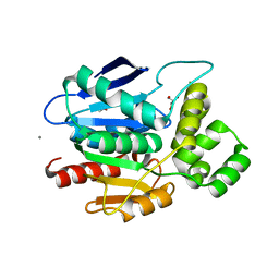
BU of 2fea by Molmil
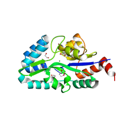
Crystal structure of MtnX phosphatase from Bacillus Subtilis at 2.00 A resolution
分子名称: 1,2-ETHANEDIOL, 2-hydroxy-3-keto-5-methylthiopentenyl-1-phosphate phosphatase, MAGNESIUM ION, ...
著者Joint Center for Structural Genomics (JCSG)
登録日2005-12-15
公開日2005-12-27
最終更新日2023-01-25
実験手法X-RAY DIFFRACTION (2 Å)
主引用文献Crystal structure of MtnX phosphatase from Bacillus subtilis at 2.0 A resolution provides a structural basis for bipartite phosphomonoester hydrolysis of 2-hydroxy-3-keto-5-methylthiopentenyl-1-phosphate.
Proteins, 69, 2007
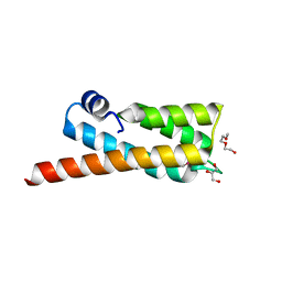
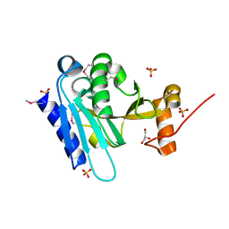
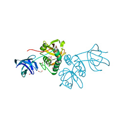
6W08
DownloadVisualize
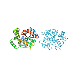
BU of 6w08 by Molmil
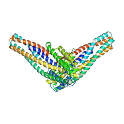
Crystal Structure of Motility Associated Killing Factor E from Vibrio cholerae
分子名称: 1,2-ETHANEDIOL, ACETIC ACID, CHLORIDE ION, ...
著者Kim, Y, Jedrzejczak, R, Joachimiak, G, Endres, M, Joachimiak, A, Center for Structural Genomics of Infectious Diseases (CSGID)
登録日2020-02-29
公開日2020-03-11
最終更新日2024-04-03
実験手法X-RAY DIFFRACTION (1.75 Å)
主引用文献A Genomic Island of Vibrio cholerae Encodes a Three-Component Cytotoxin with Monomer and Protomer Forms Structurally Similar to Alpha-Pore-Forming Toxins.
J.Bacteriol., 204, 2022
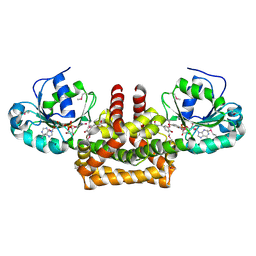
6W61
DownloadVisualize
BU of 6w61 by Molmil
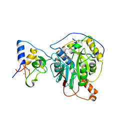
Crystal Structure of the methyltransferase-stimulatory factor complex of NSP16 and NSP10 from SARS CoV-2.
分子名称: 1,2-ETHANEDIOL, 2'-O-methyltransferase, CHLORIDE ION, ...
著者Kim, Y, Jedrzejczak, R, Maltseva, N, Endres, M, Godzik, A, Joachimiak, A, Center for Structural Genomics of Infectious Diseases (CSGID)
登録日2020-03-15
公開日2020-03-25
最終更新日2023-11-15
実験手法X-RAY DIFFRACTION (2 Å)
主引用文献The crystal structure of nsp10-nsp16 heterodimer from SARS-CoV-2 in complex with S-adenosylmethionine
Biorxiv, 2020
5CAG
DownloadVisualize
BU of 5cag by Molmil
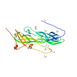
Crystal structure of a putative adhesin (BACOVA_02677) from Bacteroides ovatus ATCC 8483 at 3.00 A resolution (PSI Community Target, Nakayama)
分子名称: SULFATE ION, Uncharacterized protein
著者Joint Center for Structural Genomics (JCSG), Nakayama, K.
登録日2015-06-29
公開日2015-10-14
最終更新日2020-04-22
実験手法X-RAY DIFFRACTION (3 Å)
主引用文献A Distinct Type of Pilus from the Human Microbiome.
Cell, 165, 2016
5CXT
DownloadVisualize
BU of 5cxt by Molmil
Crystal structure of a RNA-binding protein 39 (RBM39) in complex with fragment of splicing factor (U2AF) from Unknown at 2.20 A resolution
分子名称: RNA-binding protein 39, Splicing factor U2AF 65 kDa subunit
著者Joint Center for Structural Genomics (JCSG), Partnership for T-Cell Biology (TCELL)
登録日2015-07-29
公開日2015-10-14
最終更新日2023-09-27
実験手法X-RAY DIFFRACTION (2.2 Å)
主引用文献UHM-ULM interactions in the RBM39-U2AF65 splicing-factor complex.
Acta Crystallogr D Struct Biol, 72, 2016
2OOK
DownloadVisualize
BU of 2ook by Molmil
Crystal structure of a protein with unknown function (YP_749275.1) from Shewanella Frigidimarina NCIMB 400 at 1.80 A resolution
分子名称: 1,2-ETHANEDIOL, Hypothetical protein
著者Joint Center for Structural Genomics (JCSG)
登録日2007-01-25
公開日2007-02-06
最終更新日2023-01-25
実験手法X-RAY DIFFRACTION (1.8 Å)
主引用文献Open and closed conformations of two SpoIIAA-like proteins (YP_749275.1 and YP_001095227.1) provide insights into membrane association and ligand binding.
Acta Crystallogr.,Sect.F, 66, 2010
2OOC
DownloadVisualize
BU of 2ooc by Molmil
Crystal structure of Histidine Phosphotransferase ShpA (NP_419930.1) from Caulobacter crescentus at 1.52 A resolution
分子名称: GLYCEROL, Histidine phosphotransferase, TETRAETHYLENE GLYCOL
著者Joint Center for Structural Genomics (JCSG)
登録日2007-01-25
公開日2007-02-06
最終更新日2024-10-30
実験手法X-RAY DIFFRACTION (1.52 Å)
主引用文献Crystal structure of histidine phosphotransfer protein ShpA, an essential regulator of stalk biogenesis in Caulobacter crescentus.
J.Mol.Biol., 390, 2009
2PV7
DownloadVisualize
BU of 2pv7 by Molmil
Crystal structure of chorismate mutase / prephenate dehydrogenase (tyrA) (1574749) from Haemophilus influenzae RD at 2.00 A resolution
分子名称: NICOTINAMIDE-ADENINE-DINUCLEOTIDE, T-protein [Includes: Chorismate mutase (EC 5.4.99.5) (CM) and Prephenate dehydrogenase (EC 1.3.1.12) (PDH)], TYROSINE
著者Joint Center for Structural Genomics (JCSG)
登録日2007-05-09
公開日2007-05-22
最終更新日2023-01-25
実験手法X-RAY DIFFRACTION (2 Å)
主引用文献The structure of Haemophilus influenzae prephenate dehydrogenase suggests unique features of bifunctional TyrA enzymes.
Acta Crystallogr.,Sect.F, 66, 2010
2Q3L
DownloadVisualize
BU of 2q3l by Molmil
CRYSTAL STRUCTURE OF AN UNCHARACTERIZED PROTEIN FROM DUF3478 FAMILY WITH A SPOIIAA-LIKE FOLD (SHEW_3102) FROM SHEWANELLA LOIHICA PV-4 AT 2.25 A RESOLUTION
分子名称: (4S)-2-METHYL-2,4-PENTANEDIOL, CHLORIDE ION, SODIUM ION, ...
著者Joint Center for Structural Genomics (JCSG)
登録日2007-05-30
公開日2007-06-12
最終更新日2023-01-25
実験手法X-RAY DIFFRACTION (2.25 Å)
主引用文献Open and closed conformations of two SpoIIAA-like proteins (YP_749275.1 and YP_001095227.1) provide insights into membrane association and ligand binding.
Acta Crystallogr.,Sect.F, 66, 2010
2XMQ
DownloadVisualize
BU of 2xmq by Molmil
Crystal structure of human NDRG2 protein provides insight into its role as a tumor suppressor
分子名称: ACETATE ION, PROTEIN NDRG2
著者Hwang, J, Kim, Y, Lee, H, Kim, M.H.
登録日2010-07-29
公開日2011-01-19
最終更新日2023-12-20
実験手法X-RAY DIFFRACTION (2.81 Å)
主引用文献Crystal Structure of Human Ndrg2 Protein Provides Insight Into its Role as a Tumor Suppressor.
J.Biol.Chem., 286, 2011
2XMR
DownloadVisualize
BU of 2xmr by Molmil
Crystal structure of human NDRG2 protein provides insight into its role as a tumor suppressor
分子名称: ACETATE ION, CALCIUM ION, GLYCEROL, ...
著者Hwang, J, Kim, Y, Lee, H, Kim, M.H.
登録日2010-07-29
公開日2011-01-19
最終更新日2023-12-20
実験手法X-RAY DIFFRACTION (2 Å)
主引用文献Crystal Structure of Human Ndrg2 Protein Provides Insight Into its Role as a Tumor Suppressor.
J.Biol.Chem., 286, 2011
2XMS
DownloadVisualize
BU of 2xms by Molmil
Crystal structure of human NDRG2 protein provides insight into its role as a tumor suppressor
分子名称: CHLORIDE ION, IMIDAZOLE, PROTEIN NDRG2
著者Hwang, J, Kim, Y, Lee, H, Kim, M.H.
登録日2010-07-29
公開日2011-01-19
最終更新日2023-12-20
実験手法X-RAY DIFFRACTION (2.15 Å)
主引用文献Crystal Structure of Human Ndrg2 Protein Provides Insight Into its Role as a Tumor Suppressor.
J.Biol.Chem., 286, 2011
3IRB
DownloadVisualize
BU of 3irb by Molmil
Crystal structure of protein with unknown function from DUF35 family (13815350) from SULFOLOBUS SOLFATARICUS at 1.80 A resolution
分子名称: ACETIC ACID, SULFATE ION, ZINC ION, ...
著者Joint Center for Structural Genomics (JCSG)
登録日2009-08-21
公開日2009-09-01
最終更新日2023-02-01
実験手法X-RAY DIFFRACTION (1.8 Å)
主引用文献The structure of SSO2064, the first representative of Pfam family PF01796, reveals a novel two-domain zinc-ribbon OB-fold architecture with a potential acyl-CoA-binding role.
Acta Crystallogr.,Sect.F, 66, 2010
3H41
DownloadVisualize
BU of 3h41 by Molmil
CRYSTAL STRUCTURE OF A NLPC/P60 FAMILY PROTEIN (BCE_2878) FROM BACILLUS CEREUS ATCC 10987 AT 1.79 A RESOLUTION
分子名称: ALANINE, D-GLUTAMIC ACID, NLP/P60 family protein, ...
著者Joint Center for Structural Genomics (JCSG)
登録日2009-04-17
公開日2009-05-05
最終更新日2023-02-01
実験手法X-RAY DIFFRACTION (1.79 Å)
主引用文献Structure of the gamma-D-glutamyl-L-diamino acid endopeptidase YkfC from Bacillus cereus in complex with L-Ala-[gamma]-D-Glu: insights into substrate recognition by NlpC/P60 cysteine peptidases
Acta Crystallogr.,Sect.F, 66, 2010
3H50
DownloadVisualize
BU of 3h50 by Molmil
CRYSTAL STRUCTURE OF A TETRACENOMYCIN POLYKETIDE SYNTHESIS PROTEIN (TCMJ) FROM XANTHOMONAS CAMPESTRIS PV. CAMPESTRIS AT 1.60 A RESOLUTION
分子名称: ACETATE ION, Tetracenomycin polyketide synthesis protein, ZINC ION
著者Joint Center for Structural Genomics (JCSG)
登録日2009-04-21
公開日2009-05-05
最終更新日2024-10-30
実験手法X-RAY DIFFRACTION (1.6 Å)
主引用文献Conformational changes associated with the binding of zinc acetate at the putative active site of XcTcmJ, a cupin from Xanthomonas campestris pv. campestris.
Acta Crystallogr.,Sect.F, 66, 2010
3L5O
DownloadVisualize
BU of 3l5o by Molmil
Crystal structure of protein with unknown function from DUF364 family (ZP_00559375.1) from Desulfitobacterium hafniense DCB-2 at 2.01 A resolution
分子名称: 1,2-ETHANEDIOL, CHLORIDE ION, IMIDAZOLE, ...
著者Joint Center for Structural Genomics, Joint Center for Structural Genomics (JCSG)
登録日2009-12-22
公開日2010-02-02
最終更新日2023-02-01
実験手法X-RAY DIFFRACTION (2.01 Å)
主引用文献Structure of the first representative of Pfam family PF04016 (DUF364) reveals enolase and Rossmann-like folds that combine to form a unique active site with a possible role in heavy-metal chelation.
Acta Crystallogr.,Sect.F, 66, 2010
3HSA
DownloadVisualize
BU of 3hsa by Molmil
Crystal structure of pleckstrin homology domain (YP_926556.1) from SHEWANELLA AMAZONENSIS SB2B at 1.99 A resolution
分子名称: DI(HYDROXYETHYL)ETHER, GLYCEROL, pleckstrin homology domain
著者Joint Center for Structural Genomics (JCSG)
登録日2009-06-10
公開日2009-06-23
最終更新日2023-02-01
実験手法X-RAY DIFFRACTION (1.99 Å)
主引用文献Bacterial pleckstrin homology domains: a prokaryotic origin for the PH domain.
J.Mol.Biol., 396, 2010
3H0N
DownloadVisualize
BU of 3h0n by Molmil
Crystal structure of a duf1470 family protein (jann_2411) from jannaschia sp. ccs1 at 1.45 A resolution
分子名称: ACETATE ION, GLYCEROL, NICKEL (II) ION, ...
著者Joint Center for Structural Genomics (JCSG)
登録日2009-04-09
公開日2009-04-21
最終更新日2023-02-01
実験手法X-RAY DIFFRACTION (1.45 Å)
主引用文献The structure of Jann_2411 (DUF1470) from Jannaschia sp. at 1.45  resolution reveals a new fold (the ABATE domain) and suggests its possible role as a transcription regulator.
Acta Crystallogr.,Sect.F, 66, 2010
3HBZ
DownloadVisualize
BU of 3hbz by Molmil
Crystal structure of a putative glycoside hydrolase (bt_2081) from bacteroides thetaiotaomicron vpi-5482 at 2.05 A resolution
分子名称: ACETATE ION, CACODYLATE ION, CALCIUM ION, ...
著者Joint Center for Structural Genomics (JCSG)
登録日2009-05-05
公開日2009-05-26
最終更新日2023-02-01
実験手法X-RAY DIFFRACTION (2.05 Å)
主引用文献Structure of Bacteroides thetaiotaomicron BT2081 at 2.05 A resolution: the first structural representative of a new protein family that may play a role in carbohydrate metabolism.
Acta Crystallogr.,Sect.F, 66, 2010
3K5J
DownloadVisualize
BU of 3k5j by Molmil
Crystal structure of Putative SUFU (suppressor of fused protein) homolog (YP_208451.1) from Neisseria gonorrhoeae FA 1090 at 1.40 A resolution
分子名称: GLYCEROL, SULFATE ION, Suppressor of fused family protein
著者Joint Center for Structural Genomics (JCSG)
登録日2009-10-07
公開日2010-01-26
最終更新日2019-07-17
実験手法X-RAY DIFFRACTION (1.4 Å)
主引用文献The crystal structure of a bacterial Sufu-like protein defines a novel group of bacterial proteins that are similar to the N-terminal domain of human Sufu.
Protein Sci., 19, 2010
1VR0
DownloadVisualize
BU of 1vr0 by Molmil
Crystal structure of putative 2-phosphosulfolactate phosphatase (15026306) from Clostridium acetobutylicum at 2.6 A resolution
分子名称: (2R)-3-SULFOLACTIC ACID, MAGNESIUM ION, Probable 2-phosphosulfolactate phosphatase
著者Joint Center for Structural Genomics (JCSG)
登録日2005-01-26
公開日2005-02-15
最終更新日2024-10-09
実験手法X-RAY DIFFRACTION (2.49 Å)
主引用文献Crystal structure of 2-phosphosulfolactate phosphatase (ComB) from Clostridium acetobutylicum at 2.6 A resolution reveals a new fold with a novel active site.
Proteins, 65, 2006
1VQ3
DownloadVisualize
BU of 1vq3 by Molmil
Crystal structure of Phosphoribosylformylglycinamidine synthase, purS subunit (EC 6.3.5.3) (TM1244) from Thermotoga maritima at 1.90 A resolution
分子名称: Phosphoribosylformylglycinamidine synthase, purS subunit
著者Joint Center for Structural Genomics (JCSG)
登録日2004-12-15
公開日2004-12-28
最終更新日2023-09-20
実験手法X-RAY DIFFRACTION (1.9 Å)
主引用文献Crystal structure of phosphoribosylformyl-glycinamidine synthase II, PurS subunit (TM1244) from Thermotoga maritima at 1.90 A resolution.
Proteins, 65, 2006
3KK7
DownloadVisualize
BU of 3kk7 by Molmil
Crystal structure of Putative cell invasion protein with MAC/Perforin domain (NP_812351.1) from BACTERIODES THETAIOTAOMICRON VPI-5482 at 2.46 A resolution
分子名称: (4S)-2-METHYL-2,4-PENTANEDIOL, 1,2-ETHANEDIOL, CHLORIDE ION, ...
著者Joint Center for Structural Genomics (JCSG)
登録日2009-11-04
公開日2009-11-24
最終更新日2023-02-01
実験手法X-RAY DIFFRACTION (2.46 Å)
主引用文献Structure of a membrane-attack complex/perforin (MACPF) family protein from the human gut symbiont Bacteroides thetaiotaomicron.
Acta Crystallogr.,Sect.F, 66, 2010

226707

件を2024-10-30に公開中

PDB statisticsPDBj update infoContact PDBjnumon