Loading
PDBj
メニューPDBj@FacebookPDBj@TwitterPDBj@YouTubewwPDB FoundationwwPDB
RCSB PDBPDBeBMRBAdv. SearchSearch help
Search by PDB author
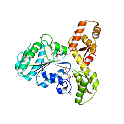
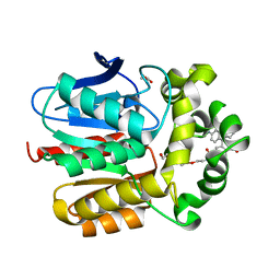
7ZIZ
DownloadVisualize
BU of 7ziz by Molmil
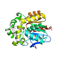
X-ray structure of the dead variant haloalkane dehalogenase HaloTag7-D106A bound to a pentanol tetramethylrhodamine ligand (TMR-Hy5)
分子名称: CHLORIDE ION, GLYCEROL, Haloalkane dehalogenase, ...
著者Tarnawski, M, Kompa, J, Johnsson, K, Hiblot, J.
登録日2022-04-08
公開日2023-02-22
最終更新日2024-02-07
実験手法X-RAY DIFFRACTION (1.5 Å)
主引用文献Exchangeable HaloTag Ligands for Super-Resolution Fluorescence Microscopy.
J.Am.Chem.Soc., 145, 2023
7ZJ0
DownloadVisualize
BU of 7zj0 by Molmil
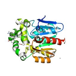
X-ray structure of the haloalkane dehalogenase HaloTag7 bound to a pentylmethanesulfonamide tetramethylrhodamine ligand (TMR-S5)
分子名称: GLYCEROL, Haloalkane dehalogenase, [9-[2-carboxy-5-[2-[2-[5-(methylsulfonylamino)pentoxy]ethoxy]ethylcarbamoyl]phenyl]-6-(dimethylamino)xanthen-3-ylidene]-dimethyl-azanium
著者Tarnawski, M, Kompa, J, Johnsson, K, Hiblot, J.
登録日2022-04-08
公開日2023-02-22
最終更新日2024-02-07
実験手法X-RAY DIFFRACTION (1.5 Å)
主引用文献Exchangeable HaloTag Ligands for Super-Resolution Fluorescence Microscopy.
J.Am.Chem.Soc., 145, 2023
7ZIY
DownloadVisualize
BU of 7ziy by Molmil
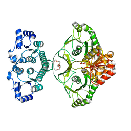
X-ray structure of the haloalkane dehalogenase HaloTag7 bound to a pentyltrifluoromethanesulfonamide tetramethylrhodamine ligand (TMR-T5)
分子名称: CALCIUM ION, Haloalkane dehalogenase, [9-[2-carboxy-5-[2-[2-[5-(trifluoromethylsulfonylamino)pentoxy]ethoxy]ethylcarbamoyl]phenyl]-6-(dimethylamino)xanthen-3-ylidene]-dimethyl-azanium
著者Tarnawski, M, Kompa, J, Johnsson, K, Hiblot, J.
登録日2022-04-08
公開日2023-02-22
最終更新日2024-02-07
実験手法X-RAY DIFFRACTION (1.7 Å)
主引用文献Exchangeable HaloTag Ligands for Super-Resolution Fluorescence Microscopy.
J.Am.Chem.Soc., 145, 2023
1ZCZ
DownloadVisualize
BU of 1zcz by Molmil
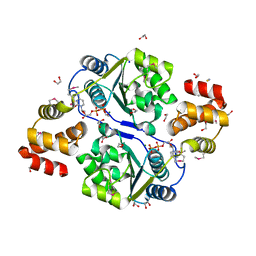
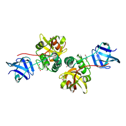
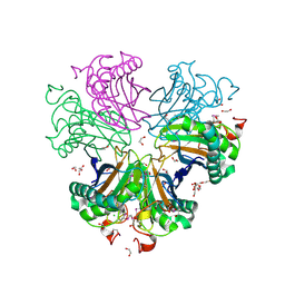
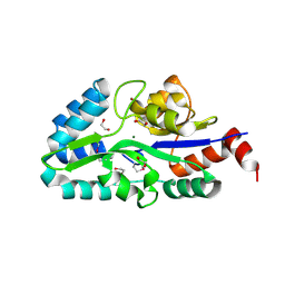
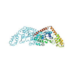
Crystal structure of Phosphoribosylaminoimidazolecarboxamide formyltransferase / IMP cyclohydrolase (TM1249) from THERMOTOGA MARITIMA at 1.88 A resolution
分子名称: Bifunctional purine biosynthesis protein purH, POTASSIUM ION, TETRAETHYLENE GLYCOL
著者Joint Center for Structural Genomics (JCSG)
登録日2005-04-13
公開日2005-04-26
最終更新日2023-09-20
実験手法X-RAY DIFFRACTION (1.88 Å)
主引用文献Crystal structure of AICAR transformylase IMP cyclohydrolase (TM1249) from Thermotoga maritima at 1.88 A resolution.
Proteins, 71, 2008
3BOS
DownloadVisualize
BU of 3bos by Molmil
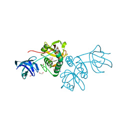
Crystal structure of a putative dna replication regulator HDA (SAMA_1916) from Shewanella amazonensis sb2b at 1.75 A resolution
分子名称: 1,2-ETHANEDIOL, CHLORIDE ION, CYTIDINE-5'-DIPHOSPHATE, ...
著者Joint Center for Structural Genomics (JCSG)
登録日2007-12-17
公開日2008-01-15
最終更新日2023-01-25
実験手法X-RAY DIFFRACTION (1.75 Å)
主引用文献A structural basis for the regulatory inactivation of DnaA.
J.Mol.Biol., 385, 2009
1ZX8
DownloadVisualize
BU of 1zx8 by Molmil
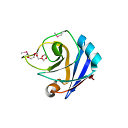
CRYSTAL STRUCTURE OF an atypical cyclophilin (peptidylprolyl cis-trans isomerase) (TM1367) FROM THERMOTOGA MARITIMA AT 1.90 A RESOLUTION
分子名称: NICKEL (II) ION, PENTAETHYLENE GLYCOL, hypothetical protein TM1367
著者Joint Center for Structural Genomics (JCSG)
登録日2005-06-07
公開日2005-07-26
最終更新日2023-01-25
実験手法X-RAY DIFFRACTION (1.9 Å)
主引用文献Crystal structure of TM1367 from Thermotoga maritima at 1.90 A resolution reveals an atypical member of the cyclophilin (peptidylprolyl isomerase) fold.
Proteins, 63, 2006
1Z9F
DownloadVisualize
BU of 1z9f by Molmil
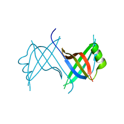
Crystal structure of single stranded DNA-binding protein (TM0604) from Thermotoga maritima at 2.60 A resolution
分子名称: Single-strand binding protein
著者Joint Center for Structural Genomics (JCSG)
登録日2005-04-01
公開日2005-04-12
最終更新日2023-09-20
実験手法X-RAY DIFFRACTION (2.3 Å)
主引用文献Crystal structure of a single-stranded DNA-binding protein (TM0604) from Thermotoga maritima at 2.60 A resolution.
Proteins, 63, 2006
2FG0
DownloadVisualize
BU of 2fg0 by Molmil
Crystal structure of a putative gamma-d-glutamyl-l-diamino acid endopeptidase (npun_r0659) from nostoc punctiforme pcc 73102 at 1.79 A resolution
分子名称: COG0791: Cell wall-associated hydrolases (invasion-associated proteins), GLYCEROL
著者Joint Center for Structural Genomics (JCSG)
登録日2005-12-20
公開日2006-01-10
最終更新日2023-01-25
実験手法X-RAY DIFFRACTION (1.79 Å)
主引用文献Structural Basis of Murein Peptide Specificity of a gamma-D-Glutamyl-L-Diamino Acid Endopeptidase.
Structure, 17, 2009
2EVR
DownloadVisualize
BU of 2evr by Molmil
CRYSTAL STRUCTURE OF A PUTATIVE GAMMA-D-GLUTAMYL-L-DIAMINO ACID ENDOPEPTIDASE (NPUN_R0659) FROM NOSTOC PUNCTIFORME PCC 73102 AT 1.60 A RESOLUTION
分子名称: 1,2-ETHANEDIOL, CHLORIDE ION, COG0791: Cell wall-associated hydrolases (invasion-associated proteins), ...
著者Joint Center for Structural Genomics (JCSG)
登録日2005-10-31
公開日2005-11-22
最終更新日2023-01-25
実験手法X-RAY DIFFRACTION (1.6 Å)
主引用文献Structural Basis of Murein Peptide Specificity of a gamma-D-Glutamyl-L-Diamino Acid Endopeptidase.
Structure, 17, 2009
1ZKG
DownloadVisualize
BU of 1zkg by Molmil
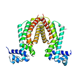
Crystal structure of Transcriptional regulator, TETR family (tm1030) from Thermotoga maritima at 2.30 A resolution
分子名称: UNKNOWN LIGAND, transcriptional regulator, TetR family
著者Joint Center for Structural Genomics (JCSG)
登録日2005-05-02
公開日2005-07-26
最終更新日2023-01-25
実験手法X-RAY DIFFRACTION (2.3 Å)
主引用文献Crystal structure of TM1030 from Thermotoga maritima at 2.3 A resolution reveals molecular details of its transcription repressor function.
Proteins, 68, 2007
4AYK
DownloadVisualize
BU of 4ayk by Molmil
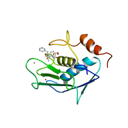
CATALYTIC FRAGMENT OF HUMAN FIBROBLAST COLLAGENASE COMPLEXED WITH CGS-27023A, NMR, 30 STRUCTURES
分子名称: CALCIUM ION, N-HYDROXY-2(R)-[[(4-METHOXYPHENYL)SULFONYL](3-PICOLYL)AMINO]-3-METHYLBUTANAMIDE HYDROCHLORIDE, PROTEIN (COLLAGENASE), ...
著者Powers, R, Moy, F.J.
登録日1999-02-01
公開日1999-06-05
最終更新日2023-12-27
実験手法SOLUTION NMR
主引用文献NMR solution structure of the catalytic fragment of human fibroblast collagenase complexed with a sulfonamide derivative of a hydroxamic acid compound.
Biochemistry, 38, 1999
2FNO
DownloadVisualize
BU of 2fno by Molmil
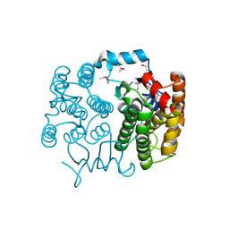
Crystal structure of a glutathione s-transferase (atu5508) from agrobacterium tumefaciens str. c58 at 2.00 A resolution
分子名称: AGR_pAT_752p, THIOCYANATE ION
著者Joint Center for Structural Genomics (JCSG)
登録日2006-01-11
公開日2006-02-14
最終更新日2023-01-25
実験手法X-RAY DIFFRACTION (2 Å)
主引用文献Comparative structural analysis of a novel glutathioneS-transferase (ATU5508) from Agrobacterium tumefaciens at 2.0 A resolution.
Proteins, 65, 2006
3BYQ
DownloadVisualize
BU of 3byq by Molmil
Crystal structure of a duf1185 family protein (bb2672) from bordetella bronchiseptica rb50 at 1.70 A resolution
分子名称: 1,2-ETHANEDIOL, CHLORIDE ION, TETRAETHYLENE GLYCOL, ...
著者Joint Center for Structural Genomics (JCSG)
登録日2008-01-16
公開日2008-01-29
最終更新日2023-02-01
実験手法X-RAY DIFFRACTION (1.7 Å)
主引用文献Structures of the first representatives of Pfam family PF06684 (DUF1185) reveal a novel variant of the Bacillus chorismate mutase fold and suggest a role in amino-acid metabolism.
Acta Crystallogr.,Sect.F, 66, 2010
2FEA
DownloadVisualize
BU of 2fea by Molmil
Crystal structure of MtnX phosphatase from Bacillus Subtilis at 2.00 A resolution
分子名称: 1,2-ETHANEDIOL, 2-hydroxy-3-keto-5-methylthiopentenyl-1-phosphate phosphatase, MAGNESIUM ION, ...
著者Joint Center for Structural Genomics (JCSG)
登録日2005-12-15
公開日2005-12-27
最終更新日2023-01-25
実験手法X-RAY DIFFRACTION (2 Å)
主引用文献Crystal structure of MtnX phosphatase from Bacillus subtilis at 2.0 A resolution provides a structural basis for bipartite phosphomonoester hydrolysis of 2-hydroxy-3-keto-5-methylthiopentenyl-1-phosphate.
Proteins, 69, 2007
1VR8
DownloadVisualize
BU of 1vr8 by Molmil
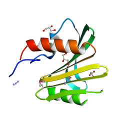
Crystal structure of GTP binding regulator (TM1622) from Thermotoga Maritima at 1.75 A resolution
分子名称: AZIDE ION, GLYCEROL, GTP binding regulator
著者Joint Center for Structural Genomics (JCSG)
登録日2005-02-16
公開日2005-03-15
最終更新日2023-01-25
実験手法X-RAY DIFFRACTION (1.75 Å)
主引用文献Crystal structure of an ORFan protein (TM1622) from Thermotoga maritima at 1.75 A resolution reveals a fold similar to the Ran-binding protein Mog1p.
Proteins, 65, 2006
1VK9
DownloadVisualize
BU of 1vk9 by Molmil
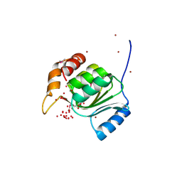
CRYSTAL STRUCTURE OF A DUF1893 family protein (TM1506) FROM THERMOTOGA MARITIMA AT 2.70 A RESOLUTION
分子名称: UNKNOWN LIGAND, ZINC ION, conserved hypothetical protein TM1506
著者Joint Center for Structural Genomics (JCSG)
登録日2004-05-06
公開日2004-05-18
最終更新日2023-01-25
実験手法X-RAY DIFFRACTION (2.7 Å)
主引用文献Crystal structure of an ADP-ribosylated protein with a cytidine deaminase-like fold, but unknown function (TM1506), from Thermotoga maritima at 2.70 A resolution.
Proteins, 71, 2008
2A6A
DownloadVisualize
BU of 2a6a by Molmil
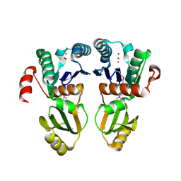
Crystal structure of Glycoprotein endopeptidase (tm0874) from THERMOTOGA MARITIMA at 2.50 A resolution
分子名称: UNKNOWN LIGAND, hypothetical protein TM0874
著者Joint Center for Structural Genomics (JCSG)
登録日2005-07-02
公開日2005-07-19
最終更新日2023-01-25
実験手法X-RAY DIFFRACTION (2.5 Å)
主引用文献Structure of an essential bacterial protein YeaZ (TM0874) from Thermotoga maritima at 2.5 A resolution.
Acta Crystallogr.,Sect.F, 66, 2010
2FNA
DownloadVisualize
BU of 2fna by Molmil
Crystal structure of an archaeal aaa+ atpase (sso1545) from sulfolobus solfataricus p2 at 2.00 A resolution
分子名称: 1,2-ETHANEDIOL, ADENOSINE-5'-DIPHOSPHATE, Conserved hypothetical protein, ...
著者Joint Center for Structural Genomics (JCSG)
登録日2006-01-10
公開日2006-02-07
最終更新日2023-01-25
実験手法X-RAY DIFFRACTION (2 Å)
主引用文献Crystal structure of a novel archaeal AAA+ ATPase SSO1545 from Sulfolobus solfataricus.
Proteins, 74, 2009
2G36
DownloadVisualize
BU of 2g36 by Molmil
Crystal structure of Tryptophanyl-tRNA synthetase (EC 6.1.1.2) (Tryptophan-tRNA ligase)(TrpRS) (tm0492) from THERMOTOGA MARITIMA at 2.50 A resolution
分子名称: IRON/SULFUR CLUSTER, TRYPTOPHAN, Tryptophanyl-tRNA synthetase
著者Joint Center for Structural Genomics (JCSG)
登録日2006-02-17
公開日2006-03-28
最終更新日2024-04-03
実験手法X-RAY DIFFRACTION (2.5 Å)
主引用文献Structure of a tryptophanyl-tRNA synthetase containing an iron-sulfur cluster.
Acta Crystallogr.,Sect.F, 66, 2010
7B8Q
DownloadVisualize
BU of 7b8q by Molmil
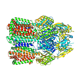
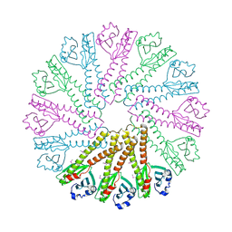
Acinetobacter baumannii multidrug transporter AdeB in L*OO state
分子名称: Efflux pump membrane transporter
著者Ornik-Cha, A, Reitz, J, Seybert, A, Frangakis, A, Pos, K.M.
登録日2020-12-13
公開日2021-10-20
最終更新日2024-07-10
実験手法ELECTRON MICROSCOPY (3.84 Å)
主引用文献Structural and functional analysis of the promiscuous AcrB and AdeB efflux pumps suggests different drug binding mechanisms.
Nat Commun, 12, 2021
7B8P
DownloadVisualize
BU of 7b8p by Molmil
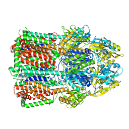
Acinetobacter baumannii multidrug transporter AdeB in OOO state
分子名称: Efflux pump membrane transporter
著者Ornik-Cha, A, Reitz, J, Seybert, A, Frangakis, A, Pos, K.M.
登録日2020-12-13
公開日2021-10-20
最終更新日2024-07-10
実験手法ELECTRON MICROSCOPY (3.54 Å)
主引用文献Structural and functional analysis of the promiscuous AcrB and AdeB efflux pumps suggests different drug binding mechanisms.
Nat Commun, 12, 2021
2ETS
DownloadVisualize
BU of 2ets by Molmil
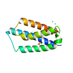
CRYSTAL STRUCTURE OF A BACTERIAL DOMAIN OF UNKNOWN FUNCTION FROM DUF1798 FAMILY (MW1337) FROM STAPHYLOCOCCUS AUREUS SUBSP. AUREUS AT 2.25 A RESOLUTION
分子名称: CHLORIDE ION, PHOSPHATE ION, hypothetical protein
著者Joint Center for Structural Genomics (JCSG)
登録日2005-10-27
公開日2005-11-08
最終更新日2023-01-25
実験手法X-RAY DIFFRACTION (2.25 Å)
主引用文献Crystal structures of MW1337R and lin2004: representatives of a novel protein family that adopt a four-helical bundle fold.
Proteins, 71, 2008
3IRB
DownloadVisualize
BU of 3irb by Molmil
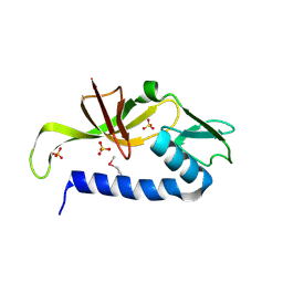
Crystal structure of protein with unknown function from DUF35 family (13815350) from SULFOLOBUS SOLFATARICUS at 1.80 A resolution
分子名称: ACETIC ACID, SULFATE ION, ZINC ION, ...
著者Joint Center for Structural Genomics (JCSG)
登録日2009-08-21
公開日2009-09-01
最終更新日2023-02-01
実験手法X-RAY DIFFRACTION (1.8 Å)
主引用文献The structure of SSO2064, the first representative of Pfam family PF01796, reveals a novel two-domain zinc-ribbon OB-fold architecture with a potential acyl-CoA-binding role.
Acta Crystallogr.,Sect.F, 66, 2010
3K5J
DownloadVisualize
BU of 3k5j by Molmil
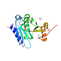
Crystal structure of Putative SUFU (suppressor of fused protein) homolog (YP_208451.1) from Neisseria gonorrhoeae FA 1090 at 1.40 A resolution
分子名称: GLYCEROL, SULFATE ION, Suppressor of fused family protein
著者Joint Center for Structural Genomics (JCSG)
登録日2009-10-07
公開日2010-01-26
最終更新日2019-07-17
実験手法X-RAY DIFFRACTION (1.4 Å)
主引用文献The crystal structure of a bacterial Sufu-like protein defines a novel group of bacterial proteins that are similar to the N-terminal domain of human Sufu.
Protein Sci., 19, 2010
3B77
DownloadVisualize
BU of 3b77 by Molmil
Crystal structure of a ph domain containing bacterial protein (exig_2160) from exiguobacterium sibiricum 255-15 at 2.42 A resolution
分子名称: Uncharacterized protein
著者Joint Center for Structural Genomics (JCSG)
登録日2007-10-30
公開日2007-11-13
最終更新日2023-01-25
実験手法X-RAY DIFFRACTION (2.42 Å)
主引用文献Bacterial pleckstrin homology domains: a prokaryotic origin for the PH domain.
J.Mol.Biol., 396, 2010

223790

件を2024-08-14に公開中

PDB statisticsPDBj update infoContact PDBjnumon