5AKO
 
 | |
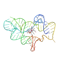
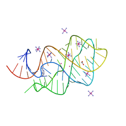
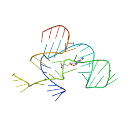
2XNW
 | | XPT-PBUX C74U RIBOSWITCH FROM B. SUBTILIS BOUND TO A TRIAZOLO- TRIAZOLE-DIAMINE LIGAND IDENTIFIED BY VIRTUAL SCREENING | | 分子名称: | 3,6-diamino-1,5-dihydro[1,2,4]triazolo[4,3-b][1,2,4]triazol-4-ium, ACETATE ION, COBALT HEXAMMINE(III), ... | | 著者 | Daldrop, P, Reyes, F.E, Robinson, D.A, Hammond, C.M, Lilley, D.M.J, Brenk, R. | | 登録日 | 2010-08-06 | | 公開日 | 2011-04-06 | | 最終更新日 | 2023-12-20 | | 実験手法 | X-RAY DIFFRACTION (1.5 Å) | | 主引用文献 | Novel ligands for a purine riboswitch discovered by RNA-ligand docking.
Chem. Biol., 18, 2011
|
|
2K1P
 | |
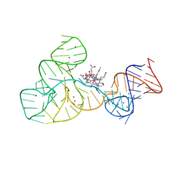
2XNZ
 | | xpt-pbuX C74U Riboswitch from B. subtilis bound to acetoguanamine identified by virtual screening | | 分子名称: | 6-METHYL-1,3,5-TRIAZINE-2,4-DIAMINE, ACETATE ION, COBALT HEXAMMINE(III), ... | | 著者 | Daldrop, P, Reyes, F.E, Robinson, D.A, Hammond, C.M, Lilley, D.M.J, Batey, R.T, Brenk, R. | | 登録日 | 2010-08-06 | | 公開日 | 2011-04-06 | | 最終更新日 | 2023-12-20 | | 実験手法 | X-RAY DIFFRACTION (1.59 Å) | | 主引用文献 | Novel ligands for a purine riboswitch discovered by RNA-ligand docking.
Chem. Biol., 18, 2011
|
|
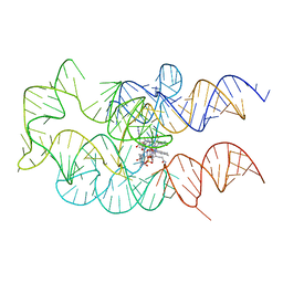
4FRN
 | | Crystal structure of the cobalamin riboswitch regulatory element | | 分子名称: | BARIUM ION, Cobalamin riboswitch aptamer domain, Hydroxocobalamin | | 著者 | Reyes, F.E, Johnson, J.E, Polaski, J.T, Batey, R.T. | | 登録日 | 2012-06-26 | | 公開日 | 2012-10-17 | | 最終更新日 | 2023-09-13 | | 実験手法 | X-RAY DIFFRACTION (3.43 Å) | | 主引用文献 | B12 cofactors directly stabilize an mRNA regulatory switch.
Nature, 492, 2012
|
|
4FRG
 | | Crystal structure of the cobalamin riboswitch aptamer domain | | 分子名称: | Hydroxocobalamin, IRIDIUM (III) ION, MAGNESIUM ION, ... | | 著者 | Reyes, F.E, Johnson, J.E, Polaski, J.T, Batey, R.T. | | 登録日 | 2012-06-26 | | 公開日 | 2012-10-17 | | 最終更新日 | 2024-02-28 | | 実験手法 | X-RAY DIFFRACTION (2.95 Å) | | 主引用文献 | B12 cofactors directly stabilize an mRNA regulatory switch.
Nature, 492, 2012
|
|
3FO6
 | | Crystal structure of guanine riboswitch bound to 6-O-methylguanine | | 分子名称: | 6-O-methylguanine, ACETATE ION, COBALT HEXAMMINE(III), ... | | 著者 | Gilbert, S.D, Reyes, F.E, Batey, R.T. | | 登録日 | 2008-12-28 | | 公開日 | 2009-06-23 | | 最終更新日 | 2023-09-06 | | 実験手法 | X-RAY DIFFRACTION (1.9 Å) | | 主引用文献 | Adaptive ligand binding by the purine riboswitch in the recognition of Guanine and adenine analogs.
Structure, 17, 2009
|
|
3FCT
 | | MATURE METAL CHELATASE CATALYTIC ANTIBODY WITH HAPTEN | | 分子名称: | CADMIUM ION, CALCIUM ION, MAGNESIUM ION, ... | | 著者 | Romesberg, F.E, Santarsiero, B.D, Barnes, D, Yin, J, Spiller, B, Schultz, P.G, Stevens, R.C. | | 登録日 | 1999-06-13 | | 公開日 | 1999-06-23 | | 最終更新日 | 2024-11-20 | | 実験手法 | X-RAY DIFFRACTION (2.4 Å) | | 主引用文献 | Structural and kinetic evidence for strain in biological catalysis.
Biochemistry, 37, 1998
|
|
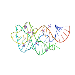
2XO1
 | | xpt-pbuX C74U Riboswitch from B. subtilis bound to N6-methyladenine | | 分子名称: | ACETATE ION, COBALT HEXAMMINE(III), Guanine riboswitch, ... | | 著者 | Daldrop, P, Reyes, F.E, Robinson, D.A, Hammond, C.M, Lilley, D.M.J, Batey, R.T, Brenk, R. | | 登録日 | 2010-08-09 | | 公開日 | 2011-04-06 | | 最終更新日 | 2024-05-08 | | 実験手法 | X-RAY DIFFRACTION (1.6 Å) | | 主引用文献 | Novel ligands for a purine riboswitch discovered by RNA-ligand docking.
Chem. Biol., 18, 2011
|
|
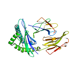
3F5H
 | | Crystal structure of fused docking domains from PikAIII and PikAIV of the pikromycin polyketide synthase | | 分子名称: | SODIUM ION, Type I polyketide synthase PikAIII, Type I polyketide synthase PikAIV fusion protein | | 著者 | Buchholz, T.J, Geders, T.W, Bartley, F.E, Reynolds, K.A, Smith, J.L, Sherman, D.H. | | 登録日 | 2008-11-03 | | 公開日 | 2009-01-27 | | 最終更新日 | 2023-12-27 | | 実験手法 | X-RAY DIFFRACTION (1.75 Å) | | 主引用文献 | Structural basis for binding specificity between subclasses of modular polyketide synthase docking domains.
Acs Chem.Biol., 4, 2009
|
|
5AMT
 | | Intracellular growth locus protein E | | 分子名称: | 1,2-ETHANEDIOL, BROMIDE ION, IGLE | | 著者 | Robb, C.S, Nano, F.E, Boraston, A.B. | | 登録日 | 2015-09-01 | | 公開日 | 2016-10-05 | | 最終更新日 | 2024-05-08 | | 実験手法 | X-RAY DIFFRACTION (1.62 Å) | | 主引用文献 | Cloning, Expression, Purification, Crystallization and Preliminary X-Ray Diffraction Analysis of Intracellular Growth Locus E (Igle) Protein from Francisella Tularensis Subsp. Novicida.
Acta Crystallogr.,Sect.F, 66, 2010
|
|
3BW9
 | | Crystal Structure of HLA B*3508 in complex with a HCMV 12-mer peptide from the pp65 protein | | 分子名称: | Beta-2-microglobulin, CPS peptide from 65 kDa lower matrix phosphoprotein, HLA class I histocompatibility antigen, ... | | 著者 | Wynn, K.K, Marland, Z, Cooper, L, Silins, S.L, Gras, S, Archbold, J.K, Tynan, F.E, Miles, J.J, McCluskey, J, Burrows, S.R, Rossjohn, J, Khanna, R. | | 登録日 | 2008-01-08 | | 公開日 | 2008-04-22 | | 最終更新日 | 2024-10-09 | | 実験手法 | X-RAY DIFFRACTION (1.75 Å) | | 主引用文献 | Impact of clonal competition for peptide-MHC complexes on the CD8+ T-cell repertoire selection in a persistent viral infection
Blood, 111, 2008
|
|
4GMA
 | | Crystal structure of the adenosylcobalamin riboswitch | | 分子名称: | Adenosylcobalamin, Adenosylcobalamin riboswitch | | 著者 | Reyes, F.E, Johnson, J.E, Polaski, J.T, Batey, R.T. | | 登録日 | 2012-08-15 | | 公開日 | 2012-10-17 | | 最終更新日 | 2024-02-28 | | 実験手法 | X-RAY DIFFRACTION (3.94 Å) | | 主引用文献 | B12 cofactors directly stabilize an mRNA regulatory switch.
Nature, 492, 2012
|
|
3BWA
 | | Crystal Structure of HLA B*3508 in complex with a HCMV 8-mer peptide from the pp65 protein | | 分子名称: | Beta-2-microglobulin, FPT peptide from 65 kDa lower matrix phosphoprotein, HLA class I histocompatibility antigen, ... | | 著者 | Wynn, K.K, Marland, Z, Cooper, L, Silins, S.L, Gras, S, Archbold, J.K, Tynan, F.E, Miles, J.J, McCluskey, J, Burrows, S.R, Rossjohn, J, Khanna, R. | | 登録日 | 2008-01-08 | | 公開日 | 2008-04-22 | | 最終更新日 | 2024-10-16 | | 実験手法 | X-RAY DIFFRACTION (1.3 Å) | | 主引用文献 | Impact of clonal competition for peptide-MHC complexes on the CD8+ T-cell repertoire selection in a persistent viral infection
Blood, 111, 2008
|
|
5AMU
 | | IglE I39A,Y40A,V44A | | 分子名称: | IGLE | | 著者 | Robb, C.S, Nano, F.E, Boraston, A.B. | | 登録日 | 2015-09-01 | | 公開日 | 2016-10-05 | | 最終更新日 | 2024-01-10 | | 実験手法 | X-RAY DIFFRACTION (2.2 Å) | | 主引用文献 | The Structure, Dimerisation and Impact on Intramacrophage Replication of Igle in Francisella Novicida
To be Published
|
|
2LI8
 | |
2LHO
 | | Solution Structure of a DNA duplex Containing an Unnatural, Hydrophobic Base Pair | | 分子名称: | DNA (5'-D(*CP*GP*TP*TP*TP*CP*(LHO)P*TP*TP*CP*TP*C)-3'), DNA (5'-D(*GP*AP*GP*AP*AP*(MM7)P*GP*AP*AP*AP*CP*G)-3') | | 著者 | Malyshev, D.A, Pfaff, D.A, Ippoliti, S.L, Hwang, G.T, Dwyer, T.J, Romesberg, F.E. | | 登録日 | 2011-08-12 | | 公開日 | 2012-07-04 | | 最終更新日 | 2024-05-01 | | 実験手法 | SOLUTION NMR | | 主引用文献 | Solution structure, mechanism of replication, and optimization of an unnatural base pair.
Chemistry, 16, 2010
|
|
1A4J
 | | DIELS ALDER CATALYTIC ANTIBODY GERMLINE PRECURSOR | | 分子名称: | IMMUNOGLOBULIN, DIELS ALDER CATALYTIC ANTIBODY (HEAVY CHAIN), DIELS ALDER CATALYTIC ANTIBODY (LIGHT CHAIN) | | 著者 | Spiller, B.W, Romesburg, F.E, Schultz, P.G, Stevens, R.C. | | 登録日 | 1998-01-30 | | 公開日 | 1998-05-13 | | 最終更新日 | 2024-10-30 | | 実験手法 | X-RAY DIFFRACTION (2.1 Å) | | 主引用文献 | Immunological origins of binding and catalysis in a Diels-Alderase antibody.
Science, 279, 1998
|
|
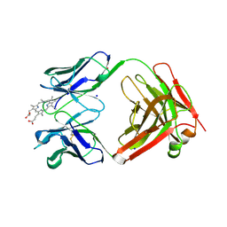
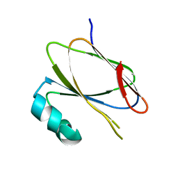
3G9Y
 | | Crystal structure of the second zinc finger from ZRANB2/ZNF265 bound to 6 nt ssRNA sequence AGGUAA | | 分子名称: | RNA (5'-R(*AP*GP*GP*UP*AP*A)-3'), ZINC ION, Zinc finger Ran-binding domain-containing protein 2 | | 著者 | Loughlin, F.E, McGrath, A.P, Lee, M, Guss, J.M, Mackay, J.P. | | 登録日 | 2009-02-15 | | 公開日 | 2009-03-03 | | 最終更新日 | 2024-03-20 | | 実験手法 | X-RAY DIFFRACTION (1.4 Å) | | 主引用文献 | The zinc fingers of the SR-like protein ZRANB2 are single-stranded RNA-binding domains that recognize 5' splice site-like sequences
Proc.Natl.Acad.Sci.USA, 106, 2009
|
|
1A4K
 | | DIELS ALDER CATALYTIC ANTIBODY WITH TRANSITION STATE ANALOGUE | | 分子名称: | ANTIBODY FAB, CADMIUM ION, [4-(4-ACETYLAMINO-PHENYL)-3,5-DIOXO-4-AZA-TRICYCLO[5.2.2.0 2,6]UNDEC-1-YLCARBAMOYLOXY]-ACETIC ACID | | 著者 | Spiller, B.W, Romesburg, F.E, Schultz, P.G, Stevens, R.C. | | 登録日 | 1998-01-30 | | 公開日 | 1998-05-13 | | 最終更新日 | 2024-10-23 | | 実験手法 | X-RAY DIFFRACTION (2.4 Å) | | 主引用文献 | Immunological origins of binding and catalysis in a Diels-Alderase antibody.
Science, 279, 1998
|
|
1B07
 | | CRK SH3 DOMAIN COMPLEXED WITH PEPTOID INHIBITOR | | 分子名称: | PHENYLETHANE, PROTEIN (PROTO-ONCOGENE CRK (CRK)), PROTEIN (SH3 PEPTOID INHIBITOR) | | 著者 | Nguyen, J.T, Turck, C.W, Cohen, F.E, Zuckermann, R.N, Lim, W.A. | | 登録日 | 1998-11-17 | | 公開日 | 1999-01-06 | | 最終更新日 | 2024-10-30 | | 実験手法 | X-RAY DIFFRACTION (2.5 Å) | | 主引用文献 | Exploiting the basis of proline recognition by SH3 and WW domains: design of N-substituted inhibitors.
Science, 282, 1998
|
|
3NPQ
 | |
3NPN
 | |
1I17
 | | NMR STRUCTURE OF MOUSE DOPPEL 51-157 | | 分子名称: | PRION-LIKE PROTEIN | | 著者 | Mo, H, Moore, R.C, Cohen, F.E, Westaway, D, Prusiner, S.B, Wright, P.E, Dyson, H.J. | | 登録日 | 2001-01-31 | | 公開日 | 2001-03-07 | | 最終更新日 | 2024-10-30 | | 実験手法 | SOLUTION NMR | | 主引用文献 | Two different neurodegenerative diseases caused by proteins with similar structures.
Proc.Natl.Acad.Sci.USA, 98, 2001
|
|
8EEX
 | | Cas7-11 in complex with Csx29 | | 分子名称: | Cas7-11, Csx29, ZINC ION, ... | | 著者 | Demircioglu, F.E, Wilkinson, M.E, Strecker, J, Li, D, Faure, G, Macrae, R.K, Zhang, F. | | 登録日 | 2022-09-07 | | 公開日 | 2022-11-16 | | 最終更新日 | 2024-06-19 | | 実験手法 | ELECTRON MICROSCOPY (2.95 Å) | | 主引用文献 | RNA-activated protein cleavage with a CRISPR-associated endopeptidase.
Science, 378, 2022
|
|


