2Y2A
 
 | | Structure of segment KLVFFA from the amyloid-beta peptide (Ab, residues 16-21), alternate polymorph I | | 分子名称: | ACETATE ION, AMYLOID BETA A4 PROTEIN | | 著者 | Colletier, J, Laganowsky, A, Sawaya, M.R, Eisenberg, D. | | 登録日 | 2010-12-14 | | 公開日 | 2011-10-26 | | 最終更新日 | 2024-05-08 | | 実験手法 | X-RAY DIFFRACTION (1.91 Å) | | 主引用文献 | Molecular Basis for Amyloid-{Beta} Polymorphism.
Proc.Natl.Acad.Sci.USA, 108, 2011
|
|
2Y3L
 | | Structure of segment MVGGVVIA from the amyloid-beta peptide (Ab, residues 35-42), alternate polymorph 2 | | 分子名称: | AMYLOID BETA A4 PROTEIN | | 著者 | Colletier, J.P, Laganowsky, A, Sawaya, M.R, Eisenberg, D. | | 登録日 | 2010-12-21 | | 公開日 | 2011-11-02 | | 最終更新日 | 2024-05-08 | | 実験手法 | X-RAY DIFFRACTION (2.1 Å) | | 主引用文献 | Molecular Basis for Amyloid-{Beta} Polymorphism.
Proc.Natl.Acad.Sci.USA, 108, 2011
|
|
2Y29
 | | Structure of segment KLVFFA from the amyloid-beta peptide (Ab, residues 16-21), alternate polymorph III | | 分子名称: | AMYLOID BETA A4 PROTEIN | | 著者 | Colletier, J, Laganowsky, A, Sawaya, M.R, Eisenberg, D. | | 登録日 | 2010-12-14 | | 公開日 | 2011-10-26 | | 最終更新日 | 2024-05-08 | | 実験手法 | X-RAY DIFFRACTION (2.3 Å) | | 主引用文献 | Molecular Basis for Amyloid-{Beta} Polymorphism.
Proc.Natl.Acad.Sci.USA, 108, 2011
|
|
5K7P
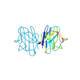
 | | MicroED structure of xylanase at 2.3 A resolution | | 分子名称: | Endo-1,4-beta-xylanase 2, IODIDE ION | | 著者 | de la Cruz, M.J, Hattne, J, Shi, D, Seidler, P, Rodriguez, J, Reyes, F.E, Sawaya, M.R, Cascio, D, Eisenberg, D, Gonen, T. | | 登録日 | 2016-05-26 | | 公開日 | 2017-04-05 | | 最終更新日 | 2024-02-28 | | 実験手法 | ELECTRON CRYSTALLOGRAPHY (2.3 Å) | | 主引用文献 | Atomic-resolution structures from fragmented protein crystals with the cryoEM method MicroED.
Nat. Methods, 14, 2017
|
|
3DG1
 | | Segment SSTNVG derived from IAPP | | 分子名称: | SSTNVG from Islet Amyloid Polypeptide | | 著者 | Wiltzius, J.J, Sievers, S.A, Sawaya, M.R, Cascio, D, Eisenberg, D. | | 登録日 | 2008-06-12 | | 公開日 | 2008-07-01 | | 最終更新日 | 2024-04-03 | | 実験手法 | X-RAY DIFFRACTION (1.66 Å) | | 主引用文献 | Atomic structure of the cross-beta spine of islet amyloid polypeptide (amylin).
Protein Sci., 17, 2008
|
|
3DGJ
 | | NNFGAIL segment from Islet Amyloid Polypeptide (IAPP or amylin) | | 分子名称: | NNFGAIL peptide | | 著者 | Wiltzius, J.J, Sievers, S.A, Sawaya, M.R, Cascio, D, Eisenberg, D. | | 登録日 | 2008-06-13 | | 公開日 | 2008-07-01 | | 最終更新日 | 2024-02-21 | | 実験手法 | X-RAY DIFFRACTION (1.8 Å) | | 主引用文献 | Atomic structure of the cross-beta spine of islet amyloid polypeptide (amylin).
Protein Sci., 17, 2008
|
|
3DBO
 | | Crystal structure of a member of the VapBC family of toxin-antitoxin systems, VapBC-5, from Mycobacterium tuberculosis | | 分子名称: | ACETATE ION, BETA-MERCAPTOETHANOL, SODIUM ION, ... | | 著者 | Miallau, L, Cascio, D, Eisenberg, D, Integrated Center for Structure and Function Innovation (ISFI), TB Structural Genomics Consortium (TBSGC) | | 登録日 | 2008-06-02 | | 公開日 | 2008-07-15 | | 最終更新日 | 2024-02-21 | | 実験手法 | X-RAY DIFFRACTION (1.76 Å) | | 主引用文献 | Structure and Proposed Activity of a Member of the VapBC Family of Toxin-Antitoxin Systems: VapBC-5 FROM MYCOBACTERIUM TUBERCULOSIS.
J.Biol.Chem., 284, 2009
|
|
5CCA
 | | Crystal structure of Mtb toxin | | 分子名称: | Endoribonuclease MazF3 | | 著者 | Cascio, D, Arbing, M, de Serrano, V, Eisenberg, D, Miallau, L, TB Structural Genomics Consortium (TBSGC) | | 登録日 | 2015-07-01 | | 公開日 | 2016-09-07 | | 最終更新日 | 2023-09-27 | | 実験手法 | X-RAY DIFFRACTION (3.2 Å) | | 主引用文献 | Crystal structure of Mtb toxin
To Be Published
|
|
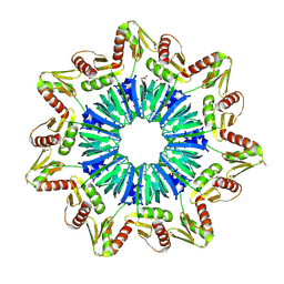
1LOJ
 | | Crystal structure of a Methanobacterial Sm-like archaeal protein (SmAP1) bound to uridine-5'-monophosphate (UMP) | | 分子名称: | (4S)-2-METHYL-2,4-PENTANEDIOL, URIDINE, URIDINE-5'-MONOPHOSPHATE, ... | | 著者 | Mura, C, Kozhukhovsky, A, Eisenberg, D. | | 登録日 | 2002-05-06 | | 公開日 | 2003-03-25 | | 最終更新日 | 2023-08-16 | | 実験手法 | X-RAY DIFFRACTION (1.9 Å) | | 主引用文献 | The oligomerization and ligand-binding properties of Sm-like archaeal proteins (SmAPs)
Protein Sci., 12, 2003
|
|
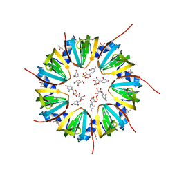
1M5Q
 | | Crystal structure of a novel Sm-like archaeal protein from Pyrobaculum aerophilum | | 分子名称: | ACETIC ACID, CADMIUM ION, GLYCEROL, ... | | 著者 | Mura, C, Phillips, M, Kozhukhovsky, A, Eisenberg, D. | | 登録日 | 2002-07-09 | | 公開日 | 2003-03-18 | | 最終更新日 | 2017-05-17 | | 実験手法 | X-RAY DIFFRACTION (2 Å) | | 主引用文献 | Structure and assembly of an augmented Sm-like archaeal protein 14-mer
Proc.Natl.Acad.Sci.USA, 100, 2003
|
|
1LNX
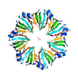 | | Crystal structure of the P.aerophilum SmAP1 heptamer in a new crystal form (C2221) | | 分子名称: | ACETIC ACID, GLYCEROL, URIDINE, ... | | 著者 | Mura, C, Kozhukhovsky, A, Eisenberg, D. | | 登録日 | 2002-05-04 | | 公開日 | 2003-03-25 | | 最終更新日 | 2023-08-16 | | 実験手法 | X-RAY DIFFRACTION (2.05 Å) | | 主引用文献 | The oligomerization and ligand-binding properties of Sm-like archaeal proteins (SmAPs)
Protein Sci., 12, 2003
|
|
1COS
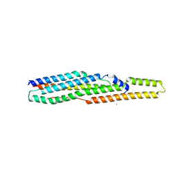
 | | CRYSTAL STRUCTURE OF A SYNTHETIC TRIPLE-STRANDED ALPHA-HELICAL BUNDLE | | 分子名称: | COILED SERINE | | 著者 | Lovejoy, B, Choe, S, Cascio, D, Mcrorie, D.K, Degrado, W, Eisenberg, D. | | 登録日 | 1993-01-22 | | 公開日 | 1993-10-31 | | 最終更新日 | 2019-08-14 | | 実験手法 | X-RAY DIFFRACTION (2.1 Å) | | 主引用文献 | Crystal structure of a synthetic triple-stranded alpha-helical bundle.
Science, 259, 1993
|
|
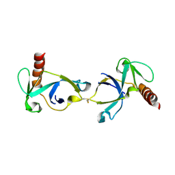
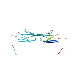
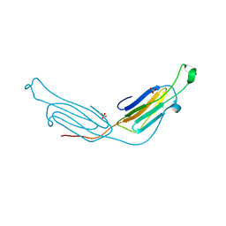
3DMK
 | | Crystal structure of Down Syndrome Cell Adhesion Molecule (DSCAM) isoform 1.30.30, N-terminal eight Ig domains | | 分子名称: | 2-acetamido-2-deoxy-beta-D-glucopyranose-(1-4)-2-acetamido-2-deoxy-beta-D-glucopyranose, 2-acetamido-2-deoxy-beta-D-glucopyranose-(1-4)-2-acetamido-2-deoxy-beta-D-glucopyranose-(1-4)-2-acetamido-2-deoxy-beta-D-glucopyranose, Down Syndrome Cell Adhesion Molecule (DSCAM) isoform 1.30.30, ... | | 著者 | Sawaya, M.R, Wojtowicz, W.M, Eisenberg, D, Zipursky, S.L. | | 登録日 | 2008-07-01 | | 公開日 | 2008-10-07 | | 最終更新日 | 2023-08-30 | | 実験手法 | X-RAY DIFFRACTION (4.19 Å) | | 主引用文献 | A double S shape provides the structural basis for the extraordinary binding specificity of Dscam isoforms.
Cell(Cambridge,Mass.), 134, 2008
|
|
3L1F
 | | Bovine AlphaA crystallin | | 分子名称: | (4S)-2-METHYL-2,4-PENTANEDIOL, Alpha-crystallin A chain | | 著者 | Laganowsky, A, Sawaya, M.R, Cascio, D, Eisenberg, D. | | 登録日 | 2009-12-11 | | 公開日 | 2010-05-12 | | 最終更新日 | 2024-02-21 | | 実験手法 | X-RAY DIFFRACTION (1.53 Å) | | 主引用文献 | Crystal structures of truncated alphaA and alphaB crystallins reveal structural mechanisms of polydispersity important for eye lens function.
Protein Sci., 19, 2010
|
|
3L1G
 | | Human AlphaB crystallin | | 分子名称: | Alpha-crystallin B chain, SULFATE ION | | 著者 | Laganowsky, A, Sawaya, M.R, Cascio, D, Eisenberg, D. | | 登録日 | 2009-12-11 | | 公開日 | 2010-05-12 | | 最終更新日 | 2024-02-21 | | 実験手法 | X-RAY DIFFRACTION (3.32 Å) | | 主引用文献 | Crystal structures of truncated alphaA and alphaB crystallins reveal structural mechanisms of polydispersity important for eye lens function.
Protein Sci., 19, 2010
|
|
3L1E
 | | Bovine AlphaA crystallin Zinc Bound | | 分子名称: | Alpha-crystallin A chain, GLYCEROL, ZINC ION | | 著者 | Laganowsky, A, Sawaya, M.R, Cascio, D, Eisenberg, D. | | 登録日 | 2009-12-11 | | 公開日 | 2010-05-12 | | 最終更新日 | 2024-02-21 | | 実験手法 | X-RAY DIFFRACTION (1.15 Å) | | 主引用文献 | Crystal structures of truncated alphaA and alphaB crystallins reveal structural mechanisms of polydispersity important for eye lens function.
Protein Sci., 19, 2010
|
|
4E0L
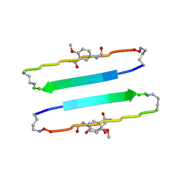 | | FYLLYYT segment from human Beta 2 Microglobulin (62-68) displayed on 54-membered macrocycle scaffold | | 分子名称: | (4S)-2-METHYL-2,4-PENTANEDIOL, Cyclic pseudo-peptide FYLLYYT(ORN)KN(HAO)SA(ORN) | | 著者 | Zhao, M, Liu, C, Michael, S.R, Eisenberg, D. | | 登録日 | 2012-03-04 | | 公開日 | 2012-12-19 | | 最終更新日 | 2023-11-15 | | 実験手法 | X-RAY DIFFRACTION (1.7 Å) | | 主引用文献 | Out-of-register beta-sheets suggest a pathway to toxic amyloid aggregates.
Proc.Natl.Acad.Sci.USA, 109, 2012
|
|
2JCW
 | | REDUCED BRIDGE-BROKEN YEAST CU/ZN SUPEROXIDE DISMUTASE ROOM TEMPERATURE (298K) STRUCTURE | | 分子名称: | COPPER (I) ION, CU/ZN SUPEROXIDE DISMUTASE, ZINC ION | | 著者 | Hart, P.J, Balbirnie, M.M, Ogihara, N.L, Nersissian, A.M, Weiss, M.S, Valentine, J.S, Eisenberg, D. | | 登録日 | 1998-12-21 | | 公開日 | 1999-06-08 | | 最終更新日 | 2023-08-09 | | 実験手法 | X-RAY DIFFRACTION (1.7 Å) | | 主引用文献 | A structure-based mechanism for copper-zinc superoxide dismutase.
Biochemistry, 38, 1999
|
|
2G38
 | | A PE/PPE Protein Complex from Mycobacterium tuberculosis | | 分子名称: | MANGANESE (II) ION, PE FAMILY PROTEIN, PPE FAMILY PROTEIN | | 著者 | Strong, M, Sawaya, M.R, Eisenberg, D, TB Structural Genomics Consortium (TBSGC) | | 登録日 | 2006-02-17 | | 公開日 | 2006-03-14 | | 最終更新日 | 2024-02-14 | | 実験手法 | X-RAY DIFFRACTION (2.2 Å) | | 主引用文献 | Toward the structural genomics of complexes: Crystal structure of a PE/PPE protein complex from Mycobacterium tuberculosis.
Proc.Natl.Acad.Sci.Usa, 103, 2006
|
|
3MML
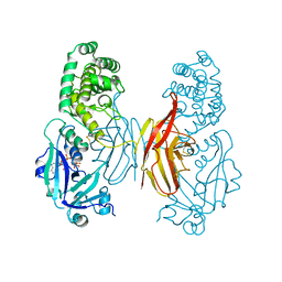
 | | Allophanate Hydrolase Complex from Mycobacterium smegmatis, Msmeg0435-Msmeg0436 | | 分子名称: | Allophanate hydrolase subunit 1, Allophanate hydrolase subunit 2, CHLORIDE ION | | 著者 | Kaufmann, M, Chernishof, I, Shin, A, Germano, D, Sawaya, M.R, Waldo, G.S, Arbing, M.A, Perry, J, Eisenberg, D, Integrated Center for Structure and Function Innovation (ISFI), TB Structural Genomics Consortium (TBSGC) | | 登録日 | 2010-04-20 | | 公開日 | 2010-04-28 | | 最終更新日 | 2017-11-08 | | 実験手法 | X-RAY DIFFRACTION (2.5 Å) | | 主引用文献 | Crystal Structure of Allphanate Hydrolase Complex from M. smegmatis, Msmeg0435-Msmeg0436
To be Published
|
|
1DDT
 | |
1JS0
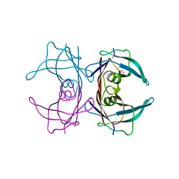
 | | Crystal Structure of 3D Domain-swapped RNase A Minor Trimer | | 分子名称: | RIBONUCLEASE A, SULFATE ION | | 著者 | Liu, Y, Gotte, G, Libonati, M, Eisenberg, D. | | 登録日 | 2001-08-15 | | 公開日 | 2002-03-13 | | 最終更新日 | 2023-08-16 | | 実験手法 | X-RAY DIFFRACTION (2.2 Å) | | 主引用文献 | Structures of the two 3D domain-swapped RNase A trimers.
Protein Sci., 11, 2002
|
|
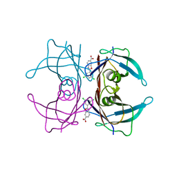
6E75
 | | Structure of Human Transthyretin Asp38Ala Mutant | | 分子名称: | ACETATE ION, Transthyretin | | 著者 | Chung, K, Saelices, L, Sawaya, M.R, Cascio, D, Eisenberg, D. | | 登録日 | 2018-07-25 | | 公開日 | 2019-07-31 | | 最終更新日 | 2024-03-13 | | 実験手法 | X-RAY DIFFRACTION (1.5 Å) | | 主引用文献 | Structural Variants of Transthyretin
To Be Published
|
|
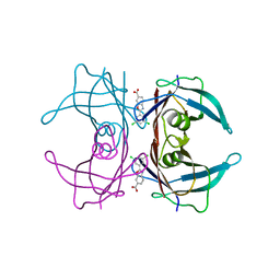
6E6Z
 | | Structure of Wild Type Human Transthyretin in Complex with Tafamidis | | 分子名称: | 2-(3,5-dichlorophenyl)-1,3-benzoxazole-6-carboxylic acid, Transthyretin | | 著者 | Chung, K, Saelices, L, Sawaya, M.R, Cascio, D, Eisenberg, D. | | 登録日 | 2018-07-25 | | 公開日 | 2019-07-31 | | 最終更新日 | 2024-03-13 | | 実験手法 | X-RAY DIFFRACTION (1.75 Å) | | 主引用文献 | Structural Variants of Transthyretin
To Be Published
|
|
6E77
 | | Structure of Human Transthyretin Asp38Ala Mutant in Complex with Tafamidis | | 分子名称: | 2-(3,5-dichlorophenyl)-1,3-benzoxazole-6-carboxylic acid, Transthyretin | | 著者 | Saelices, L, Chung, K, Sawaya, M.R, Cascio, D, Eisenberg, D. | | 登録日 | 2018-07-25 | | 公開日 | 2019-07-31 | | 最終更新日 | 2024-03-13 | | 実験手法 | X-RAY DIFFRACTION (1.6 Å) | | 主引用文献 | Structural Variants of Transthyretin
To Be Published
|
|


