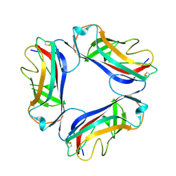
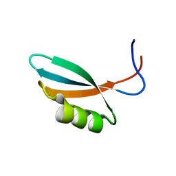
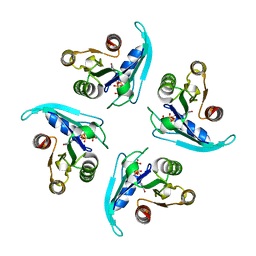
2XSK
 
 | | E. coli curli protein CsgC - SeCys | | 分子名称: | ACETATE ION, CSGC | | 著者 | Salgado, P.S, Taylor, J.D, Cota, E, Matthews, S.J. | | 登録日 | 2010-09-29 | | 公開日 | 2010-12-29 | | 最終更新日 | 2014-01-29 | | 実験手法 | X-RAY DIFFRACTION (1.7 Å) | | 主引用文献 | Extending the Usability of the Phasing Power of Diselenide Bonds: Secys Sad Phasing of Csgc Using a Non-Auxotrophic Strain.
Acta Crystallogr.,Sect.D, 67, 2011
|
|
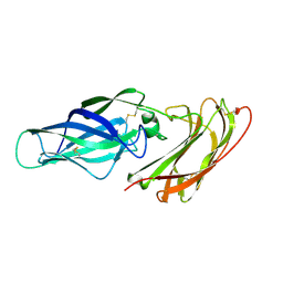
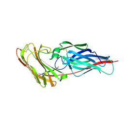
2VER
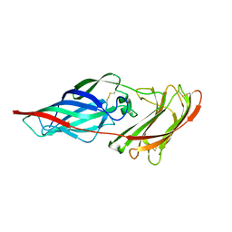
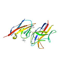 | | Structural model for the complex between the Dr adhesins and carcinoembryonic antigen (CEA) | | 分子名称: | AFIMBRIAL ADHESIN AFA-III, ARCINOEMBRYONIC ANTIGEN-RELATED CELL ADHESION MOLECULE 5, S-[(1-oxyl-2,2,5,5-tetramethyl-2,5-dihydro-1H-pyrrol-3-yl)methyl] methanesulfonothioate | | 著者 | Korotkova, N, Yang, Y, Le Trong, I, Cota, E, Demeler, B, Marchant, J, Thomas, W.E, Stenkamp, R.E, Moseley, S.L, Matthews, S. | | 登録日 | 2007-10-26 | | 公開日 | 2008-01-08 | | 最終更新日 | 2011-07-13 | | 実験手法 | SOLUTION NMR | | 主引用文献 | Binding of Dr Adhesins of Escherichia Coli to Carcinoembryonic Antigen Triggers Receptor Dissociation.
Mol.Microbiol., 67, 2008
|
|
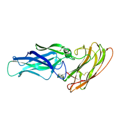
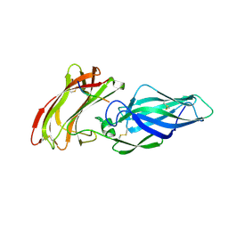
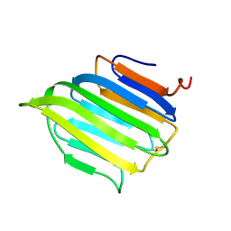
1UT2
 | | AfaE-3 adhesin from Escherichia Coli | | 分子名称: | AFIMBRIAL ADHESIN AFA-III, SULFATE ION | | 著者 | Anderson, K.L, Billington, J, Pettigrew, D, Cota, E, Roversi, P, Simpson, P, Chen, H.A, Urvil, P, Dumerle, L, Barlow, P, Medof, E, Smith, R.A.G, Nowicki, B, Le Bouguenec, C, Lea, S.M, Matthews, S. | | 登録日 | 2003-12-02 | | 公開日 | 2004-08-31 | | 最終更新日 | 2023-12-13 | | 実験手法 | X-RAY DIFFRACTION (3.3 Å) | | 主引用文献 | High Resolution Studies of the Afa/Dr Adhesin Drae and its Interaction with Chloramphenicol
J.Biol.Chem., 279, 2004
|
|
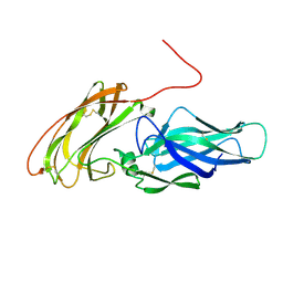
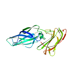
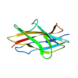
1UT1
 | | DraE adhesin from Escherichia Coli | | 分子名称: | 1,2-ETHANEDIOL, DR HEMAGGLUTININ STRUCTURAL SUBUNIT, SULFATE ION | | 著者 | Anderson, K.L, Billington, J, Pettigrew, D, Cota, E, Roversi, P, Simpson, P, Chen, H.A, Urvil, P, Dumerle, L, Barlow, P, Medof, E, Smith, R.A.G, Nowicki, B, Le Bouguenec, C, Lea, S.M, Matthews, S. | | 登録日 | 2003-12-02 | | 公開日 | 2004-08-31 | | 最終更新日 | 2023-12-13 | | 実験手法 | X-RAY DIFFRACTION (1.7 Å) | | 主引用文献 | High Resolution Studies of the Afa/Dr Adhesin Drae and its Interaction with Chloramphenicol
J.Biol.Chem., 279, 2004
|
|
1USZ
 | | SeMet AfaE-3 adhesin from Escherichia Coli | | 分子名称: | AFIMBRIAL ADHESIN AFA-III, CHLORIDE ION, SULFATE ION | | 著者 | Anderson, K.L, Billington, J, Pettigrew, D, Cota, E, Roversi, P, Simpson, P, Chen, H.A, Urvil, P, Dumerle, L, Barlow, P, Medof, E, Smith, R.A.G, Nowicki, B, Le Bouguenec, C, Lea, S.M, Matthews, S. | | 登録日 | 2003-12-02 | | 公開日 | 2004-08-31 | | 最終更新日 | 2011-07-13 | | 実験手法 | X-RAY DIFFRACTION (3.28 Å) | | 主引用文献 | High Resolution Studies of the Afa/Dr Adhesin Drae and its Interaction with Chloramphenicol
J.Biol.Chem., 279, 2004
|
|
1USQ
 | | Complex of E. Coli DraE adhesin with Chloramphenicol | | 分子名称: | 1,2-ETHANEDIOL, CHLORAMPHENICOL, DR HEMAGGLUTININ STRUCTURAL SUBUNIT, ... | | 著者 | Anderson, K.L, Billington, J, Pettigrew, D, Cota, E, Roversi, P, Simpson, P, Chen, H.A, Urvil, P, Dumerle, L, Barlow, P, Medof, E, Smith, R.A.G, Nowicki, B, Le Bouguenec, C, Lea, S.M, Matthews, S. | | 登録日 | 2003-11-27 | | 公開日 | 2004-08-31 | | 最終更新日 | 2023-12-13 | | 実験手法 | X-RAY DIFFRACTION (1.9 Å) | | 主引用文献 | High Resolution Studies of the Afa/Dr Adhesin Drae and its Interaction with Chloramphenicol
J.Biol.Chem., 279, 2004
|
|
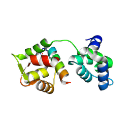
2WNM
 | | Solution structure of Gp2 | | 分子名称: | GENE 2 | | 著者 | Camara, B, Liu, M, Shadrinc, A, Liu, B, Simpson, P, Weinzierl, R, Severinovc, K, Cota, E, Matthews, S, Wigneshweraraj, S.R. | | 登録日 | 2009-07-13 | | 公開日 | 2010-02-16 | | 最終更新日 | 2024-05-15 | | 実験手法 | SOLUTION NMR | | 主引用文献 | T7 Phage Protein Gp2 Inhibits the Escherichia Coli RNA Polymerase by Antagonizing Stable DNA Strand Separation Near the Transcription Start Site.
Proc.Natl.Acad.Sci.USA, 107, 2010
|
|
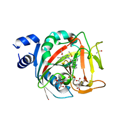
8FYZ
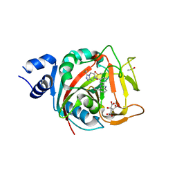 | | Crystal structure of human PARP1 ART domain bound to inhibitor UKTT10 (compound 13) | | 分子名称: | (2P)-2-{3-[(4R)-3-(trifluoromethyl)-5,6-dihydro[1,2,4]triazolo[4,3-a]pyrazine-7(8H)-carbonyl]phenyl}-1H-benzimidazole-4-carboxamide, CITRIC ACID, DIMETHYL SULFOXIDE, ... | | 著者 | Rouleau-Turcotte, E, Pascal, J.M. | | 登録日 | 2023-01-27 | | 公開日 | 2024-02-07 | | 最終更新日 | 2024-03-20 | | 実験手法 | X-RAY DIFFRACTION (3.4 Å) | | 主引用文献 | Novel modifications of PARP inhibitor veliparib increase PARP1 binding to DNA breaks.
Biochem.J., 481, 2024
|
|
8FYY
 | |
8G0H
 | |
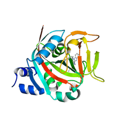
8FZ1
 | | Crystal structure of human PARP1 ART domain bound to inhibitor UKTT22 (compound 14) | | 分子名称: | (2P)-2-{3-[(2-amino-4,5-dimethylphenyl)carbamoyl]phenyl}-1H-benzimidazole-4-carboxamide, 1,2-ETHANEDIOL, CITRIC ACID, ... | | 著者 | Rouleau-Turcotte, E, Pascal, J.M. | | 登録日 | 2023-01-27 | | 公開日 | 2024-02-07 | | 最終更新日 | 2024-03-20 | | 実験手法 | X-RAY DIFFRACTION (2.7 Å) | | 主引用文献 | Novel modifications of PARP inhibitor veliparib increase PARP1 binding to DNA breaks.
Biochem.J., 481, 2024
|
|
2Y7M
 | |
2Y7O
 | |
2Y7N
 | |
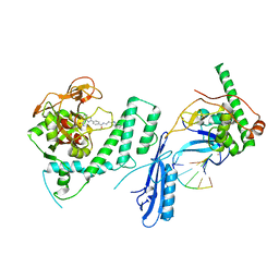
6HUF
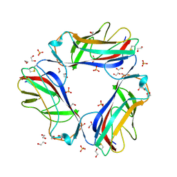
 | | Coping with strong translational non-crystallographic symmetry and extreme anisotropy in molecular replacement with Phaser: human Rab27a | | 分子名称: | MAGNESIUM ION, PHOSPHOAMINOPHOSPHONIC ACID-GUANYLATE ESTER, Ras-related protein Rab-27A | | 著者 | Jamshidiha, M, Perez-Dorado, I, Murray, J.W, Tate, E.W, Cota, E, Read, R.J. | | 登録日 | 2018-10-08 | | 公開日 | 2019-03-27 | | 最終更新日 | 2024-01-24 | | 実験手法 | X-RAY DIFFRACTION (2.82 Å) | | 主引用文献 | Coping with strong translational noncrystallographic symmetry and extreme anisotropy in molecular replacement with Phaser: human Rab27a.
Acta Crystallogr D Struct Biol, 75, 2019
|
|
2Y7L
 | |
2YLH
 | |
4LEB
 | |
4LE8
 | |
4LEE
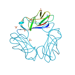
 | | Structure of the Als3 adhesin from Candida albicans, residues 1-313 (mature sequence), triple mutant in the binding cavity: K59M, A116V, Y301F | | 分子名称: | Agglutinin-like protein 3 | | 著者 | Lin, J, Garnett, J.A, Cota, E. | | 登録日 | 2013-06-25 | | 公開日 | 2014-05-14 | | 最終更新日 | 2023-09-20 | | 実験手法 | X-RAY DIFFRACTION (3 Å) | | 主引用文献 | The Peptide-binding Cavity Is Essential for Als3-mediated Adhesion of Candida albicans to Human Cells.
J.Biol.Chem., 289, 2014
|
|
2BVB
 | | The C-terminal domain from Micronemal Protein 1 (MIC1) from Toxoplasma Gondii | | 分子名称: | MICRONEMAL PROTEIN 1 | | 著者 | Saouros, S, Edwards-Jones, B, Reiss, M, Sawmynaden, K, Cota, E, Simpson, P, Dowse, T.J, Jakle, U, Ramboarina, S, Shivarattan, T, Matthews, S, Soldati-Favre, D. | | 登録日 | 2005-06-23 | | 公開日 | 2005-10-12 | | 最終更新日 | 2021-06-23 | | 実験手法 | SOLUTION NMR | | 主引用文献 | A Novel Galectin-Like Domain from Toxoplasma Gondll Micronemal Protein 1 Assists the Folding, Assembly,and Transport of a Cell-Adhesion Complex.
J.Biol.Chem., 280, 2005
|
|
1RXL
 | | Solution structure of the engineered protein Afae-dsc | | 分子名称: | Afimbrial adhesin AFA-III | | 著者 | Anderson, K.L, Billington, J, Pettigrew, D, Cota, E, Roversi, P, Simpson, P, Chen, H.A, Urvil, P, du Merle, L, Barlow, P.N, Medof, M.E, Smith, R.A, Nowicki, B, Le Bouguenec, C, Lea, S.M, Matthews, S. | | 登録日 | 2003-12-18 | | 公開日 | 2005-01-11 | | 最終更新日 | 2020-02-05 | | 実験手法 | SOLUTION NMR | | 主引用文献 | An atomic resolution model for assembly, architecture, and function of the Dr adhesins.
Mol.Cell, 15, 2004
|
|
4MZJ
 | | Crystal Structure of MTIP from Plasmodium falciparum in complex with pGly[801,805], a stapled myoA tail peptide | | 分子名称: | Myosin A tail domain interacting protein, Myosin-A | | 著者 | Douse, C.H, Garnett, J.A, Maas, S.J, Cota, E, Tate, E.W. | | 登録日 | 2013-09-30 | | 公開日 | 2013-11-06 | | 最終更新日 | 2023-12-06 | | 実験手法 | X-RAY DIFFRACTION (1.474 Å) | | 主引用文献 | Crystal Structures of Stapled and Hydrogen Bond Surrogate Peptides Targeting a Fully Buried Protein-Helix Interaction.
Acs Chem.Biol., 8, 2014
|
|
4MZK
 | | Crystal Structure of MTIP from Plasmodium falciparum in complex with pGly[807,811], a stapled myoA tail peptide | | 分子名称: | Myosin A tail domain interacting protein, pGly[807,811], a stapled myoA tail peptide | | 著者 | Douse, C.H, Garnett, J.A, Maas, S.J, Cota, E, Tate, E.W. | | 登録日 | 2013-09-30 | | 公開日 | 2013-11-06 | | 最終更新日 | 2023-12-06 | | 実験手法 | X-RAY DIFFRACTION (1.82 Å) | | 主引用文献 | Crystal Structures of Stapled and Hydrogen Bond Surrogate Peptides Targeting a Fully Buried Protein-Helix Interaction.
Acs Chem.Biol., 8, 2014
|
|
4MZL
 | | Crystal Structure of MTIP from Plasmodium falciparum in complex with HBS myoA, a hydrogen bond surrogate myoA helix mimetic | | 分子名称: | Myosin A tail domain interacting protein, hydrogen bond surrogate (HBS) myoA helix mimetic | | 著者 | Douse, C.H, Garnett, J.A, Maas, S.J, Cota, E, Tate, E.W. | | 登録日 | 2013-09-30 | | 公開日 | 2013-11-06 | | 最終更新日 | 2023-06-07 | | 実験手法 | X-RAY DIFFRACTION (2.01 Å) | | 主引用文献 | Crystal Structures of Stapled and Hydrogen Bond Surrogate Peptides Targeting a Fully Buried Protein-Helix Interaction.
Acs Chem.Biol., 8, 2014
|
|


