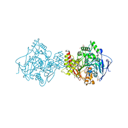
4JTA
 
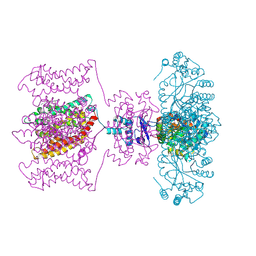 | | Crystal structure of Kv1.2-2.1 paddle chimera channel in complex with Charybdotoxin | | 分子名称: | (1R)-2-{[(S)-{[(2S)-2,3-dihydroxypropyl]oxy}(hydroxy)phosphoryl]oxy}-1-[(hexadecanoyloxy)methyl]ethyl (9Z)-octadec-9-enoate, NADP NICOTINAMIDE-ADENINE-DINUCLEOTIDE PHOSPHATE, POTASSIUM ION, ... | | 著者 | MacKinnon, R, Banerjee, A, Lee, A, Campbell, E. | | 登録日 | 2013-03-23 | | 公開日 | 2013-06-12 | | 最終更新日 | 2019-12-25 | | 実験手法 | X-RAY DIFFRACTION (2.5 Å) | | 主引用文献 | Structure of a pore-blocking toxin in complex with a eukaryotic voltage-dependent K(+) channel.
Elife, 2, 2013
|
|
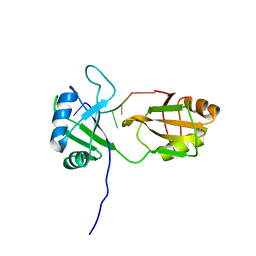
5AF0
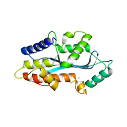 | | MAEL domain from Bombyx mori Maelstrom | | 分子名称: | MAELSTROM, ZINC ION | | 著者 | Chen, K, Campbell, E, Pandey, R.R, Yang, Z, McCarthy, A.A, Pillai, R.S. | | 登録日 | 2015-01-13 | | 公開日 | 2015-04-01 | | 最終更新日 | 2024-05-08 | | 実験手法 | X-RAY DIFFRACTION (2.401 Å) | | 主引用文献 | Metazoan Maelstrom is an RNA-Binding Protein that Has Evolved from an Ancient Nuclease Active in Protists.
RNA, 21, 2015
|
|
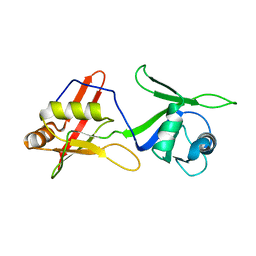
4JTC
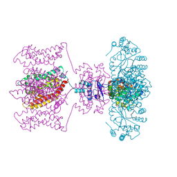 | | Crystal structure of Kv1.2-2.1 paddle chimera channel in complex with Charybdotoxin in Cs+ | | 分子名称: | (1R)-2-{[(S)-{[(2S)-2,3-dihydroxypropyl]oxy}(hydroxy)phosphoryl]oxy}-1-[(hexadecanoyloxy)methyl]ethyl (9Z)-octadec-9-enoate, CESIUM ION, NADP NICOTINAMIDE-ADENINE-DINUCLEOTIDE PHOSPHATE, ... | | 著者 | Banerjee, A, Lee, A, Campbell, E, MacKinnon, R. | | 登録日 | 2013-03-23 | | 公開日 | 2013-06-12 | | 最終更新日 | 2019-12-25 | | 実験手法 | X-RAY DIFFRACTION (2.56 Å) | | 主引用文献 | Structure of a pore-blocking toxin in complex with a eukaryotic voltage-dependent K(+) channel.
Elife, 2, 2013
|
|
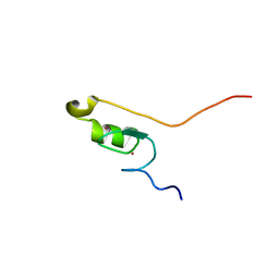
4JTD
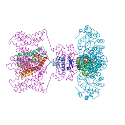 | | Crystal structure of Kv1.2-2.1 paddle chimera channel in complex with Lys27Met mutant of Charybdotoxin | | 分子名称: | (1R)-2-{[(S)-{[(2S)-2,3-dihydroxypropyl]oxy}(hydroxy)phosphoryl]oxy}-1-[(hexadecanoyloxy)methyl]ethyl (9Z)-octadec-9-enoate, NADP NICOTINAMIDE-ADENINE-DINUCLEOTIDE PHOSPHATE, POTASSIUM ION, ... | | 著者 | Banerjee, A, Lee, A, Campbell, E, MacKinnon, R. | | 登録日 | 2013-03-23 | | 公開日 | 2013-06-12 | | 最終更新日 | 2019-12-25 | | 実験手法 | X-RAY DIFFRACTION (2.54 Å) | | 主引用文献 | Structure of a pore-blocking toxin in complex with a eukaryotic voltage-dependent K(+) channel.
Elife, 2, 2013
|
|
2CEK
 | | Conformational Flexibility in the Peripheral Site of Torpedo californica Acetylcholinesterase Revealed by the Complex Structure with a Bifunctional Inhibitor | | 分子名称: | 2-(N-MORPHOLINO)-ETHANESULFONIC ACID, 2-acetamido-2-deoxy-beta-D-glucopyranose, 2-acetamido-2-deoxy-beta-D-glucopyranose-(1-4)-2-acetamido-2-deoxy-beta-D-glucopyranose, ... | | 著者 | Sanson, B, Colletier, J.P, Nachon, F, Gabellieri, E, Fattorusso, C, Campiani, G, Weik, M. | | 登録日 | 2006-02-08 | | 公開日 | 2006-04-10 | | 最終更新日 | 2023-12-13 | | 実験手法 | X-RAY DIFFRACTION (2.2 Å) | | 主引用文献 | Conformational flexibility in the peripheral site of Torpedo californica acetylcholinesterase revealed by the complex structure with a bifunctional inhibitor.
J. Am. Chem. Soc., 128, 2006
|
|
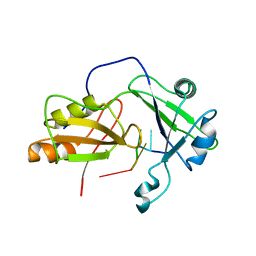
2MKI
 | | Solution structure of tandem RRM domains of cytoplasmic polyadenylation element binding protein 4 (CPEB4) in complex with RNA | | 分子名称: | Cytoplasmic polyadenylation element-binding protein 4, RNA (5'-R(*CP*UP*UP*UP*A)-3') | | 著者 | Afroz, T, Skrisovska, L, Belloc, E, Boixet, J.G, Mendez, R, Allain, F.H.-T. | | 登録日 | 2014-02-07 | | 公開日 | 2014-07-23 | | 最終更新日 | 2024-05-01 | | 実験手法 | SOLUTION NMR | | 主引用文献 | A fly trap mechanism provides sequence-specific RNA recognition by CPEB proteins
Genes Dev., 28, 2014
|
|
2MKH
 | | Solution structure of tandem RRM domains of cytoplasmic polyadenylation element binding protein 1 (CPEB1) in free state | | 分子名称: | Cytoplasmic polyadenylation element-binding protein 1 | | 著者 | Afroz, T, Skrisovska, L, Belloc, E, Boixet, J.G, Mendez, R, Allain, F.H.-T. | | 登録日 | 2014-02-07 | | 公開日 | 2014-07-23 | | 最終更新日 | 2024-05-01 | | 実験手法 | SOLUTION NMR | | 主引用文献 | A fly trap mechanism provides sequence-specific RNA recognition by CPEB proteins
Genes Dev., 28, 2014
|
|
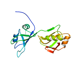
2MKE
 | | Solution structure of CPEB1 ZZ domain in the free state | | 分子名称: | Cytoplasmic polyadenylation element-binding protein 1, ZINC ION | | 著者 | Afroz, T, Skrisovska, L, Belloc, E, Boixet, J.G, Mendez, R, Allain, F.H.-T. | | 登録日 | 2014-02-06 | | 公開日 | 2014-07-23 | | 最終更新日 | 2024-05-01 | | 実験手法 | SOLUTION NMR | | 主引用文献 | A fly trap mechanism provides sequence-specific RNA recognition by CPEB proteins
Genes Dev., 28, 2014
|
|
2MKK
 | | Structural model of tandem RRM domains of cytoplasmic polyadenylation element binding protein 1 (CPEB1) in complex with RNA | | 分子名称: | Cytoplasmic polyadenylation element-binding protein 1, RNA (5'-R(*UP*UP*UP*UP*A)-3') | | 著者 | Afroz, T, Skrisovska, L, Belloc, E, Boixet, J.G, Mendez, R, Allain, F.H.-T. | | 登録日 | 2014-02-07 | | 公開日 | 2014-07-23 | | 最終更新日 | 2024-05-01 | | 実験手法 | SOLUTION NMR | | 主引用文献 | A fly trap mechanism provides sequence-specific RNA recognition by CPEB proteins
Genes Dev., 28, 2014
|
|
2MKJ
 | | Solution structure of tandem RRM domains of cytoplasmic polyadenylation element binding protein 4 (CPEB4) in free state | | 分子名称: | Cytoplasmic polyadenylation element-binding protein 4 | | 著者 | Afroz, T, Skrisovska, L, Belloc, E, Boixet, J.G, Mendez, R, Allain, F.H.-T. | | 登録日 | 2014-02-07 | | 公開日 | 2014-07-23 | | 最終更新日 | 2024-05-15 | | 実験手法 | SOLUTION NMR | | 主引用文献 | A fly trap mechanism provides sequence-specific RNA recognition by CPEB proteins
Genes Dev., 28, 2014
|
|
6HQF
 | | Structure of Phenylalanine ammonia-lyase from Petroselinum crispum in complex with (R)-APEP | | 分子名称: | Phenylalanine ammonia-lyase 1, [(1R)-1-amino-2-phenylethyl]phosphonic acid | | 著者 | Bata, Z, Molnar, B, Leveles, I, Poppe, L, Vertessy, G.B. | | 登録日 | 2018-09-24 | | 公開日 | 2019-10-09 | | 最終更新日 | 2024-01-24 | | 実験手法 | X-RAY DIFFRACTION (1.76 Å) | | 主引用文献 | Substrate Tunnel Engineering Aided by X-ray Crystallography and Functional Dynamics Swaps the Function of MIO-Enzymes
Acs Catalysis, 2021
|
|
6F6T
 | |
6H2O
 | | APO structure of Phenylalanine ammonia-lyase from Petroselinum crispum | | 分子名称: | Phenylalanine ammonia-lyase 1 | | 著者 | Molnar, B, Bata, Z, Leveles, I, Poppe, L, Vertessy, G.B. | | 登録日 | 2018-07-14 | | 公開日 | 2019-07-31 | | 最終更新日 | 2024-01-17 | | 実験手法 | X-RAY DIFFRACTION (1.9 Å) | | 主引用文献 | Substrate Tunnel Engineering Aided by X-ray Crystallography and Functional Dynamics Swaps the Function of MIO-Enzymes
Acs Catalysis, 2021
|
|
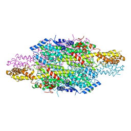
6DHN
 | | Bovine glutamate dehydrogenase complexed with Eu3+ | | 分子名称: | 1,4-DIHYDRONICOTINAMIDE ADENINE DINUCLEOTIDE, GLUTAMIC ACID, GUANOSINE-5'-TRIPHOSPHATE, ... | | 著者 | Smith, T.J. | | 登録日 | 2018-05-20 | | 公開日 | 2018-07-25 | | 実験手法 | X-RAY DIFFRACTION (3.3 Å) | | 主引用文献 | A novel mechanism of V-type zinc inhibition of glutamate dehydrogenase results from disruption of subunit interactions necessary for efficient catalysis.
FEBS J., 278, 2011
|
|
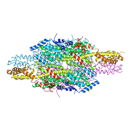
6DHM
 | | Bovine glutamate dehydrogenase complexed with zinc | | 分子名称: | GLUTAMIC ACID, GUANOSINE-5'-TRIPHOSPHATE, Glutamate dehydrogenase 1, ... | | 著者 | Smith, T.J. | | 登録日 | 2018-05-20 | | 公開日 | 2018-07-25 | | 最終更新日 | 2024-03-13 | | 実験手法 | X-RAY DIFFRACTION (3 Å) | | 主引用文献 | A novel mechanism of V-type zinc inhibition of glutamate dehydrogenase results from disruption of subunit interactions necessary for efficient catalysis.
FEBS J., 278, 2011
|
|
7P2B
 | |
6EGR
 | | Crystal structure of Citrobacter freundii methionine gamma-lyase with V358Y replacement | | 分子名称: | DI(HYDROXYETHYL)ETHER, Methionine gamma-lyase, PYRIDOXAL-5'-PHOSPHATE, ... | | 著者 | Revtovich, S.V, Demitri, N, Raboni, S, Nikulin, A.D, Morozova, E.A, Demidkina, T.V, Storici, P, Mozzarelli, A. | | 登録日 | 2017-09-12 | | 公開日 | 2018-10-10 | | 最終更新日 | 2024-01-17 | | 実験手法 | X-RAY DIFFRACTION (1.45 Å) | | 主引用文献 | Engineering methionine gamma-lyase from Citrobacter freundii for anticancer activity.
Biochim Biophys Acta Proteins Proteom, 1866, 2018
|
|
4BFD
 | | CRYSTAL STRUCTURE OF BACE-1 IN COMPLEX WITH CHEMICAL LIGAND | | 分子名称: | BETA-SECRETASE 1, DIMETHYL SULFOXIDE, N-[3-[(1S,3S,6S)-5-azanyl-3-methyl-4-azabicyclo[4.1.0]hept-4-en-3-yl]-4-fluoranyl-phenyl]-5-chloranyl-pyridine-2-carbox amide, ... | | 著者 | Banner, D.W, Benz, J, Stihle, M. | | 登録日 | 2013-03-18 | | 公開日 | 2013-06-19 | | 最終更新日 | 2023-12-20 | | 実験手法 | X-RAY DIFFRACTION (2.3 Å) | | 主引用文献 | Bace1 Inhibitors: A Head Group Scan on a Series of Amides.
Bioorg.Med.Chem.Lett., 23, 2013
|
|
4BEK
 | | CRYSTAL STRUCTURE OF BACE-1 IN COMPLEX WITH CHEMICAL LIGAND | | 分子名称: | (4S)-4-(4-methoxyphenyl)-4-methyl-5,6-dihydro-1,3-thiazin-2-amine, BETA-SECRETASE 1, DIMETHYL SULFOXIDE, ... | | 著者 | Banner, D.W, Benz, J, Stihle, M. | | 登録日 | 2013-03-11 | | 公開日 | 2013-06-19 | | 最終更新日 | 2023-12-20 | | 実験手法 | X-RAY DIFFRACTION (2.39 Å) | | 主引用文献 | Bace1 Inhibitors: A Head Group Scan on a Series of Amides.
Bioorg.Med.Chem.Lett., 23, 2013
|
|
6MNX
 | |
4YBI
 | | Crystal structure of BACE with amino thiazine inhibitor LY2811376 | | 分子名称: | (4S)-4-[2,4-difluoro-5-(pyrimidin-5-yl)phenyl]-4-methyl-5,6-dihydro-4H-1,3-thiazin-2-amine, Beta-secretase 1, GLYCEROL | | 著者 | Timm, D.E. | | 登録日 | 2015-02-18 | | 公開日 | 2015-04-01 | | 実験手法 | X-RAY DIFFRACTION (1.84 Å) | | 主引用文献 | Robust central reduction of amyloid-beta in humans with an orally available, non-peptidic beta-secretase inhibitor.
J.Neurosci., 31, 2011
|
|
1G6Y
 | | CRYSTAL STRUCTURE OF THE GLOBULAR REGION OF THE PRION PROTEIN URE2 FROM YEAST SACCHAROMYCES CEREVISIAE | | 分子名称: | URE2 PROTEIN | | 著者 | Bousset, L, Belrhali, H, Janin, J, Melki, R, Morera, S. | | 登録日 | 2000-11-08 | | 公開日 | 2001-02-21 | | 最終更新日 | 2024-02-07 | | 実験手法 | X-RAY DIFFRACTION (2.8 Å) | | 主引用文献 | Structure of the globular region of the prion protein Ure2 from the yeast Saccharomyces cerevisiae.
Structure, 9, 2001
|
|
1G6W
 | | CRYSTAL STRUCTURE OF THE GLOBULAR REGION OF THE PRION PROTEIN URE2 FROM THE YEAST SACCAROMYCES CEREVISIAE | | 分子名称: | URE2 PROTEIN | | 著者 | Bousset, L, Belrhali, H, Janin, J, Melki, R, Morera, S. | | 登録日 | 2000-11-08 | | 公開日 | 2001-02-21 | | 最終更新日 | 2024-02-07 | | 実験手法 | X-RAY DIFFRACTION (2.5 Å) | | 主引用文献 | Structure of the globular region of the prion protein Ure2 from the yeast Saccharomyces cerevisiae.
Structure, 9, 2001
|
|
3GTU
 | |


