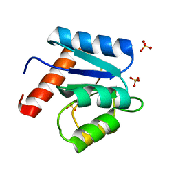
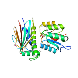
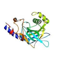
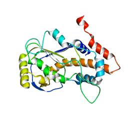
1OOH
 
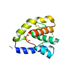 | | Complex of Drosophila odorant binding protein LUSH with butanol | | 分子名称: | 1-BUTANOL, ACETATE ION, odorant binding protein LUSH | | 著者 | Kruse, S.W, Zhao, R, Smith, D.P, Jones, D.N.M. | | 登録日 | 2003-03-03 | | 公開日 | 2003-09-02 | | 最終更新日 | 2023-08-16 | | 実験手法 | X-RAY DIFFRACTION (1.25 Å) | | 主引用文献 | Structure of a specific alcohol-binding site defined by the odorant binding protein LUSH from Drosophila melanogaster
Nat.Struct.Biol., 10, 2003
|
|
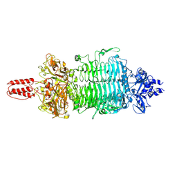
1SVL
 | | Co-crystal structure of SV40 large T antigen helicase domain and ADP | | 分子名称: | ADENOSINE-5'-DIPHOSPHATE, MAGNESIUM ION, ZINC ION, ... | | 著者 | Gai, D, Zhao, R, Finkielstein, C.V, Chen, X.S. | | 登録日 | 2004-03-29 | | 公開日 | 2004-10-19 | | 最終更新日 | 2024-02-14 | | 実験手法 | X-RAY DIFFRACTION (1.95 Å) | | 主引用文献 | Mechanisms of conformational change for a replicative hexameric helicase of SV40 large tumor antigen.
Cell(Cambridge,Mass.), 119, 2004
|
|
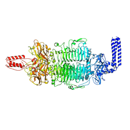
1SVM
 | | Co-crystal structure of SV40 large T antigen helicase domain and ATP | | 分子名称: | ADENOSINE-5'-TRIPHOSPHATE, MAGNESIUM ION, ZINC ION, ... | | 著者 | Gai, D, Zhao, R, Finkielstein, C.V, Chen, X.S. | | 登録日 | 2004-03-29 | | 公開日 | 2004-10-19 | | 最終更新日 | 2024-02-14 | | 実験手法 | X-RAY DIFFRACTION (1.94 Å) | | 主引用文献 | Mechanisms of conformational change for a replicative hexameric helicase of SV40 large tumor antigen.
Cell(Cambridge,Mass.), 119, 2004
|
|
1SVO
 | | Structure of SV40 large T antigen helicase domain | | 分子名称: | ZINC ION, large T antigen | | 著者 | Gai, D, Zhao, R, Finkielstein, C.V, Chen, X.S. | | 登録日 | 2004-03-29 | | 公開日 | 2004-10-19 | | 最終更新日 | 2011-07-13 | | 実験手法 | X-RAY DIFFRACTION (2.6 Å) | | 主引用文献 | Mechanisms of conformational change for a replicative hexameric helicase of SV40 large tumor antigen.
Cell(Cambridge,Mass.), 119, 2004
|
|
1D4Z
 | | CRYSTAL STRUCTURE OF CHEY-95IV, A HYPERACTIVE CHEY MUTANT | | 分子名称: | CHEMOTAXIS PROTEIN CHEY, SULFATE ION | | 著者 | Schuster, M, Zhao, R, Bourret, R.B, Collins, E.J. | | 登録日 | 1999-10-06 | | 公開日 | 1999-10-14 | | 最終更新日 | 2024-02-07 | | 実験手法 | X-RAY DIFFRACTION (1.9 Å) | | 主引用文献 | Correlated switch binding and signaling in bacterial chemotaxis.
J.Biol.Chem., 275, 2000
|
|
3HXG
 | | Crystal structure of Schistsome eIF4E complexed with m7GpppA and 4E-BP | | 分子名称: | Eukaryotic Translation Initiation Factor 4E, Eukaryotic translation initiation factor 4E-binding protein 1, P1-7-METHYLGUANOSINE-P3-ADENOSINE-5',5'-TRIPHOSPHATE | | 著者 | Liu, W, Zhao, R, Jones, D.N.M, Davis, R.E. | | 登録日 | 2009-06-20 | | 公開日 | 2009-08-25 | | 最終更新日 | 2023-09-06 | | 実験手法 | X-RAY DIFFRACTION (2.1 Å) | | 主引用文献 | Structural insights into parasite EIF4E binding specificity for m7G and m2,2,7G mRNA cap.
J.Biol.Chem., 284, 2009
|
|
3HIB
 | |
3HXI
 | | Crystal structure of Schistosome eIF4E complexed with m7GpppG and 4E-BP | | 分子名称: | 7-METHYL-GUANOSINE-5'-TRIPHOSPHATE-5'-GUANOSINE, Eukaryotic Translation Initiation 4E, Eukaryotic translation initiation factor 4E-binding protein 1 | | 著者 | Liu, W, Zhao, R, Jones, D.N.M, Davis, R.E. | | 登録日 | 2009-06-20 | | 公開日 | 2009-08-25 | | 最終更新日 | 2023-09-06 | | 実験手法 | X-RAY DIFFRACTION (1.8 Å) | | 主引用文献 | Structural insights into parasite EIF4E binding specificity for m7G and m2,2,7G mRNA cap.
J.Biol.Chem., 284, 2009
|
|
3HZH
 | | Crystal structure of the CheX-CheY-BeF3-Mg+2 complex from Borrelia burgdorferi | | 分子名称: | Chemotaxis operon protein (CheX), Chemotaxis response regulator (CheY-3), MAGNESIUM ION | | 著者 | Pazy, Y, Silversmith, R.E, Guarinari, M, Zhao, R. | | 登録日 | 2009-06-23 | | 公開日 | 2010-02-16 | | 最終更新日 | 2011-07-13 | | 実験手法 | X-RAY DIFFRACTION (1.96 Å) | | 主引用文献 | Identical phosphatase mechanisms achieved through distinct modes of binding phosphoprotein substrate.
Proc.Natl.Acad.Sci.USA, 107, 2010
|
|
8W2O
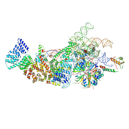 | | Yeast U1 snRNP with humanized U1C Zinc-Finger domain | | 分子名称: | 56 kDa U1 small nuclear ribonucleoprotein component, ACT1 pre-mRNA, Pre-mRNA-processing factor 39, ... | | 著者 | Shi, S.S, Kuang, Z.L, Zhao, R. | | 登録日 | 2024-02-20 | | 公開日 | 2024-05-15 | | 最終更新日 | 2024-07-24 | | 実験手法 | ELECTRON MICROSCOPY (3.49 Å) | | 主引用文献 | Selected humanization of yeast U1 snRNP leads to global suppression of pre-mRNA splicing and mitochondrial dysfunction in the budding yeast.
Rna, 30, 2024
|
|
5WWP
 | | Crystal structure of Middle East respiratory syndrome coronavirus helicase (MERS-CoV nsp13) | | 分子名称: | ORF1ab, SULFATE ION, ZINC ION | | 著者 | Hao, W, Wojdyla, J.A, Zhao, R, Han, R, Das, R, Zlatev, I, Manoharan, M, Wang, M, Cui, S. | | 登録日 | 2017-01-03 | | 公開日 | 2017-07-05 | | 最終更新日 | 2021-09-15 | | 実験手法 | X-RAY DIFFRACTION (3 Å) | | 主引用文献 | Crystal structure of Middle East respiratory syndrome coronavirus helicase
PLoS Pathog., 13, 2017
|
|
2G6Q
 | |
6BK8
 | | S. cerevisiae spliceosomal post-catalytic P complex | | 分子名称: | GUANOSINE-5'-TRIPHOSPHATE, INOSITOL HEXAKISPHOSPHATE, Lea1, ... | | 著者 | Liu, S, Li, X, Zhou, Z.H, Zhao, R. | | 登録日 | 2017-11-07 | | 公開日 | 2018-02-21 | | 最終更新日 | 2020-10-14 | | 実験手法 | ELECTRON MICROSCOPY (3.3 Å) | | 主引用文献 | Structure of the yeast spliceosomal postcatalytic P complex.
Science, 358, 2017
|
|
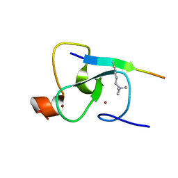
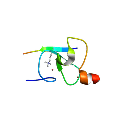
2L6J
 | | Tah1 complexed by MEEVD | | 分子名称: | C-terminus Hsp90 chaperone peptide MEEVD, TPR repeat-containing protein associated with Hsp90 | | 著者 | Jimenez, B, Ugwu, F, Zhao, R, Orti, L, Houry, W.A, Pineda-Lucena, A. | | 登録日 | 2010-11-22 | | 公開日 | 2011-12-28 | | 最終更新日 | 2024-05-01 | | 実験手法 | SOLUTION NMR | | 主引用文献 | Structure of minimal tetratricopeptide repeat domain protein Tah1 reveals mechanism of its interaction with Pih1 and Hsp90.
J.Biol.Chem., 287, 2012
|
|
2P8R
 | | Crystal structure of the C-terminal domain of C. elegans pre-mRNA splicing factor Prp8 carrying R2303K mutant | | 分子名称: | Pre-mRNA-splicing factor Prp8 | | 著者 | Zhang, L, Shen, J, Guarnieri, M.T, Heroux, A, Yang, K, Zhao, R. | | 登録日 | 2007-03-22 | | 公開日 | 2007-05-22 | | 最終更新日 | 2024-04-03 | | 実験手法 | X-RAY DIFFRACTION (2.1 Å) | | 主引用文献 | Crystal structure of the C-terminal domain of splicing factor Prp8 carrying retinitis pigmentosa mutants
Protein Sci., 16, 2007
|
|
2P87
 | | Crystal structure of the C-terminal domain of C. elegans pre-mRNA splicing factor Prp8 | | 分子名称: | Pre-mRNA-splicing factor Prp8 | | 著者 | Zhang, L, Shen, J, Guarnieri, M.T, Heroux, A, Yang, K, Zhao, R. | | 登録日 | 2007-03-21 | | 公開日 | 2007-05-22 | | 最終更新日 | 2024-02-21 | | 実験手法 | X-RAY DIFFRACTION (2.3 Å) | | 主引用文献 | Crystal structure of the C-terminal domain of splicing factor Prp8 carrying retinitis pigmentosa mutants
Protein Sci., 16, 2007
|
|
2QIC
 | | Crystal Structure of the ING1 PHD Finger in complex with a Histone H3K4ME3 peptide | | 分子名称: | H3K4ME3 PEPTIDE, Inhibitor of growth protein 1, ZINC ION | | 著者 | Pena, P.V, Champagne, K, Zhao, R, Kutateladze, T.G. | | 登録日 | 2007-07-03 | | 公開日 | 2008-05-13 | | 最終更新日 | 2023-08-30 | | 実験手法 | X-RAY DIFFRACTION (2.1 Å) | | 主引用文献 | Histone H3K4me3 binding is required for the DNA repair and apoptotic activities of ING1 tumor suppressor.
J.Mol.Biol., 380, 2008
|
|
6N7P
 | | S. cerevisiae spliceosomal E complex (UBC4) | | 分子名称: | 56 kDa U1 small nuclear ribonucleoprotein component, Nuclear cap-binding protein complex subunit 1, Nuclear cap-binding protein subunit 2, ... | | 著者 | Liu, S, Li, X, Zhou, Z.H, Zhao, R. | | 登録日 | 2018-11-27 | | 公開日 | 2019-09-18 | | 最終更新日 | 2024-03-20 | | 実験手法 | ELECTRON MICROSCOPY (3.6 Å) | | 主引用文献 | A unified mechanism for intron and exon definition and back-splicing.
Nature, 573, 2019
|
|
6N7R
 | | Saccharomyces cerevisiae spliceosomal E complex (ACT1) | | 分子名称: | 56 kDa U1 small nuclear ribonucleoprotein component, ACT1 pre-mRNA, Pre-mRNA-processing factor 39, ... | | 著者 | Liu, S, Li, X, Zhou, Z.H, Zhao, R. | | 登録日 | 2018-11-28 | | 公開日 | 2019-09-18 | | 最終更新日 | 2024-03-20 | | 実験手法 | ELECTRON MICROSCOPY (3.2 Å) | | 主引用文献 | A unified mechanism for intron and exon definition and back-splicing.
Nature, 573, 2019
|
|
8JED
 | | Crystal structure of mRNA cap (guanine-N7) methyltransferase E12 subunit from monkeypox virus and discovery of its inhibitors | | 分子名称: | mRNA-capping enzyme regulatory subunit OPG124 | | 著者 | Wang, D, Zhao, R, Shu, W, Hu, W, Wang, M, Cao, J, Zhou, X. | | 登録日 | 2023-05-15 | | 公開日 | 2023-12-06 | | 実験手法 | X-RAY DIFFRACTION (2.16 Å) | | 主引用文献 | Crystal structure of mRNA cap (guanine-N7) methyltransferase E12 subunit from monkeypox virus and discovery of its inhibitors.
Int.J.Biol.Macromol., 253, 2023
|
|
7VYV
 | | Cryo-EM structure of Depo32, a Klebsiella phage depolymerase targets the K2 serotype K. pneumoniae | | 分子名称: | Depolymerase | | 著者 | Cai, R, Ren, Z, Zhao, R, Wang, X, Guo, Z, Du, R, Han, W, Ru, H, Gu, J. | | 登録日 | 2021-11-15 | | 公開日 | 2023-08-30 | | 最終更新日 | 2024-09-11 | | 実験手法 | ELECTRON MICROSCOPY (2.32 Å) | | 主引用文献 | Structural biology and functional features of phage-derived depolymerase Depo32 on Klebsiella pneumoniae with K2 serotype capsular polysaccharides.
Microbiol Spectr, 11, 2023
|
|
7VZ3
 | | Cryo-EM structure of Depo32, a Klebsiella phage depolymerase targets the K2 serotype K. pneumoniae | | 分子名称: | Depolymerase | | 著者 | Cai, R, Ren, Z, Zhao, R, Wang, X, Guo, Z, Du, R, Han, W, Ru, H, Gu, J. | | 登録日 | 2021-11-15 | | 公開日 | 2023-08-30 | | 最終更新日 | 2024-09-11 | | 実験手法 | ELECTRON MICROSCOPY (2.46 Å) | | 主引用文献 | Structural biology and functional features of phage-derived depolymerase Depo32 on Klebsiella pneumoniae with K2 serotype capsular polysaccharides.
Microbiol Spectr, 11, 2023
|
|
6N7X
 | | S. cerevisiae U1 snRNP | | 分子名称: | 56 kDa U1 small nuclear ribonucleoprotein component, Pre-mRNA-processing factor 39, Protein NAM8, ... | | 著者 | Li, X, Liu, S, Jiang, J, Zhang, L, Espinosa, S, Hill, R.C, Hansen, K.C, Zhou, Z.H, Zhao, R. | | 登録日 | 2018-11-28 | | 公開日 | 2019-07-24 | | 最終更新日 | 2024-03-13 | | 実験手法 | ELECTRON MICROSCOPY (3.6 Å) | | 主引用文献 | CryoEM structure of Saccharomyces cerevisiae U1 snRNP offers insight into alternative splicing.
Nat Commun, 8, 2017
|
|
8J5J
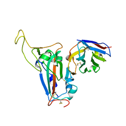 | | The crystal structure of bat coronavirus RsYN04 RBD bound to the antibody S43 | | 分子名称: | 2-acetamido-2-deoxy-beta-D-glucopyranose, Spike protein S1, nano antibody S43 | | 著者 | Zhao, R.C, Niu, S, Han, P, Qi, J.X, Gao, G.F, Wang, Q.H. | | 登録日 | 2023-04-23 | | 公開日 | 2023-11-22 | | 実験手法 | X-RAY DIFFRACTION (3 Å) | | 主引用文献 | Cross-species recognition of bat coronavirus RsYN04 and cross-reaction of SARS-CoV-2 antibodies against the virus.
Zool.Res., 44, 2023
|
|
8XS3
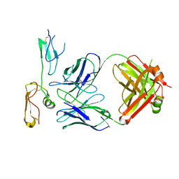 | | Structure of MPXV B6 and D68 fab complex | | 分子名称: | 2-acetamido-2-deoxy-beta-D-glucopyranose, 2-acetamido-2-deoxy-beta-D-glucopyranose-(1-4)-2-acetamido-2-deoxy-beta-D-glucopyranose, D68_heavy chain, ... | | 著者 | wu, L.L, Sun, J.Q. | | 登録日 | 2024-01-08 | | 公開日 | 2024-06-12 | | 最終更新日 | 2024-06-19 | | 実験手法 | ELECTRON MICROSCOPY (3.46 Å) | | 主引用文献 | Two noncompeting human neutralizing antibodies targeting MPXV B6 show protective effects against orthopoxvirus infections.
Nat Commun, 15, 2024
|
|


