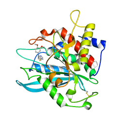
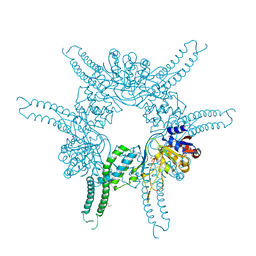
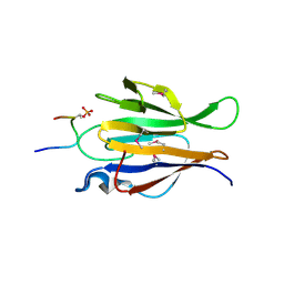
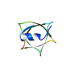
2AFW
 
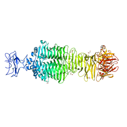
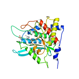 | | Crystal structure of human glutaminyl cyclase in complex with N-acetylhistamine | | 分子名称: | Glutaminyl-peptide cyclotransferase, N-[2-(1H-IMIDAZOL-4-YL)ETHYL]ACETAMIDE, SULFATE ION, ... | | 著者 | Huang, K.F, Liu, Y.L, Cheng, W.J, Ko, T.P, Wang, A.H.J. | | 登録日 | 2005-07-26 | | 公開日 | 2005-08-23 | | 最終更新日 | 2024-03-13 | | 実験手法 | X-RAY DIFFRACTION (1.56 Å) | | 主引用文献 | Crystal structures of human glutaminyl cyclase, an enzyme responsible for protein N-terminal pyroglutamate formation
Proc.Natl.Acad.Sci.Usa, 102, 2005
|
|
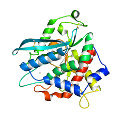
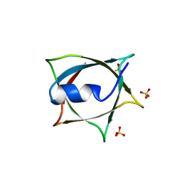
2AFS
 | | Crystal structure of the genetic mutant R54W of human glutaminyl cyclase | | 分子名称: | Glutaminyl-peptide cyclotransferase, SULFATE ION, ZINC ION | | 著者 | Huang, K.F, Liu, Y.L, Cheng, W.J, Ko, T.P, Wang, A.H.J. | | 登録日 | 2005-07-26 | | 公開日 | 2005-08-23 | | 最終更新日 | 2024-05-29 | | 実験手法 | X-RAY DIFFRACTION (2.22 Å) | | 主引用文献 | Crystal structures of human glutaminyl cyclase, an enzyme responsible for protein N-terminal pyroglutamate formation
Proc.Natl.Acad.Sci.Usa, 102, 2005
|
|
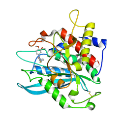
2AFU
 | | Crystal structure of human glutaminyl cyclase in complex with glutamine t-butyl ester | | 分子名称: | Glutaminyl-peptide cyclotransferase, TERT-BUTYL D-ALPHA-GLUTAMINATE, ZINC ION | | 著者 | Huang, K.F, Liu, Y.L, Cheng, W.J, Ko, T.P, Wang, A.H.J. | | 登録日 | 2005-07-26 | | 公開日 | 2005-08-23 | | 最終更新日 | 2024-05-29 | | 実験手法 | X-RAY DIFFRACTION (2.22 Å) | | 主引用文献 | Crystal structures of human glutaminyl cyclase, an enzyme responsible for protein N-terminal pyroglutamate formation
Proc.Natl.Acad.Sci.Usa, 102, 2005
|
|
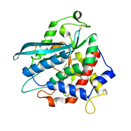
4MHN
 | | Crystal structure of a glutaminyl cyclase from Ixodes scapularis | | 分子名称: | Glutaminyl cyclase, putative, ZINC ION | | 著者 | Huang, K.F, Hsu, H.L, Wang, A.H.J. | | 登録日 | 2013-08-30 | | 公開日 | 2014-03-12 | | 最終更新日 | 2023-11-08 | | 実験手法 | X-RAY DIFFRACTION (1.15 Å) | | 主引用文献 | Structural and functional analyses of a glutaminyl cyclase from Ixodes scapularis reveal metal-independent catalysis and inhibitor binding.
Acta Crystallogr.,Sect.D, 70, 2014
|
|
4MHY
 | | Crystal structure of a glutaminyl cyclase from Ixodes scapularis in complex with PBD150 | | 分子名称: | 1-(3,4-dimethoxyphenyl)-3-[3-(1H-imidazol-1-yl)propyl]thiourea, Glutaminyl cyclase, putative, ... | | 著者 | Huang, K.F, Hsu, H.L, Wang, A.H.J. | | 登録日 | 2013-08-30 | | 公開日 | 2014-03-12 | | 最終更新日 | 2023-11-08 | | 実験手法 | X-RAY DIFFRACTION (1.38 Å) | | 主引用文献 | Structural and functional analyses of a glutaminyl cyclase from Ixodes scapularis reveal metal-independent catalysis and inhibitor binding.
Acta Crystallogr.,Sect.D, 70, 2014
|
|
4MHP
 | |
4MHZ
 | | Crystal structure of apo-form glutaminyl cyclase from Ixodes scapularis in complex with PBD150 | | 分子名称: | 1-(3,4-dimethoxyphenyl)-3-[3-(1H-imidazol-1-yl)propyl]thiourea, Glutaminyl cyclase, putative | | 著者 | Huang, K.F, Hsu, H.L, Wang, A.H.J. | | 登録日 | 2013-08-30 | | 公開日 | 2014-03-12 | | 最終更新日 | 2023-11-08 | | 実験手法 | X-RAY DIFFRACTION (1.95 Å) | | 主引用文献 | Structural and functional analyses of a glutaminyl cyclase from Ixodes scapularis reveal metal-independent catalysis and inhibitor binding.
Acta Crystallogr.,Sect.D, 70, 2014
|
|
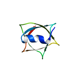
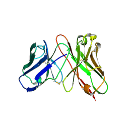
3VA1
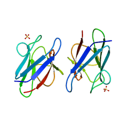 | | Crystal structure of the mammalian MDC1 FHA domain | | 分子名称: | Mediator of DNA damage checkpoint protein 1, SULFATE ION | | 著者 | Wu, H.H, Wu, P.Y, Huang, K.F, Kao, Y.Y, Tsai, M.D. | | 登録日 | 2011-12-28 | | 公開日 | 2012-02-01 | | 最終更新日 | 2024-03-20 | | 実験手法 | X-RAY DIFFRACTION (1.74 Å) | | 主引用文献 | Structural Delineation of MDC1-FHA Domain Binding with CHK2-pThr68.
Biochemistry, 2012
|
|
4FW9
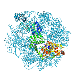 | | Crystal structure of the Lon-like protease MtaLonC | | 分子名称: | PHOSPHATE ION, TTC1975 peptidase | | 著者 | Chang, C.I, Ihara, K, Kuo, C.I, Huang, K.F, Wakatsuki, S. | | 登録日 | 2012-06-30 | | 公開日 | 2013-06-26 | | 最終更新日 | 2013-09-11 | | 実験手法 | X-RAY DIFFRACTION (2 Å) | | 主引用文献 | Structures of an ATP-independent Lon-like protease and its complexes with covalent inhibitors
Acta Crystallogr.,Sect.D, 69, 2013
|
|
4FWH
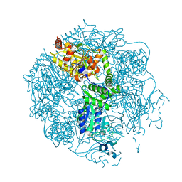 | | Crystal structure of the Lon-like protease MtaLonC in complex with MG262 | | 分子名称: | N-[(benzyloxy)carbonyl]-L-leucyl-N-[(1R)-1-(dihydroxyboranyl)-3-methylbutyl]-L-leucinamide, PHOSPHATE ION, TTC1975 peptidase | | 著者 | Chang, C.I, Kuo, C.I, Huang, K.F. | | 登録日 | 2012-07-01 | | 公開日 | 2013-06-26 | | 最終更新日 | 2023-11-08 | | 実験手法 | X-RAY DIFFRACTION (2.19 Å) | | 主引用文献 | Structures of an ATP-independent Lon-like protease and its complexes with covalent inhibitors
Acta Crystallogr.,Sect.D, 69, 2013
|
|
4FWV
 | | Crystal structure of the N-terminal domain of the Lon-like protease MtaLonC | | 分子名称: | SULFATE ION, TTC1975 peptidase | | 著者 | Chang, C.I, Li, J.K, Kuo, C.I, Huang, K.F. | | 登録日 | 2012-07-02 | | 公開日 | 2013-06-26 | | 最終更新日 | 2014-03-12 | | 実験手法 | X-RAY DIFFRACTION (2.4 Å) | | 主引用文献 | The N-terminal substrate-recognition domain of a LonC protease exhibits structural and functional similarity to cytosolic chaperones
Acta Crystallogr.,Sect.D, 69, 2013
|
|
8W68
 | | Crystal structure of Q9PR55 at pH 6.0 (use NMR model) | | 分子名称: | Uncharacterized protein UU089.1 | | 著者 | Hsu, M.F, Ko, T.P, Huang, K.F, Chen, Y.R, Huang, J.S, Hsu, S.T.D. | | 登録日 | 2023-08-28 | | 公開日 | 2024-02-07 | | 実験手法 | X-RAY DIFFRACTION (2.3 Å) | | 主引用文献 | Structure, dynamics, and stability of the smallest and most complex 7 1 protein knot.
J.Biol.Chem., 300, 2023
|
|
8IQ9
 | | Crystal structure of trimeric K2-2 TSP in complex with tetrasaccharide and octasaccharide | | 分子名称: | 1,2-ETHANEDIOL, ACETYL GROUP, K2-2 TSP, ... | | 著者 | Ye, T.J, Ko, T.P, Huang, K.F, Wu, S.H. | | 登録日 | 2023-03-16 | | 公開日 | 2024-02-21 | | 最終更新日 | 2024-04-03 | | 実験手法 | X-RAY DIFFRACTION (1.58 Å) | | 主引用文献 | Klebsiella pneumoniae K2 capsular polysaccharide degradation by a bacteriophage depolymerase does not require trimer formation.
Mbio, 15, 2024
|
|
8IQE
 | | Crystal structure of tetrameric K2-2 TSP | | 分子名称: | GLYCEROL, K2-VCL6 TSP | | 著者 | Ye, T.J, Huang, K.F, Tu, I.F, Lee, I.M, Chang, Y.P, Wu, S.H. | | 登録日 | 2023-03-16 | | 公開日 | 2024-02-21 | | 最終更新日 | 2024-04-03 | | 実験手法 | X-RAY DIFFRACTION (2.17 Å) | | 主引用文献 | Klebsiella pneumoniae K2 capsular polysaccharide degradation by a bacteriophage depolymerase does not require trimer formation.
Mbio, 15, 2024
|
|
4YM4
 | | Truncated Human TIFA in complex with its Thr9 phosphorylated N-terminal peptide 1-15 | | 分子名称: | TRAF-interacting protein with FHA domain-containing protein A | | 著者 | Weng, J.H, Wei, T.Y.W, Hsieh, Y.C, Huang, C.C.F, Wu, P.Y.G, Chen, E.S.W, Huang, K.F, Chen, C.J, Tsai, M.D. | | 登録日 | 2015-03-06 | | 公開日 | 2015-10-21 | | 最終更新日 | 2015-10-28 | | 実験手法 | X-RAY DIFFRACTION (3.12 Å) | | 主引用文献 | Uncovering the Mechanism of Forkhead-Associated Domain-Mediated TIFA Oligomerization That Plays a Central Role in Immune Responses.
Biochemistry, 54, 2015
|
|
4ZGI
 | | Structure of Truncated Human TIFA | | 分子名称: | TRAF-interacting protein with FHA domain-containing protein A | | 著者 | Weng, J.H, Wei, T.Y.W, Hsieh, Y.C, Huang, C.C.F, Wu, P.Y.G, Chen, E.S.W, Huang, K.F, Chen, C.J, Tsai, M.D. | | 登録日 | 2015-04-23 | | 公開日 | 2015-10-14 | | 最終更新日 | 2015-10-28 | | 実験手法 | X-RAY DIFFRACTION (2.701 Å) | | 主引用文献 | Uncovering the Mechanism of Forkhead-Associated Domain-Mediated TIFA Oligomerization That Plays a Central Role in Immune Responses.
Biochemistry, 54, 2015
|
|
4FWG
 | | Crystal structure of the Lon-like protease MtaLonC in complex with lactacystin | | 分子名称: | Omuralide, open form, PHOSPHATE ION, ... | | 著者 | Chang, C.I, Kuo, C.I, Huang, K.F. | | 登録日 | 2012-07-01 | | 公開日 | 2013-06-26 | | 最終更新日 | 2023-12-06 | | 実験手法 | X-RAY DIFFRACTION (1.99 Å) | | 主引用文献 | Structures of an ATP-independent Lon-like protease and its complexes with covalent inhibitors
Acta Crystallogr.,Sect.D, 69, 2013
|
|
4FWD
 | | Crystal structure of the Lon-like protease MtaLonC in complex with bortezomib | | 分子名称: | N-[(1R)-1-(DIHYDROXYBORYL)-3-METHYLBUTYL]-N-(PYRAZIN-2-YLCARBONYL)-L-PHENYLALANINAMIDE, PHOSPHATE ION, TTC1975 peptidase | | 著者 | Chang, C.I, Kuo, C.I, Huang, K.F. | | 登録日 | 2012-06-30 | | 公開日 | 2013-06-26 | | 最終更新日 | 2023-11-08 | | 実験手法 | X-RAY DIFFRACTION (2.03 Å) | | 主引用文献 | Structures of an ATP-independent Lon-like protease and its complexes with covalent inhibitors
Acta Crystallogr.,Sect.D, 69, 2013
|
|
8IWC
 | | Crystal structure of Q9PR55 at pH 6.0 | | 分子名称: | Uncharacterized protein UU089.1 | | 著者 | Hsu, M.F, Ko, T.P, Huang, K.F, Chen, Y.R, Huang, J.S, Hsu, S.T.D. | | 登録日 | 2023-03-29 | | 公開日 | 2024-02-07 | | 実験手法 | X-RAY DIFFRACTION (2.43 Å) | | 主引用文献 | Structure, dynamics, and stability of the smallest and most complex 7 1 protein knot.
J.Biol.Chem., 300, 2023
|
|
8IWA
 | | Crystal structure of Q9PR55 at pH 6.5 | | 分子名称: | SULFATE ION, Uncharacterized protein UU089.1 | | 著者 | Hsu, M.F, Ko, T.P, Huang, K.F, Chen, Y.R, Huang, J.S, Hsu, S.T.D. | | 登録日 | 2023-03-29 | | 公開日 | 2024-02-07 | | 実験手法 | X-RAY DIFFRACTION (2.51 Å) | | 主引用文献 | Structure, dynamics, and stability of the smallest and most complex 7 1 protein knot.
J.Biol.Chem., 300, 2023
|
|
8IWB
 | | Crystal structure of Q9PR55 at pH 7.5 | | 分子名称: | Uncharacterized protein UU089.1 | | 著者 | Hsu, M.F, Ko, T.P, Huang, K.F, Chen, Y.R, Huang, J.S, Hsu, S.T.D. | | 登録日 | 2023-03-29 | | 公開日 | 2024-02-07 | | 実験手法 | X-RAY DIFFRACTION (2.42 Å) | | 主引用文献 | Structure, dynamics, and stability of the smallest and most complex 7 1 protein knot.
J.Biol.Chem., 300, 2023
|
|
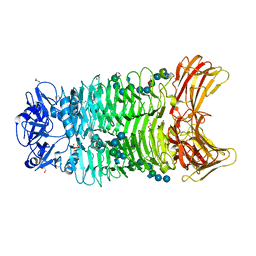
5JSD
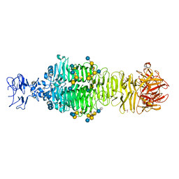 | | Crystal structure of phiAB6 tailspike in complex with five-repeated oligosaccharides of Acinetobacter baumannii surface polysaccharide | | 分子名称: | ACETIC ACID, MALONIC ACID, beta-D-galactopyranose-(1-3)-2-amino-2-deoxy-beta-D-galactopyranose-(1-3)-[5,7-bisacetamido-3,5,7,9-tetradeoxy-L-glycero-alpha-L-manno-non-2-ulopyranosonic acid-(2-6)-beta-D-glucopyranose-(1-6)]beta-D-galactopyranose-(1-3)-2-amino-2-deoxy-beta-D-galactopyranose-(1-3)-[beta-D-glucopyranose-(1-6)]beta-D-galactopyranose, ... | | 著者 | Lee, I.M, Tu, I.F, Huang, K.F, Wu, S.H. | | 登録日 | 2016-05-08 | | 公開日 | 2017-03-08 | | 最終更新日 | 2024-03-20 | | 実験手法 | X-RAY DIFFRACTION (1.48 Å) | | 主引用文献 | Structural basis for fragmenting the exopolysaccharide of Acinetobacter baumannii by bacteriophage Phi AB6 tailspike protein
Sci Rep, 7, 2017
|
|
5JSE
 | | Crystal structure of phiAB6 tailspike in complex with three-repeated oligosaccharides of Acinetobacter baumannii surface polysaccharide | | 分子名称: | ACETIC ACID, MALONIC ACID, beta-D-galactopyranose-(1-3)-2-amino-2-deoxy-beta-D-galactopyranose-(1-3)-[5,7-bisacetamido-3,5,7,9-tetradeoxy-L-glycero-alpha-L-manno-non-2-ulopyranosonic acid-(2-6)-beta-D-glucopyranose-(1-6)]beta-D-galactopyranose-(1-3)-2-amino-2-deoxy-beta-D-galactopyranose-(1-3)-[beta-D-glucopyranose-(1-6)]beta-D-galactopyranose, ... | | 著者 | Lee, I.M, Tu, I.F, Huang, K.F, Wu, S.H. | | 登録日 | 2016-05-08 | | 公開日 | 2017-03-08 | | 最終更新日 | 2024-03-20 | | 実験手法 | X-RAY DIFFRACTION (1.89 Å) | | 主引用文献 | Structural basis for fragmenting the exopolysaccharide of Acinetobacter baumannii by bacteriophage Phi AB6 tailspike protein
Sci Rep, 7, 2017
|
|
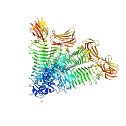
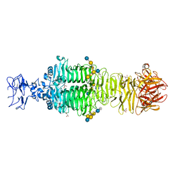
5JS4
 | | Crystal structure of phiAB6 tailspike | | 分子名称: | MALONIC ACID, phiAB6 tailspike | | 著者 | Lee, I.M, Tu, I.F, Huang, K.F, Wu, S.H. | | 登録日 | 2016-05-07 | | 公開日 | 2017-03-08 | | 最終更新日 | 2024-03-20 | | 実験手法 | X-RAY DIFFRACTION (1.48 Å) | | 主引用文献 | Structural basis for fragmenting the exopolysaccharide of Acinetobacter baumannii by bacteriophage Phi AB6 tailspike protein
Sci Rep, 7, 2017
|
|
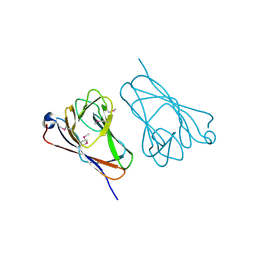
3AUV
 | | Predicting Amino Acid Preferences in the Complementarity Determining Regions of an Antibody-Antigen Recognition Interface | | 分子名称: | sc-dsFv derived from the G6-Fab | | 著者 | Yu, C.M, Peng, H.P, Chen, I.C, Lee, Y.C, Chen, J.B, Tsai, K.C, Chen, C.T, Chang, J.Y, Yang, E.W, Hsu, P.C, Jian, J.W, Hsu, H.J, Chang, H.J, Hsu, W.L, Huang, K.F, Ma, A.C, Yang, A.S. | | 登録日 | 2011-02-16 | | 公開日 | 2012-02-22 | | 最終更新日 | 2012-04-25 | | 実験手法 | X-RAY DIFFRACTION (2.4 Å) | | 主引用文献 | Rationalization and design of the complementarity determining region sequences in an antibody-antigen recognition interface
Plos One, 7, 2012
|
|


