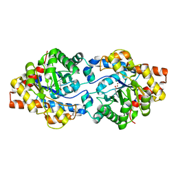
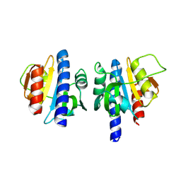
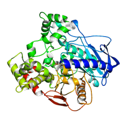
4XD6
 
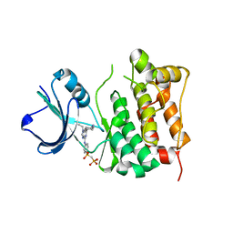
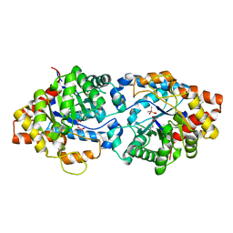 | | Phosphotriesterase Variant E2a | | 分子名称: | (4S)-2-METHYL-2,4-PENTANEDIOL, CACODYLATE ION, Phosphotriesterase variant PTE-E2, ... | | 著者 | Jackson, C.J, Campbell, E, Kaltenbach, M, Tokuriki, N. | | 登録日 | 2014-12-19 | | 公開日 | 2015-12-23 | | 最終更新日 | 2023-11-15 | | 実験手法 | X-RAY DIFFRACTION (1.75 Å) | | 主引用文献 | The role of protein dynamics in the evolution of new enzyme function.
Nat.Chem.Biol., 12, 2016
|
|
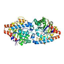
4XD5
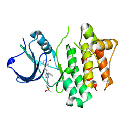
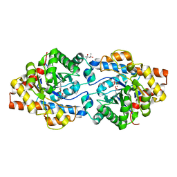 | | Phosphotriesterase variant R2 | | 分子名称: | (4S)-2-METHYL-2,4-PENTANEDIOL, CACODYLATE ION, Phosphotriesterase variant PTE-R2, ... | | 著者 | Campbell, E, Kaltenbach, M, Tokuriki, N, Jackson, C.J. | | 登録日 | 2014-12-19 | | 公開日 | 2015-12-23 | | 最終更新日 | 2023-11-15 | | 実験手法 | X-RAY DIFFRACTION (1.85 Å) | | 主引用文献 | The role of protein dynamics in the evolution of new enzyme function.
Nat.Chem.Biol., 12, 2016
|
|
4XAF
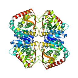
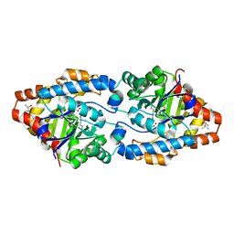 | | Cycles of destabilization and repair underlie evolutionary transitions in enzymes | | 分子名称: | (4S)-2-METHYL-2,4-PENTANEDIOL, CACODYLATE ION, Phosphotriesterase variant PTE-R1, ... | | 著者 | Jackson, C.J, Campbell, E, Kaltenbach, M, Tokuriki, N. | | 登録日 | 2014-12-14 | | 公開日 | 2015-12-16 | | 最終更新日 | 2023-11-15 | | 実験手法 | X-RAY DIFFRACTION (1.66 Å) | | 主引用文献 | The role of protein dynamics in the evolution of new enzyme function.
Nat.Chem.Biol., 12, 2016
|
|
3SR2
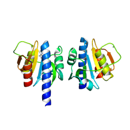
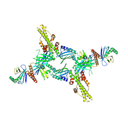 | | Crystal Structure of Human XLF-XRCC4 Complex | | 分子名称: | DNA repair protein XRCC4, Non-homologous end-joining factor 1 | | 著者 | Hammel, M, Classen, S, Tainer, J.A. | | 登録日 | 2011-07-06 | | 公開日 | 2011-07-20 | | 最終更新日 | 2023-09-13 | | 実験手法 | X-RAY DIFFRACTION (3.9708 Å) | | 主引用文献 | XRCC4 Protein Interactions with XRCC4-like Factor (XLF) Create an Extended Grooved Scaffold for DNA Ligation and Double Strand Break Repair.
J.Biol.Chem., 286, 2011
|
|
4XAZ
 | | Cycles of destabilization and repair underlie evolutionary transitions in enzymes | | 分子名称: | (4S)-2-METHYL-2,4-PENTANEDIOL, Phosphotriesterase variant PTE-R18, ZINC ION | | 著者 | Jackson, C.J, Campbell, E, Kaltenbach, M, Tokuriki, N. | | 登録日 | 2014-12-16 | | 公開日 | 2015-12-16 | | 最終更新日 | 2023-11-15 | | 実験手法 | X-RAY DIFFRACTION (1.55 Å) | | 主引用文献 | The role of protein dynamics in the evolution of new enzyme function.
Nat.Chem.Biol., 12, 2016
|
|
4XD4
 | | Phosphotriesterase variant E2b | | 分子名称: | (4S)-2-METHYL-2,4-PENTANEDIOL, CACODYLATE ION, Phosphotriesterase variant PTE-R3, ... | | 著者 | Jackson, C.J, Campbell, E, Kaltenbach, M, Tokuriki, N. | | 登録日 | 2014-12-19 | | 公開日 | 2015-12-23 | | 最終更新日 | 2023-11-15 | | 実験手法 | X-RAY DIFFRACTION (1.9 Å) | | 主引用文献 | The role of protein dynamics in the evolution of new enzyme function.
Nat.Chem.Biol., 12, 2016
|
|
6I3T
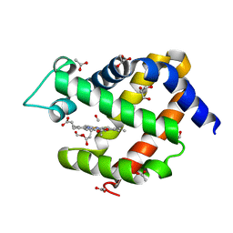 | | Crystal structure of murine neuroglobin bound to CO at 40 K. | | 分子名称: | ACETATE ION, CARBON MONOXIDE, FORMIC ACID, ... | | 著者 | Savino, C, Montemiglio, L.C, Ardiccioni, C, Exertier, C. | | 登録日 | 2018-11-07 | | 公開日 | 2019-09-11 | | 最終更新日 | 2024-01-24 | | 実験手法 | X-RAY DIFFRACTION (2 Å) | | 主引用文献 | Ligand pathways in neuroglobin revealed by low-temperature photodissociation and docking experiments.
Iucrj, 6, 2019
|
|
6I40
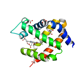 | | Crystal structure of murine neuroglobin bound to CO at 15K under illumination using optical fiber | | 分子名称: | ACETATE ION, CARBON MONOXIDE, FORMIC ACID, ... | | 著者 | Savino, C, Montemiglio, L.C, Ardiccioni, C, Exertier, C, Vallone, B. | | 登録日 | 2018-11-08 | | 公開日 | 2019-09-11 | | 最終更新日 | 2024-01-24 | | 実験手法 | X-RAY DIFFRACTION (1.9 Å) | | 主引用文献 | Ligand pathways in neuroglobin revealed by low-temperature photodissociation and docking experiments.
Iucrj, 6, 2019
|
|
4YHU
 | | Yeast Prp3 C-terminal fragment 296-469 | | 分子名称: | U4/U6 small nuclear ribonucleoprotein PRP3, YTTRIUM (III) ION | | 著者 | Liu, S, Wahl, M.C. | | 登録日 | 2015-02-27 | | 公開日 | 2015-07-22 | | 最終更新日 | 2024-05-08 | | 実験手法 | X-RAY DIFFRACTION (2.7 Å) | | 主引用文献 | A composite double-/single-stranded RNA-binding region in protein Prp3 supports tri-snRNP stability and splicing.
Elife, 4, 2015
|
|
4XAY
 | | Cycles of destabilization and repair underlie evolutionary transitions in enzymes | | 分子名称: | (4S)-2-METHYL-2,4-PENTANEDIOL, CACODYLATE ION, Phosphotriesterase variant PTE-R8, ... | | 著者 | Jackson, C.J, Campbell, E, Kaltenbach, M, Tokuriki, N. | | 登録日 | 2014-12-16 | | 公開日 | 2015-12-16 | | 最終更新日 | 2023-11-15 | | 実験手法 | X-RAY DIFFRACTION (1.84 Å) | | 主引用文献 | The role of protein dynamics in the evolution of new enzyme function.
Nat.Chem.Biol., 12, 2016
|
|
4KJU
 | | Crystal structure of XIAP-Bir2 with a bound benzodiazepinone inhibitor. | | 分子名称: | E3 ubiquitin-protein ligase XIAP, N-{(3S)-5-(4-aminobenzoyl)-1-[(2-methoxynaphthalen-1-yl)methyl]-2-oxo-2,3,4,5-tetrahydro-1H-1,5-benzodiazepin-3-yl}-N~2~-methyl-L-alaninamide, ZINC ION | | 著者 | Lukacs, C.M, Janson, C.A. | | 登録日 | 2013-05-03 | | 公開日 | 2013-11-27 | | 最終更新日 | 2023-09-20 | | 実験手法 | X-RAY DIFFRACTION (1.6 Å) | | 主引用文献 | Optimization of Benzodiazepinones as Selective Inhibitors of the X-Linked Inhibitor of Apoptosis Protein (XIAP) Second Baculovirus IAP Repeat (BIR2) Domain.
J.Med.Chem., 56, 2013
|
|
4KJV
 | | Crystal structure of XIAP-Bir2 with a bound spirocyclic benzoxazepinone inhibitor. | | 分子名称: | 6-methoxy-5-({(3S)-3-[(N-methyl-L-alanyl)amino]-4-oxo-2',3,3',4,5',6'-hexahydro-5H-spiro[1,5-benzoxazepine-2,4'-pyran]-5-yl}methyl)naphthalene-2-carboxylic acid, E3 ubiquitin-protein ligase XIAP, ZINC ION | | 著者 | Lukacs, C.M, Janson, C.A. | | 登録日 | 2013-05-03 | | 公開日 | 2013-11-27 | | 最終更新日 | 2023-09-20 | | 実験手法 | X-RAY DIFFRACTION (1.7 Å) | | 主引用文献 | Optimization of Benzodiazepinones as Selective Inhibitors of the X-Linked Inhibitor of Apoptosis Protein (XIAP) Second Baculovirus IAP Repeat (BIR2) Domain.
J.Med.Chem., 56, 2013
|
|
3TJP
 | | Crystal Structure of PI3K gamma with N6-(3,4-dimethoxyphenyl)-2-morpholino-[4,5'-bipyrimidine]-2',6-diamine | | 分子名称: | N~6~-(3,4-dimethoxyphenyl)-2-(morpholin-4-yl)-4,5'-bipyrimidine-2',6-diamine, Phosphatidylinositol-4,5-bisphosphate 3-kinase catalytic subunit gamma isoform, SULFATE ION | | 著者 | Knapp, M.S, Elling, R.A, Ornelas, E. | | 登録日 | 2011-08-24 | | 公開日 | 2012-08-29 | | 最終更新日 | 2023-09-13 | | 実験手法 | X-RAY DIFFRACTION (2.7 Å) | | 主引用文献 | The Identification of 5-(2,4-dimorpholinopyrimidin-6-yl)-4-(trifluoromethyl)pyridin-2-amine (NVP-BKM120) as a Potent, Selective and Orally Bioavailable Class I PI3 Kinase Inhibitor for the Treatment of Cancer
To be Published
|
|
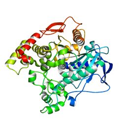
2VK1
 | | Crystal structure of the Saccharomyces cerevisiae pyruvate decarboxylase variant D28A in complex with its substrate | | 分子名称: | MAGNESIUM ION, PYRUVATE DECARBOXYLASE ISOZYME 1, PYRUVIC ACID, ... | | 著者 | Kutter, S, Weik, M, Weiss, M.S, Konig, S. | | 登録日 | 2007-12-16 | | 公開日 | 2009-01-27 | | 最終更新日 | 2023-12-13 | | 実験手法 | X-RAY DIFFRACTION (1.71 Å) | | 主引用文献 | Covalently Bound Substrate at the Regulatory Site of Yeast Pyruvate Decarboxylases Triggers Allosteric Enzyme Activation.
J.Biol.Chem., 284, 2009
|
|
2VK8
 | | Crystal structure of the Saccharomyces cerevisiae pyruvate decarboxylase variant E477Q in complex with its substrate | | 分子名称: | (2S)-2-HYDROXYPROPANOIC ACID, MAGNESIUM ION, PYRUVATE DECARBOXYLASE ISOZYME 1, ... | | 著者 | Kutter, S, Weik, M, Weiss, M.S, Konig, S. | | 登録日 | 2007-12-17 | | 公開日 | 2009-01-27 | | 最終更新日 | 2023-12-13 | | 実験手法 | X-RAY DIFFRACTION (1.42 Å) | | 主引用文献 | Covalently Bound Substrate at the Regulatory Site of Yeast Pyruvate Decarboxylases Triggers Allosteric Enzyme Activation.
J.Biol.Chem., 284, 2009
|
|
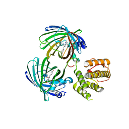
4L1D
 | | Voltage-gated sodium channel beta3 subunit Ig domain | | 分子名称: | Sodium channel subunit beta-3 | | 著者 | Namadurai, S, Weimhofer, M, Rajappa, R, Stott, K, Klingauf, J, Chirgadze, D.Y, Jackson, A.P. | | 登録日 | 2013-06-03 | | 公開日 | 2014-03-05 | | 最終更新日 | 2023-09-20 | | 実験手法 | X-RAY DIFFRACTION (2.5 Å) | | 主引用文献 | Crystal Structure and Molecular Imaging of the Nav Channel beta 3 Subunit Indicates a Trimeric Assembly.
J.Biol.Chem., 289, 2014
|
|
2BDF
 | | Src kinase in complex with inhibitor AP23451 | | 分子名称: | Proto-oncogene tyrosine-protein kinase Src, {[(4-{[2-(4-AMINOCYCLOHEXYL)-9-ETHYL-9H-PURIN-6-YL]AMINO}PHENYL)(HYDROXY)PHOSPHORYL]METHYL}PHOSPHONIC ACID | | 著者 | Dalgarno, D, Stehle, T, Schelling, P, Sawyer, T, Narula, S. | | 登録日 | 2005-10-20 | | 公開日 | 2006-10-24 | | 最終更新日 | 2023-08-23 | | 実験手法 | X-RAY DIFFRACTION (2.1 Å) | | 主引用文献 | Structural basis of Src tyrosine kinase inhibition with a new class of potent and selective trisubstituted purine-based compounds.
Chem.Biol.Drug Des., 67, 2006
|
|
2BDJ
 | | Src kinase in complex with inhibitor AP23464 | | 分子名称: | 3-[2-(2-CYCLOPENTYL-6-{[4-(DIMETHYLPHOSPHORYL)PHENYL]AMINO}-9H-PURIN-9-YL)ETHYL]PHENOL, Proto-oncogene tyrosine-protein kinase Src | | 著者 | Dalgarno, D, Stehle, T, Schelling, P, Narula, S, Sawyer, T. | | 登録日 | 2005-10-20 | | 公開日 | 2006-10-24 | | 最終更新日 | 2023-08-23 | | 実験手法 | X-RAY DIFFRACTION (2.5 Å) | | 主引用文献 | Structural basis of Src tyrosine kinase inhibition with a new class of potent and selective trisubstituted purine-based compounds.
Chem.Biol.Drug Des., 67, 2006
|
|
4JCO
 | |
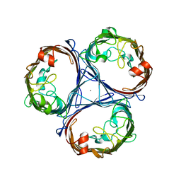
8OGN
 | | PanDDA analysis group deposition -- CdaA in complex with fragment F2X-Entry A09 | | 分子名称: | Cyclic di-AMP synthase CdaA, MAGNESIUM ION, [1,2]thiazolo[5,4-b]pyridin-3-amine | | 著者 | Garbers, T.B, Neumann, P, Wollenhaupt, J, Weiss, M.S, Ficner, R. | | 登録日 | 2023-03-20 | | 公開日 | 2024-03-27 | | 実験手法 | X-RAY DIFFRACTION (1.29 Å) | | 主引用文献 | CdaA in complex with fragment F2X-Entry A09
To Be Published
|
|
8OGM
 | |
5IVD
 | |
5J3N
 | | C-terminal domain of EcoR124I HsdR subunit fused with the pH-sensitive GFP variant ratiometric pHluorin | | 分子名称: | Green fluorescent protein,HsdR | | 著者 | Grinkevich, P, Iermak, I, Luedtke, N, Mesters, J.R, Ettrich, R, Ludwig, J. | | 登録日 | 2016-03-31 | | 公開日 | 2017-04-12 | | 最終更新日 | 2024-01-10 | | 実験手法 | X-RAY DIFFRACTION (2.45 Å) | | 主引用文献 | Crystal structure of a novel domain of the motor subunit of the Type I restriction enzyme EcoR124 involved in complex assembly and DNA binding.
J. Biol. Chem., 293, 2018
|
|
5NXN
 | |
5IVK
 | |


