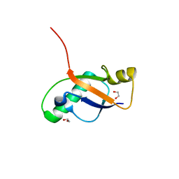
6OE7
 
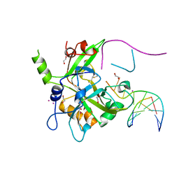 | | Crystal structure of HMCES cross-linked to DNA abasic site | | 分子名称: | 1,2-ETHANEDIOL, DNA (5'-D(*CP*CP*AP*GP*AP*CP*GP*TP*(DRZ)P*GP*TP*T)-3'), DNA (5'-D(*GP*TP*CP*TP*GP*G)-3'), ... | | 著者 | Halabelian, L, Li, Y, Zeng, H, Bountra, C, Edwards, A.M, Arrowsmith, C.H, Structural Genomics Consortium (SGC) | | 登録日 | 2019-03-27 | | 公開日 | 2019-04-24 | | 最終更新日 | 2019-07-17 | | 実験手法 | X-RAY DIFFRACTION (2.2 Å) | | 主引用文献 | Structural basis of HMCES interactions with abasic DNA and multivalent substrate recognition.
Nat.Struct.Mol.Biol., 26, 2019
|
|
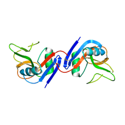
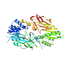
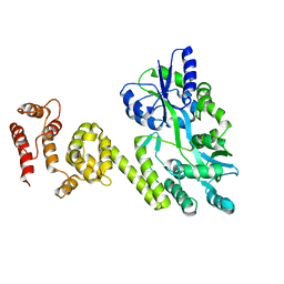
1K75
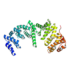
 | | The L-histidinol dehydrogenase (hisD) structure implicates domain swapping and gene duplication. | | 分子名称: | GLYCEROL, L-histidinol dehydrogenase, SULFATE ION | | 著者 | Barbosa, J.A.R.G, Sivaraman, J, Li, Y, Larocque, R, Matte, A, Schrag, J, Cygler, M, Montreal-Kingston Bacterial Structural Genomics Initiative (BSGI) | | 登録日 | 2001-10-18 | | 公開日 | 2002-02-27 | | 最終更新日 | 2014-11-12 | | 実験手法 | X-RAY DIFFRACTION (1.75 Å) | | 主引用文献 | Mechanism of action and NAD+-binding mode revealed by the crystal structure of L-histidinol dehydrogenase.
Proc.Natl.Acad.Sci.USA, 99, 2002
|
|
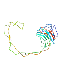
1KAH
 | | L-HISTIDINOL DEHYDROGENASE (HISD) STRUCTURE COMPLEXED WITH L-HISTIDINE (PRODUCT), ZN AND NAD (COFACTOR) | | 分子名称: | HISTIDINE, Histidinol dehydrogenase, ZINC ION | | 著者 | Barbosa, J.A.R.G, Sivaraman, J, Li, Y, Larocque, R, Matte, A, Schrag, J.D, Cygler, M. | | 登録日 | 2001-11-02 | | 公開日 | 2002-06-12 | | 最終更新日 | 2023-11-15 | | 実験手法 | X-RAY DIFFRACTION (2.1 Å) | | 主引用文献 | Mechanism of action and NAD+-binding mode revealed by the crystal structure of L-histidinol dehydrogenase.
Proc.Natl.Acad.Sci.USA, 99, 2002
|
|
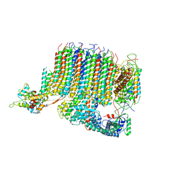
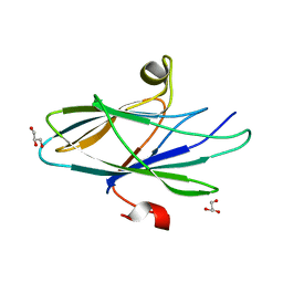
1JA3
 | | Crystal Structure of the Murine NK Cell Inhibitory Receptor Ly-49I | | 分子名称: | MHC class I recognition receptor Ly49I | | 著者 | Dimasi, N, Sawicki, W.M, Reineck, L.A, Li, Y, Natarajan, K, Murgulies, D.H, Mariuzza, A.R. | | 登録日 | 2001-05-29 | | 公開日 | 2002-07-17 | | 最終更新日 | 2023-08-16 | | 実験手法 | X-RAY DIFFRACTION (3 Å) | | 主引用文献 | Crystal structure of the Ly49I natural killer cell receptor reveals variability in dimerization mode within the Ly49 family.
J.Mol.Biol., 320, 2002
|
|
1JHN
 | | Crystal Structure of the Lumenal Domain of Calnexin | | 分子名称: | CALCIUM ION, calnexin | | 著者 | Schrag, J.D, Bergeron, J.M, Li, Y, Borisova, S, Hahn, M, Thomas, D.Y, Cygler, M. | | 登録日 | 2001-06-28 | | 公開日 | 2001-10-10 | | 最終更新日 | 2011-07-13 | | 実験手法 | X-RAY DIFFRACTION (2.9 Å) | | 主引用文献 | The Structure of calnexin, an ER chaperone involved in quality control of protein folding.
Mol.Cell, 8, 2001
|
|
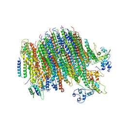
6PE4
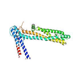
 | | Yeast Vo motor in complex with 1 VopQ molecule | | 分子名称: | Cation transporter, Uncharacterized protein YPR170W-B, V-type proton ATPase subunit a, ... | | 著者 | Peng, W, Li, Y, Tomchick, D.R, Orth, K. | | 登録日 | 2019-06-20 | | 公開日 | 2020-05-20 | | 最終更新日 | 2024-03-20 | | 実験手法 | ELECTRON MICROSCOPY (3.1 Å) | | 主引用文献 | A distinct inhibitory mechanism of the V-ATPase by Vibrio VopQ revealed by cryo-EM.
Nat.Struct.Mol.Biol., 27, 2020
|
|
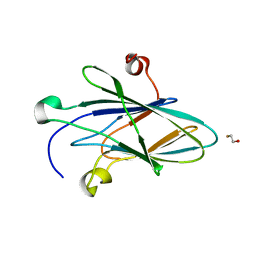
6P0Q
 | | Crystal Structure of Ubiquitin-like Domain of Human WDR12 | | 分子名称: | 1,2-ETHANEDIOL, Ribosome biogenesis protein WDR12 | | 著者 | Halabelian, L, Dong, A, Zeng, H, Li, Y, Bountra, C, Edwards, A.M, Arrowsmith, C.H, Structural Genomics Consortium (SGC) | | 登録日 | 2019-05-17 | | 公開日 | 2019-05-29 | | 最終更新日 | 2023-10-11 | | 実験手法 | X-RAY DIFFRACTION (1.72 Å) | | 主引用文献 | Crystal Structure of Ubiquitin-like Domain of Human WDR12
to be published
|
|
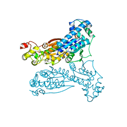
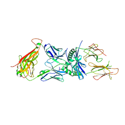
6PDM
 | | Crystal structure of Human Protein Arginine Methyltransferase 9 (PRMT9) | | 分子名称: | Protein arginine N-methyltransferase 9, UNKNOWN ATOM OR ION | | 著者 | Halabelian, L, Tempel, W, Zeng, H, Li, Y, Seitova, A, Hutchinson, A, Bountra, C, Edwards, A.M, Arrowsmith, C.H, Structural Genomics Consortium (SGC) | | 登録日 | 2019-06-19 | | 公開日 | 2019-07-31 | | 最終更新日 | 2023-10-11 | | 実験手法 | X-RAY DIFFRACTION (2.45 Å) | | 主引用文献 | Crystal structure of Human Protein Arginine Methyltransferase 9 (PRMT9)
To Be Published
|
|
6PE5
 | | Yeast Vo motor in complex with 2 VopQ molecules | | 分子名称: | Cation transporter, Uncharacterized protein YPR170W-B, V-type proton ATPase subunit a, ... | | 著者 | Peng, W, Li, Y, Tomchick, D.R, Orth, K. | | 登録日 | 2019-06-20 | | 公開日 | 2020-05-20 | | 最終更新日 | 2024-03-20 | | 実験手法 | ELECTRON MICROSCOPY (3.2 Å) | | 主引用文献 | A distinct inhibitory mechanism of the V-ATPase by Vibrio VopQ revealed by cryo-EM.
Nat.Struct.Mol.Biol., 27, 2020
|
|
1KAR
 | | L-HISTIDINOL DEHYDROGENASE (HISD) STRUCTURE COMPLEXED WITH HISTAMINE (INHIBITOR), ZINC AND NAD (COFACTOR) | | 分子名称: | HISTAMINE, Histidinol dehydrogenase, ZINC ION | | 著者 | Barbosa, J.A.R.G, Sivaraman, J, Li, Y, Larocque, R, Matte, A, Schrag, J.D, Cygler, M. | | 登録日 | 2001-11-02 | | 公開日 | 2002-06-12 | | 最終更新日 | 2023-11-15 | | 実験手法 | X-RAY DIFFRACTION (2.1 Å) | | 主引用文献 | Mechanism of action and NAD+-binding mode revealed by the crystal structure of L-histidinol dehydrogenase.
Proc.Natl.Acad.Sci.USA, 99, 2002
|
|
1K6M
 | | Crystal Structure of Human Liver 6-Phosphofructo-2-Kinase/Fructose-2,6-Bisphosphatase | | 分子名称: | 6-phosphofructo-2-kinase/fructose-2,6-biphosphatase 2-phosphatase, PHOSPHATE ION, PHOSPHOTHIOPHOSPHORIC ACID-ADENYLATE ESTER | | 著者 | Lee, Y.H, Li, Y, Uyeda, K, Hasemann, C.A. | | 登録日 | 2001-10-16 | | 公開日 | 2002-12-11 | | 最終更新日 | 2023-08-16 | | 実験手法 | X-RAY DIFFRACTION (2.4 Å) | | 主引用文献 | Tissue-specific structure/function differentiation of the liver isoform of 6-phosphofructo-2-kinase/fructose-2,6-bisphosphatase.
J.Biol.Chem., 278, 2003
|
|
5JQE
 | | Crystal structure of caspase8 tDED | | 分子名称: | Sugar ABC transporter substrate-binding protein,Caspase-8 chimera | | 著者 | Fu, T, Li, Y, Lu, A, Wu, H. | | 登録日 | 2016-05-04 | | 公開日 | 2016-10-26 | | 最終更新日 | 2024-04-03 | | 実験手法 | X-RAY DIFFRACTION (3.157 Å) | | 主引用文献 | Cryo-EM Structure of Caspase-8 Tandem DED Filament Reveals Assembly and Regulation Mechanisms of the Death-Inducing Signaling Complex.
Mol. Cell, 64, 2016
|
|
1IJI
 | | Crystal Structure of L-Histidinol Phosphate Aminotransferase with PLP | | 分子名称: | Histidinol Phosphate Aminotransferase, PYRIDOXAL-5'-PHOSPHATE | | 著者 | Sivaraman, J, Li, Y, Larocque, R, Schrag, J.D, Cygler, M, Matte, A. | | 登録日 | 2001-04-26 | | 公開日 | 2001-08-29 | | 最終更新日 | 2017-10-04 | | 実験手法 | X-RAY DIFFRACTION (2.2 Å) | | 主引用文献 | Crystal structure of histidinol phosphate aminotransferase (HisC) from Escherichia coli, and its covalent complex with pyridoxal-5'-phosphate and l-histidinol phosphate.
J.Mol.Biol., 311, 2001
|
|
5CYW
 | | Crystal Structure of Vaccinia Virus C7 | | 分子名称: | GLYCEROL, Interferon antagonist C7 | | 著者 | Krumm, B.E, Meng, X, Li, Y, Xiang, Y, Deng, J. | | 登録日 | 2015-07-30 | | 公開日 | 2015-11-18 | | 最終更新日 | 2024-03-06 | | 実験手法 | X-RAY DIFFRACTION (2 Å) | | 主引用文献 | Structural basis for antagonizing a host restriction factor by C7 family of poxvirus host-range proteins.
Proc.Natl.Acad.Sci.USA, 112, 2015
|
|
5CZ3
 | | Crystal Structure of Myxoma Virus M64 | | 分子名称: | BETA-MERCAPTOETHANOL, M64R | | 著者 | Krumm, B.E, Meng, X, Li, Y, Xiang, Y, Deng, J. | | 登録日 | 2015-07-31 | | 公開日 | 2015-11-18 | | 最終更新日 | 2023-09-27 | | 実験手法 | X-RAY DIFFRACTION (2.5 Å) | | 主引用文献 | Structural basis for antagonizing a host restriction factor by C7 family of poxvirus host-range proteins.
Proc.Natl.Acad.Sci.USA, 112, 2015
|
|
3O6F
 | | Crystal structure of a human autoimmune TCR MS2-3C8 bound to MHC class II self-ligand MBP/HLA-DR4 | | 分子名称: | HLA class II histocompatibility antigen, DR alpha chain, DRB1-4 beta chain, ... | | 著者 | Yin, Y, Li, Y, Martin, R, Mariuzza, R.A. | | 登録日 | 2010-07-29 | | 公開日 | 2011-03-02 | | 最終更新日 | 2023-09-06 | | 実験手法 | X-RAY DIFFRACTION (2.8 Å) | | 主引用文献 | Structure of a TCR with high affinity for self-antigen reveals basis for escape from negative selection.
Embo J., 30, 2011
|
|
4NFB
 | | Structure of paired immunoglobulin-like type 2 receptor (PILR ) | | 分子名称: | Paired immunoglobulin-like type 2 receptor alpha | | 著者 | Lu, Q, Lu, G, Qi, J, Li, Y, Zhang, Y, Wang, H, Fan, Z, Yan, J, Gao, G. | | 登録日 | 2013-10-31 | | 公開日 | 2014-05-28 | | 最終更新日 | 2024-03-20 | | 実験手法 | X-RAY DIFFRACTION (1.6 Å) | | 主引用文献 | PILR alpha and PILR beta have a siglec fold and provide the basis of binding to sialic acid
Proc.Natl.Acad.Sci.USA, 111, 2014
|
|
4NFD
 | | Structure of PILR L108W mutant in complex with sialic acid | | 分子名称: | N-acetyl-alpha-neuraminic acid, Paired immunoglobulin-like type 2 receptor beta | | 著者 | Lu, Q, Lu, G, Qi, J, Li, Y, Zhang, Y, Wang, H, Fan, Z, Yan, J, Gao, G.F. | | 登録日 | 2013-10-31 | | 公開日 | 2014-05-28 | | 最終更新日 | 2023-11-08 | | 実験手法 | X-RAY DIFFRACTION (1.708 Å) | | 主引用文献 | PILR alpha and PILR beta have a siglec fold and provide the basis of binding to sialic acid
Proc.Natl.Acad.Sci.USA, 111, 2014
|
|
4NFC
 | | Structure of paired immunoglobulin-like type 2 receptor (PILR ) | | 分子名称: | Paired immunoglobulin-like type 2 receptor beta | | 著者 | Lu, Q, Lu, G, Qi, J, Li, Y, Zhang, Y, Wang, H, Fan, Z, Yan, J, Gao, G.F. | | 登録日 | 2013-10-31 | | 公開日 | 2014-05-28 | | 最終更新日 | 2023-11-08 | | 実験手法 | X-RAY DIFFRACTION (2.2 Å) | | 主引用文献 | PILR alpha and PILR beta have a siglec fold and provide the basis of binding to sialic acid
Proc.Natl.Acad.Sci.USA, 111, 2014
|
|
3PP2
 | | Crystal structure of the pleckstrin homology domain of ArhGAP27 | | 分子名称: | CITRIC ACID, GLYCEROL, Rho GTPase-activating protein 27, ... | | 著者 | Shen, L, Tempel, W, Tong, Y, Nedyalkova, L, Li, Y, Wernimont, A.K, Arrowsmith, C.H, Edwards, A.M, Bountra, C, Weigelt, J, Park, H, Structural Genomics Consortium (SGC) | | 登録日 | 2010-11-23 | | 公開日 | 2010-12-08 | | 最終更新日 | 2024-02-21 | | 実験手法 | X-RAY DIFFRACTION (1.421 Å) | | 主引用文献 | Crystal structure of the pleckstrin homology domain of ArhGAP27
to be published
|
|
4QN1
 | | Crystal Structure of a Functionally Uncharacterized Domain of E3 Ubiquitin Ligase SHPRH | | 分子名称: | E3 ubiquitin-protein ligase SHPRH, SULFATE ION, UNKNOWN ATOM OR ION, ... | | 著者 | Dong, A, Zhang, Q, Li, Y, Walker, J.R, Guan, X, Bountra, C, Arrowsmith, C.H, Edwards, A.M, Tong, Y, Structural Genomics Consortium (SGC) | | 登録日 | 2014-06-17 | | 公開日 | 2014-08-13 | | 最終更新日 | 2017-11-22 | | 実験手法 | X-RAY DIFFRACTION (2.48 Å) | | 主引用文献 | Crystal structure of a Function Uncharacterized Domain of E3 Ubiquitin Ligase SHPRH
To be Published
|
|
3RCO
 | | Crystal structure of a conserved motif in human TDRD7 | | 分子名称: | CHLORIDE ION, Tudor domain-containing protein 7 | | 著者 | Dong, A, Xu, C, Walker, J.R, Lam, R, Guo, Y, Bian, C, Li, Y, Bountra, C, Weigelt, J, Arrowsmith, C.H, Edwards, A.M, Min, J, Structural Genomics Consortium (SGC) | | 登録日 | 2011-03-31 | | 公開日 | 2012-04-04 | | 最終更新日 | 2024-02-21 | | 実験手法 | X-RAY DIFFRACTION (1.8 Å) | | 主引用文献 | Crystal structure of a conserved motif in human TDRD7
To be Published
|
|
4QT6
 | | Crystal structure of the SPRY domain of human HERC1 | | 分子名称: | FORMAMIDE, Probable E3 ubiquitin-protein ligase HERC1, UNKNOWN ATOM OR ION | | 著者 | Dong, A, Hu, J, Guan, X, Wernimont, A, Li, Y, Bountra, C, Arrowsmith, C.H, Edwards, A.M, Tong, Y, Structural Genomics Consortium (SGC) | | 登録日 | 2014-07-07 | | 公開日 | 2015-01-07 | | 最終更新日 | 2017-11-22 | | 実験手法 | X-RAY DIFFRACTION (1.64 Å) | | 主引用文献 | Crystal structure of the SPRY domain of human HERC1
To be Published
|
|
3S4Y
 | | Crystal structure of human thiamin pyrophosphokinase 1 | | 分子名称: | CALCIUM ION, SULFATE ION, THIAMINE DIPHOSPHATE, ... | | 著者 | Shen, L, Tempel, W, Tong, Y, Li, Y, Walker, J.R, Arrowsmith, C.H, Edwards, A.M, Bountra, C, Weigelt, J, Park, H, Structural Genomics Consortium (SGC) | | 登録日 | 2011-05-20 | | 公開日 | 2011-06-08 | | 最終更新日 | 2024-02-28 | | 実験手法 | X-RAY DIFFRACTION (1.8 Å) | | 主引用文献 | Crystal structure of human thiamin pyrophosphokinase 1
to be published
|
|
4RXX
 | | Crystal Structure of the N-terminal Domain of Human Ubiquitin Specific Protease 38 | | 分子名称: | 1,2-ETHANEDIOL, CHLORIDE ION, UNKNOWN ATOM OR ION, ... | | 著者 | Dong, A, Shen, L, Hu, J, Li, Y, Tempel, W, Bountra, C, Arrowsmith, C.H, Edwards, A.M, Tong, Y, Structural Genomics Consortium (SGC) | | 登録日 | 2014-12-12 | | 公開日 | 2015-01-21 | | 最終更新日 | 2017-11-22 | | 実験手法 | X-RAY DIFFRACTION (2.06 Å) | | 主引用文献 | Crystal Structure of the N-terminal Domain of Human Ubiquitin Specific Protease 38
to be published
|
|


