7RE1
 
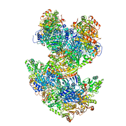
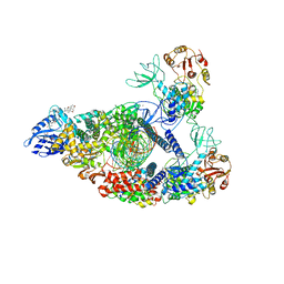 | | SARS-CoV-2 replication-transcription complex bound to nsp13 helicase - nsp13(2)-RTC (composite) | | 分子名称: | ADENOSINE-5'-DIPHOSPHATE, ALUMINUM FLUORIDE, CHAPSO, ... | | 著者 | Chen, J, Malone, B, Campbell, E.A, Darst, S.A. | | 登録日 | 2021-07-12 | | 公開日 | 2021-12-01 | | 最終更新日 | 2024-06-05 | | 実験手法 | ELECTRON MICROSCOPY (2.91 Å) | | 主引用文献 | Ensemble cryo-EM reveals conformational states of the nsp13 helicase in the SARS-CoV-2 helicase replication-transcription complex.
Nat.Struct.Mol.Biol., 29, 2022
|
|
6H3R
 | |
8YLZ
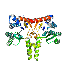
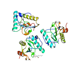
 | | Structure of a cis-Geranylfarnesyl Diphosphate Synthase from Streptomyces clavuligerus | | 分子名称: | Isoprenyl transferase | | 著者 | Li, F.R, Wang, Q.L, Pan, X.M, Dong, L.B. | | 登録日 | 2024-03-07 | | 公開日 | 2024-05-08 | | 実験手法 | X-RAY DIFFRACTION (1.6 Å) | | 主引用文献 | Discovery, Structure, and Engineering of a cis-Geranylfarnesyl Diphosphate Synthase.
Angew.Chem.Int.Ed.Engl., 2024
|
|
8WU7
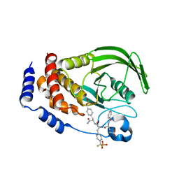
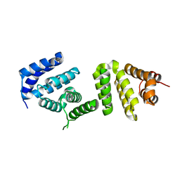
 | | Structure of a cis-Geranylfarnesyl Diphosphate Synthase from Streptomyces clavuligerus | | 分子名称: | Isoprenyl transferase | | 著者 | Li, F.R, Wang, Q.L, Pan, X.M, Dong, L.B. | | 登録日 | 2023-10-20 | | 公開日 | 2024-05-08 | | 実験手法 | X-RAY DIFFRACTION (2.09 Å) | | 主引用文献 | Discovery, Structure, and Engineering of a cis-Geranylfarnesyl Diphosphate Synthase.
Angew.Chem.Int.Ed.Engl., 2024
|
|
8WU6
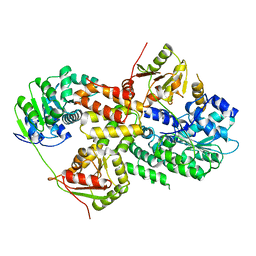
 | | Structure of a Nerylneryl Diphosphate Synthase from Solanum lycopersicum | | 分子名称: | Nerylneryl diphosphate synthase CPT2, chloroplastic | | 著者 | Li, F.R, Wang, Q.L, Pan, X.M, Dong, L.B. | | 登録日 | 2023-10-20 | | 公開日 | 2024-05-08 | | 実験手法 | X-RAY DIFFRACTION (1.81 Å) | | 主引用文献 | Discovery, Structure, and Engineering of a cis-Geranylfarnesyl Diphosphate Synthase.
Angew.Chem.Int.Ed.Engl., 2024
|
|
1LQF
 | | Structure of PTP1b in Complex with a Peptidic Bisphosphonate Inhibitor | | 分子名称: | N-BENZOYL-L-GLUTAMYL-[4-PHOSPHONO(DIFLUOROMETHYL)]-L-PHENYLALANINE-[4-PHOSPHONO(DIFLUORO-METHYL)]-L-PHENYLALANINEAMIDE, protein-tyrosine phosphatase, non-receptor type 1 | | 著者 | Asante-Appiah, E, Patel, S, Dufresne, C, Scapin, G. | | 登録日 | 2002-05-10 | | 公開日 | 2002-07-24 | | 最終更新日 | 2023-08-16 | | 実験手法 | X-RAY DIFFRACTION (2.5 Å) | | 主引用文献 | The structure of PTP-1B in complex with a peptide inhibitor reveals an alternative binding mode for bisphosphonates.
Biochemistry, 41, 2002
|
|
4RSU
 | | Crystal structure of the light and hvem complex | | 分子名称: | 2-acetamido-2-deoxy-beta-D-glucopyranose, CHLORIDE ION, GLYCEROL, ... | | 著者 | Liu, W, Ramagoal, U.A, Himmel, D, Bonanno, J.B, Nathenson, S.G, Almo, S.C, Atoms-to-Animals: The Immune Function Network (IFN), New York Structural Genomics Research Consortium (NYSGRC) | | 登録日 | 2014-11-11 | | 公開日 | 2015-02-04 | | 最終更新日 | 2023-09-20 | | 実験手法 | X-RAY DIFFRACTION (2.3 Å) | | 主引用文献 | HVEM structures and mutants reveal distinct functions of binding to LIGHT and BTLA/CD160.
J.Exp.Med., 218, 2021
|
|
6L8Q
 | | Complex structure of bat CD26 and MERS-RBD | | 分子名称: | 2-acetamido-2-deoxy-beta-D-glucopyranose, 2-acetamido-2-deoxy-beta-D-glucopyranose-(1-4)-2-acetamido-2-deoxy-beta-D-glucopyranose, Dipeptidyl peptidase 4, ... | | 著者 | Yuan, Y. | | 登録日 | 2019-11-07 | | 公開日 | 2019-12-04 | | 最終更新日 | 2023-11-22 | | 実験手法 | X-RAY DIFFRACTION (3.1 Å) | | 主引用文献 | Molecular Basis of Binding between Middle East Respiratory Syndrome Coronavirus and CD26 from Seven Bat Species.
J.Virol., 94, 2020
|
|
6LK8
 | | Structure of Xenopus laevis Cytoplasmic Ring subunit. | | 分子名称: | GATOR complex protein SEC13, MGC154553 protein, MGC83295 protein, ... | | 著者 | Shi, Y, Huang, G, Yan, C, Zhang, Y. | | 登録日 | 2019-12-18 | | 公開日 | 2021-07-21 | | 最終更新日 | 2024-05-29 | | 実験手法 | ELECTRON MICROSCOPY (5.5 Å) | | 主引用文献 | Structure of the cytoplasmic ring of the Xenopus laevis nuclear pore complex by cryo-electron microscopy single particle analysis.
Cell Res., 30, 2020
|
|
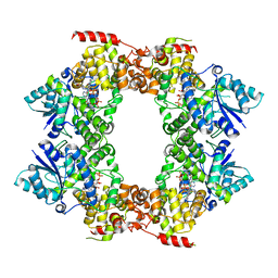
6U9H
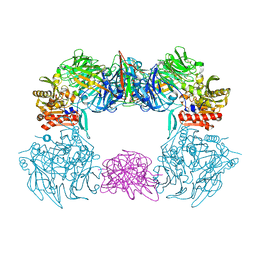
 | | Arabidopsis thaliana acetohydroxyacid synthase complex | | 分子名称: | Acetolactate synthase small subunit 2, chloroplastic, Acetolactate synthase, ... | | 著者 | Guddat, L.W, Low, Y.S, Rao, Z. | | 登録日 | 2019-09-08 | | 公開日 | 2020-07-15 | | 最終更新日 | 2024-03-20 | | 実験手法 | ELECTRON MICROSCOPY (3.8 Å) | | 主引用文献 | Structures of fungal and plant acetohydroxyacid synthases.
Nature, 586, 2020
|
|
2EAV
 | |
2XKB
 | |
2XKA
 | |
4RAY
 | |
4RB3
 | |
4RB0
 | |
4RB2
 | |
4RB1
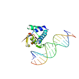
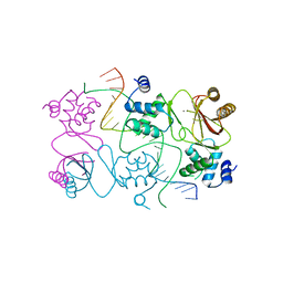 | | Crystal structure of Magnetospirillum gryphiswaldense MSR-1 Fur-Mn2+-E. coli Fur box | | 分子名称: | DNA (5'-D(*CP*GP*CP*GP*AP*TP*AP*AP*TP*GP*AP*TP*AP*AP*TP*CP*AP*TP*TP*AP*TP*CP*CP*GP*C)-3'), DNA-binding transcriptional dual regulator of siderophore biosynthesis and transport(Fur family), MANGANESE (II) ION | | 著者 | Deng, Z, Chen, Z. | | 登録日 | 2014-09-12 | | 公開日 | 2015-07-15 | | 最終更新日 | 2023-09-20 | | 実験手法 | X-RAY DIFFRACTION (2.75 Å) | | 主引用文献 | Mechanistic insights into metal ion activation and operator recognition by the ferric uptake regulator.
Nat Commun, 6
|
|
2EAX
 | | Crystal structure of human PGRP-IBETAC in complex with glycosamyl muramyl pentapeptide | | 分子名称: | 2-acetamido-2-deoxy-beta-D-glucopyranose-(1-4)-methyl 2-acetamido-3-O-[(1R)-1-carboxyethyl]-2-deoxy-beta-D-glucopyranoside, GLYCOSAMYL MURAMYL PENTAPEPTIDE, Peptidoglycan recognition protein-I-beta | | 著者 | Cho, S. | | 登録日 | 2007-02-03 | | 公開日 | 2007-10-02 | | 最終更新日 | 2023-10-25 | | 実験手法 | X-RAY DIFFRACTION (2.1 Å) | | 主引用文献 | Structural insights into the bactericidal mechanism of human peptidoglycan recognition proteins
Proc.Natl.Acad.Sci.Usa, 104, 2007
|
|
3EIJ
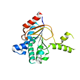
 | | Crystal structure of Pdcd4 | | 分子名称: | Programmed cell death protein 4 | | 著者 | Loh, P.G. | | 登録日 | 2008-09-16 | | 公開日 | 2009-02-17 | | 最終更新日 | 2024-03-20 | | 実験手法 | X-RAY DIFFRACTION (2.8 Å) | | 主引用文献 | Structural basis for translational inhibition by the tumour suppressor Pdcd4
Embo J., 28, 2009
|
|
3EIQ
 | | Crystal structure of Pdcd4-eIF4A | | 分子名称: | Eukaryotic initiation factor 4A-I, Programmed cell death protein 4 | | 著者 | Loh, P.G, Cheng, Z, Song, H. | | 登録日 | 2008-09-17 | | 公開日 | 2009-02-24 | | 最終更新日 | 2023-11-01 | | 実験手法 | X-RAY DIFFRACTION (3.5 Å) | | 主引用文献 | Structural basis for translational inhibition by the tumour suppressor Pdcd4
Embo J., 28, 2009
|
|
6M79
 | |
6LNA
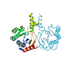
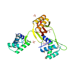
 | | YdiU complex with AMPNPP and Mn2+ | | 分子名称: | CALCIUM ION, MANGANESE (II) ION, PHOSPHOAMINOPHOSPHONIC ACID-ADENYLATE ESTER, ... | | 著者 | Li, B, Yang, Y, Ma, Y. | | 登録日 | 2019-12-28 | | 公開日 | 2020-12-30 | | 最終更新日 | 2023-11-22 | | 実験手法 | X-RAY DIFFRACTION (1.701 Å) | | 主引用文献 | The YdiU Domain Modulates Bacterial Stress Signaling through Mn 2+ -Dependent UMPylation.
Cell Rep, 32, 2020
|
|
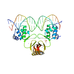
7AMT
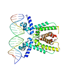 | | Structure of LuxR with DNA (activation) | | 分子名称: | DNA (5'-D(P*AP*TP*AP*AP*TP*GP*AP*CP*AP*TP*TP*AP*CP*TP*GP*TP*AP*TP*AP*TP*A)-3'), DNA (5'-D(P*TP*AP*TP*AP*TP*AP*CP*AP*GP*TP*AP*AP*TP*GP*TP*CP*AP*TP*TP*AP*T)-3'), HTH-type transcriptional regulator LuxR | | 著者 | Liu, B, Reverter, D. | | 登録日 | 2020-10-09 | | 公開日 | 2021-03-31 | | 最終更新日 | 2024-01-31 | | 実験手法 | X-RAY DIFFRACTION (2.6 Å) | | 主引用文献 | Binding site profiles and N-terminal minor groove interactions of the master quorum-sensing regulator LuxR enable flexible control of gene activation and repression.
Nucleic Acids Res., 49, 2021
|
|
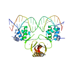
7AMN
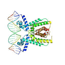 | | Structure of LuxR with DNA (repression) | | 分子名称: | DNA (5'-D(P*TP*AP*TP*TP*GP*AP*TP*AP*AP*AP*AP*TP*TP*AP*TP*CP*AP*AP*TP*AP*A)-3'), DNA (5'-D(P*TP*TP*AP*TP*TP*GP*AP*TP*AP*AP*TP*TP*TP*TP*AP*TP*CP*AP*AP*TP*A)-3'), HTH-type transcriptional regulator LuxR | | 著者 | Liu, B, Reverter, D. | | 登録日 | 2020-10-09 | | 公開日 | 2021-03-31 | | 最終更新日 | 2021-04-14 | | 実験手法 | X-RAY DIFFRACTION (2.3 Å) | | 主引用文献 | Binding site profiles and N-terminal minor groove interactions of the master quorum-sensing regulator LuxR enable flexible control of gene activation and repression.
Nucleic Acids Res., 49, 2021
|
|


