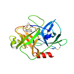
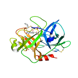
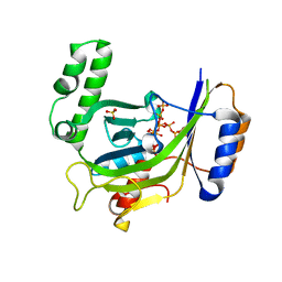
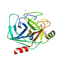
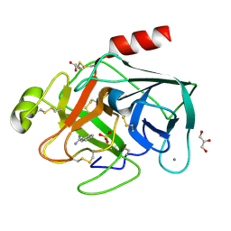
2PTK
 
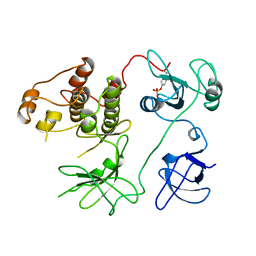 | | CHICKEN SRC TYROSINE KINASE | | 分子名称: | TYROSINE-PROTEIN KINASE TRANSFORMING PROTEIN SRC | | 著者 | Williams, J.C, Wierenga, R. | | 登録日 | 1997-06-17 | | 公開日 | 1997-12-24 | | 最終更新日 | 2018-04-11 | | 実験手法 | X-RAY DIFFRACTION (2.35 Å) | | 主引用文献 | The 2.35 A crystal structure of the inactivated form of chicken Src: a dynamic molecule with multiple regulatory interactions
J.Mol.Biol., 274, 1997
|
|
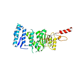
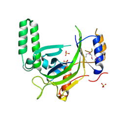
6TE6
 | | Crystal structure of Dot1L in complex with an inhibitor (compound 3). | | 分子名称: | Histone-lysine N-methyltransferase, H3 lysine-79 specific, ~{N}1-[(~{S})-(3-chlorophenyl)-pyridin-2-yl-methyl]-4-methylsulfonyl-~{N}2-pyrimidin-2-yl-benzene-1,2-diamine | | 著者 | Scheufler, C, Stauffer, F, Be, C, Moebitz, H. | | 登録日 | 2019-11-11 | | 公開日 | 2019-12-11 | | 最終更新日 | 2024-01-24 | | 実験手法 | X-RAY DIFFRACTION (1.98 Å) | | 主引用文献 | New Potent DOT1L Inhibitors forin VivoEvaluation in Mouse.
Acs Med.Chem.Lett., 10, 2019
|
|
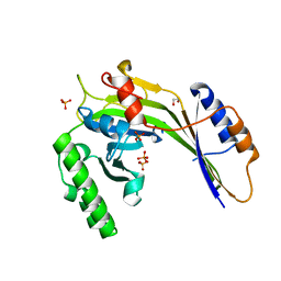
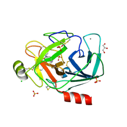
7Q6Y
 | | The X-ray crystal structure of CbTan2, a tannase enzyme from Clostridium butyricum | | 分子名称: | 1,2-ETHANEDIOL, Alpha/beta hydrolase, DI(HYDROXYETHYL)ETHER | | 著者 | Coleman, T, Mazurkewich, S, Larsbrink, J. | | 登録日 | 2021-11-09 | | 公開日 | 2022-03-02 | | 最終更新日 | 2024-01-31 | | 実験手法 | X-RAY DIFFRACTION (2.22 Å) | | 主引用文献 | Structural diversity and substrate preferences of three tannase enzymes encoded by the anaerobic bacterium Clostridium butyricum.
J.Biol.Chem., 298, 2022
|
|
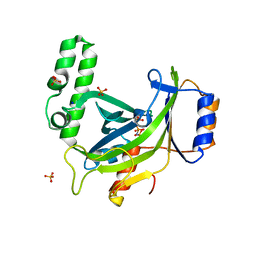
1W10
 | | Urokinase type plasminogen activator | | 分子名称: | N-(ISOBUTOXYCARBONYL)-D-SERYL-N-((1S)-4-{[AMINO(IMINO)METHYL]AMINO}-1-FORMYLBUTYL)-L-ALANINAMIDE, SULFATE ION, UROKINASE-TYPE PLASMINOGEN ACTIVATOR | | 著者 | Jacob, U. | | 登録日 | 2004-06-15 | | 公開日 | 2008-05-20 | | 最終更新日 | 2019-09-18 | | 実験手法 | X-RAY DIFFRACTION (2 Å) | | 主引用文献 | Crystals of urokinase type plasminogen activator complexes reveal the binding mode of peptidomimetic inhibitors.
J.Mol.Biol., 328, 2003
|
|
1W11
 | | UROKINASE TYPE PLASMINOGEN ACTIVATOR | | 分子名称: | N-(BENZYLSULFONYL)-D-SERYL-N-{4-[AMINO(IMINO)METHYL]BENZYL}-L-ALANINAMIDE, SULFATE ION, UROKINASE-TYPE PLASMINOGEN ACTIVATOR | | 著者 | Jacob, U. | | 登録日 | 2004-06-15 | | 公開日 | 2008-05-20 | | 最終更新日 | 2019-10-09 | | 実験手法 | X-RAY DIFFRACTION (2 Å) | | 主引用文献 | Crystals of Urokinase Type Plasminogen Activator Complexes Reveal the Binding Mode of Peptidomimetic Inhibitors.
J.Mol.Biol., 328, 2003
|
|
1W13
 | | UROKINASE TYPE PLASMINOGEN ACTIVATOR | | 分子名称: | N-(BENZYLSULFONYL)-D-SERYL-N-(4-{[AMINO(IMINO)METHYL]AMINO}BENZYL)-L-ALANINAMIDE, SULFATE ION, UROKINASE-TYPE PLASMINOGEN ACTIVATOR | | 著者 | Jacob, U. | | 登録日 | 2004-06-15 | | 公開日 | 2008-05-20 | | 最終更新日 | 2019-05-22 | | 実験手法 | X-RAY DIFFRACTION (2 Å) | | 主引用文献 | Crystals of Urokinase Type Plasminogen Activator Complexes Reveal the Binding Mode of Peptidomimetic Inhibitors.
J.Mol.Biol., 328, 2003
|
|
1LKT
 | |
1W12
 | | UROKINASE TYPE PLASMINOGEN ACTIVATOR | | 分子名称: | N-((1S)-4-{[AMINO(IMINO)METHYL]AMINO}-1-FORMYLBUTYL)-2-{(3R)-3-[(BENZYLSULFONYL)AMINO]-2-OXO-5-PHENYL-2,3-DIHYDRO-1H-1,4-BENZODIAZEPIN-1-YL}ACETAMIDE, UROKINASE-TYPE PLASMINOGEN ACTIVATOR | | 著者 | Jacob, U. | | 登録日 | 2004-06-15 | | 公開日 | 2008-05-20 | | 最終更新日 | 2019-05-22 | | 実験手法 | X-RAY DIFFRACTION (2.4 Å) | | 主引用文献 | Crystals of Urokinase Type Plasminogen Activator Complexes Reveal the Binding Mode of Peptidomimetic Inhibitors.
J.Mol.Biol., 328, 2003
|
|
1W0Z
 | | Urokinase type plasminogen activator | | 分子名称: | N-(BUTYLSULFONYL)-D-SERYL-N-{4-[AMINO(IMINO)METHYL]BENZYL}-L-ALANINAMIDE, SULFATE ION, UROKINASE-TYPE PLASMINOGEN ACTIVATOR | | 著者 | Jacob, U. | | 登録日 | 2004-06-15 | | 公開日 | 2008-05-20 | | 最終更新日 | 2019-09-18 | | 実験手法 | X-RAY DIFFRACTION (1.9 Å) | | 主引用文献 | Crystals of urokinase type plasminogen activator complexes reveal the binding mode of peptidomimetic inhibitors.
J.Mol.Biol., 328, 2003
|
|
1W14
 | | UROKINASE TYPE PLASMINOGEN ACTIVATOR | | 分子名称: | N-[(2-PHENYLETHYL)SULFONYL]-D-SERYL-N-[(1S)-4-[(DIAMINOMETHYLENE)AMINO]-1-(HYDROXYMETHYL)BUTYL]-L-ALANINAMIDE, SULFATE ION, UROKINASE-TYPE PLASMINOGEN ACTIVATOR | | 著者 | Jacob, U. | | 登録日 | 2004-06-15 | | 公開日 | 2008-05-20 | | 最終更新日 | 2019-05-22 | | 実験手法 | X-RAY DIFFRACTION (2.2 Å) | | 主引用文献 | Crystals of Urokinase Type Plasminogen Activator Complexes Reveal the Binding Mode of Peptidomimetic Inhibitors.
J.Mol.Biol., 328, 2003
|
|
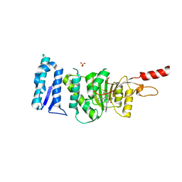
6TEL
 | | Crystal structure of Dot1L in complex with an inhibitor (compound 10). | | 分子名称: | Histone-lysine N-methyltransferase, H3 lysine-79 specific, POTASSIUM ION, ... | | 著者 | Scheufler, C, Stauffer, F, Be, C, Moebitz, H. | | 登録日 | 2019-11-12 | | 公開日 | 2019-12-11 | | 最終更新日 | 2024-01-24 | | 実験手法 | X-RAY DIFFRACTION (2.19 Å) | | 主引用文献 | New Potent DOT1L Inhibitors forin VivoEvaluation in Mouse.
Acs Med.Chem.Lett., 10, 2019
|
|
6TEN
 | | Crystal structure of Dot1L in complex with an inhibitor (compound 11). | | 分子名称: | 3-[(4-azanyl-6-methoxy-1,3,5-triazin-2-yl)amino]-4-[[(~{S})-[2,2-bis(fluoranyl)-1,3-benzodioxol-4-yl]-(3-chloranylpyridin-2-yl)methyl]amino]benzenesulfonamide, Histone-lysine N-methyltransferase, H3 lysine-79 specific, ... | | 著者 | Scheufler, C, Stauffer, F, Be, C, Moebitz, H. | | 登録日 | 2019-11-12 | | 公開日 | 2019-12-11 | | 最終更新日 | 2024-01-24 | | 実験手法 | X-RAY DIFFRACTION (2.21 Å) | | 主引用文献 | New Potent DOT1L Inhibitors forin VivoEvaluation in Mouse.
Acs Med.Chem.Lett., 10, 2019
|
|
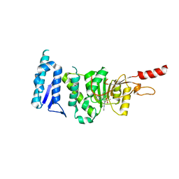
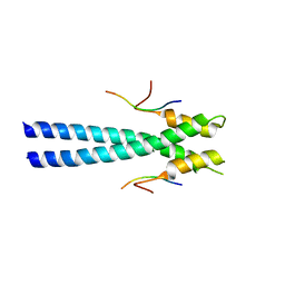
2LXH
 | | NMR structure of the RING domain in ubiquitin ligase gp78 | | 分子名称: | E3 ubiquitin-protein ligase AMFR, ZINC ION | | 著者 | Das, R, Linag, Y, Mariano, J, Li, J, Huang, T, King, A, Weissman, A, Ji, X, Byrd, R. | | 登録日 | 2012-08-27 | | 公開日 | 2013-08-28 | | 最終更新日 | 2024-05-15 | | 実験手法 | SOLUTION NMR | | 主引用文献 | Allosteric regulation of E2:E3 interactions promote a processive ubiquitination machine.
Embo J., 32, 2013
|
|
2LXP
 | | NMR structure of two domains in ubiquitin ligase gp78, RING and G2BR, bound to its conjugating enzyme Ube2g | | 分子名称: | E3 ubiquitin-protein ligase AMFR, Ubiquitin-conjugating enzyme E2 G2, ZINC ION | | 著者 | Das, R, Linag, Y, Mariano, J, Li, J, Huang, T, King, A, Weissman, A, Ji, X, Byrd, R. | | 登録日 | 2012-08-30 | | 公開日 | 2013-08-28 | | 最終更新日 | 2024-05-15 | | 実験手法 | SOLUTION NMR | | 主引用文献 | Allosteric regulation of E2:E3 interactions promote a processive ubiquitination machine.
Embo J., 32, 2013
|
|
3GJO
 | |
3G3Q
 | |
3G3O
 | |
3G3U
 | |
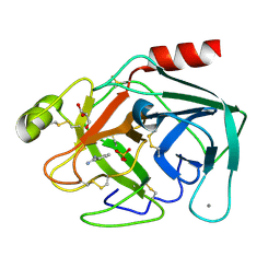
3G3T
 | | Crystal structure of a eukaryotic polyphosphate polymerase in complex with orthophosphate | | 分子名称: | 1,2-ETHANEDIOL, PHOSPHATE ION, Vacuolar transporter chaperone 4 | | 著者 | Lenherr, E.D, Hothorn, M, Scheffzek, K. | | 登録日 | 2009-02-02 | | 公開日 | 2009-05-05 | | 最終更新日 | 2023-09-06 | | 実験手法 | X-RAY DIFFRACTION (1.85 Å) | | 主引用文献 | Catalytic core of a membrane-associated eukaryotic polyphosphate polymerase.
Science, 324, 2009
|
|
3G3R
 | |
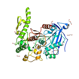
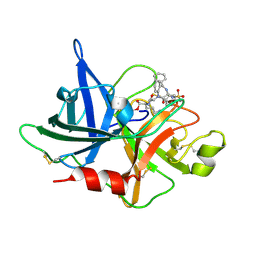
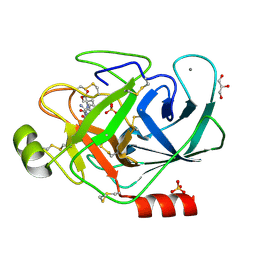
3PYH
 | | Bovine trypsin variant X(tripleGlu217Ile227) in complex with small molecule inhibitor | | 分子名称: | 3-(3-carbamimidoylphenyl)-N-(2'-sulfamoylbiphenyl-4-yl)-1,2-oxazole-4-carboxamide, CALCIUM ION, CHLORIDE ION, ... | | 著者 | Tziridis, A, Neumann, P, Kolenko, P, Stubbs, M.T. | | 登録日 | 2010-12-13 | | 公開日 | 2011-12-21 | | 最終更新日 | 2023-09-13 | | 実験手法 | X-RAY DIFFRACTION (2 Å) | | 主引用文献 | Correlating structure and ligand affinity in drug discovery: a cautionary tale involving second shell residues.
Biol.Chem., 395, 2014
|
|
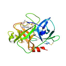
3PMJ
 | | Bovine trypsin variant X(tripleIle227) in complex with small molecule inhibitor | | 分子名称: | CALCIUM ION, CHLORIDE ION, Cationic trypsin, ... | | 著者 | Tziridis, A, Neumann, P, Kolenko, P, Stubbs, M.T. | | 登録日 | 2010-11-17 | | 公開日 | 2011-11-23 | | 最終更新日 | 2023-09-06 | | 実験手法 | X-RAY DIFFRACTION (1.45 Å) | | 主引用文献 | Correlating structure and ligand affinity in drug discovery: a cautionary tale involving second shell residues.
Biol.Chem., 395, 2014
|
|
3PWB
 | | Bovine trypsin variant X(tripleGlu217Ile227) in complex with small molecule inhibitor | | 分子名称: | BENZAMIDINE, CALCIUM ION, Cationic trypsin, ... | | 著者 | Tziridis, A, Neumann, P, Kolenko, P, Stubbs, M.T. | | 登録日 | 2010-12-08 | | 公開日 | 2011-12-21 | | 最終更新日 | 2023-09-13 | | 実験手法 | X-RAY DIFFRACTION (1.63 Å) | | 主引用文献 | Correlating structure and ligand affinity in drug discovery: a cautionary tale involving second shell residues.
Biol.Chem., 395, 2014
|
|
3PWC
 | | Bovine trypsin variant X(tripleGlu217Ile227) in complex with small molecule inhibitor | | 分子名称: | CALCIUM ION, Cationic trypsin, GLYCEROL, ... | | 著者 | Tziridis, A, Neumann, P, Kolenko, P, Stubbs, M.T. | | 登録日 | 2010-12-08 | | 公開日 | 2011-12-21 | | 最終更新日 | 2023-09-13 | | 実験手法 | X-RAY DIFFRACTION (1.6 Å) | | 主引用文献 | Correlating structure and ligand affinity in drug discovery: a cautionary tale involving second shell residues.
Biol.Chem., 395, 2014
|
|
3PLB
 | | Bovine trypsin variant X(tripleIle227) in complex with small molecule inhibitor | | 分子名称: | BENZAMIDINE, CALCIUM ION, Cationic trypsin, ... | | 著者 | Tziridis, A, Neumann, P, Kolenko, P, Stubbs, M.T. | | 登録日 | 2010-11-15 | | 公開日 | 2011-12-07 | | 最終更新日 | 2023-09-06 | | 実験手法 | X-RAY DIFFRACTION (1.18 Å) | | 主引用文献 | Correlating structure and ligand affinity in drug discovery: a cautionary tale involving second shell residues.
Biol.Chem., 395, 2014
|
|


