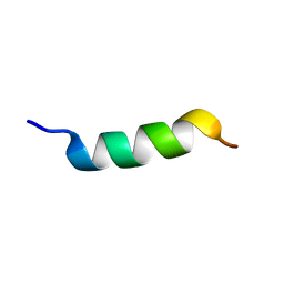
5XJO
 
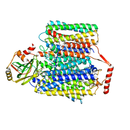
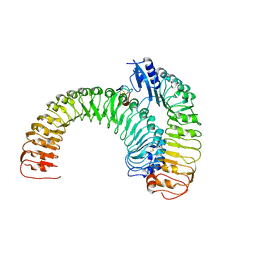 | | Plant receptor ERL1-TMM in complex with peptide EPF1 | | 分子名称: | LRR receptor-like serine/threonine-protein kinase ERL1, Protein EPIDERMAL PATTERNING FACTOR 1, Protein TOO MANY MOUTHS | | 著者 | Chai, J, Lin, G, Zhang, L, Han, Z, Shpak, E.D, Yang, X. | | 登録日 | 2017-05-03 | | 公開日 | 2019-01-23 | | 実験手法 | X-RAY DIFFRACTION (2.626 Å) | | 主引用文献 | A receptor-like protein acts as a specificity switch for the regulation of stomatal development.
Genes Dev., 31, 2017
|
|
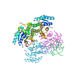
2RJG
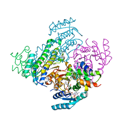
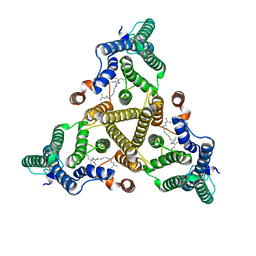
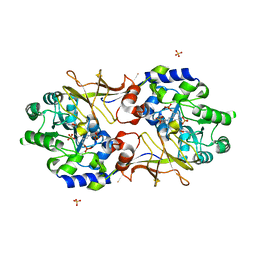 | | Crystal structure of biosynthetic alaine racemase from Escherichia coli | | 分子名称: | Alanine racemase, PYRIDOXAL-5'-PHOSPHATE, SULFATE ION | | 著者 | Wu, D, Hu, T, Zhang, L, Jiang, H, Shen, X. | | 登録日 | 2007-10-15 | | 公開日 | 2008-07-08 | | 最終更新日 | 2023-11-15 | | 実験手法 | X-RAY DIFFRACTION (2.4 Å) | | 主引用文献 | Residues Asp164 and Glu165 at the substrate entryway function potently in substrate orientation of alanine racemase from E. coli: Enzymatic characterization with crystal structure analysis
Protein Sci., 17, 2008
|
|
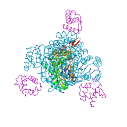
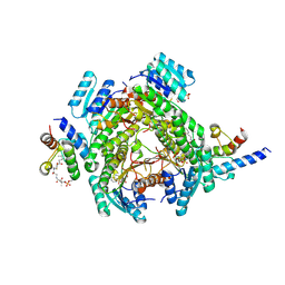
4ZA1
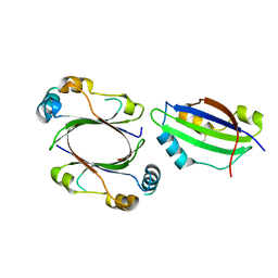 | | Crystal Structure of NosA Involved in Nosiheptide Biosynthesis | | 分子名称: | 2,3-DIHYDROXY-1,4-DITHIOBUTANE, NosA | | 著者 | Liu, S, Guo, H, Zhang, T, Han, L, Yao, P, Zhang, Y, Rong, N, Yu, Y, Lan, W, Wang, C, Ding, J, Wang, R, Liu, W, Cao, C. | | 登録日 | 2015-04-13 | | 公開日 | 2015-08-19 | | 最終更新日 | 2024-03-20 | | 実験手法 | X-RAY DIFFRACTION (2.5 Å) | | 主引用文献 | Structure-based Mechanistic Insights into Terminal Amide Synthase in Nosiheptide-Represented Thiopeptides Biosynthesis
Sci Rep, 5, 2015
|
|
2FXZ
 | |
2FXY
 | |
8JFG
 | |
8JFN
 | |
8JFM
 | |
8JFJ
 | |
8JFA
 | |
8JFI
 | |
8JFH
 | |
8JF9
 | |
6WYH
 | |
6WYG
 | |
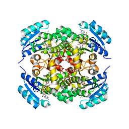
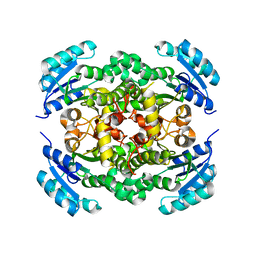
5BOF
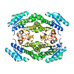
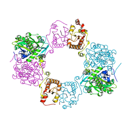
 | | Crystal Structure of Staphylococcus aureus Enolase | | 分子名称: | Enolase, MAGNESIUM ION, SULFATE ION | | 著者 | Wu, Y.F, Wang, C.L, Wu, M.H, Han, L, Zhang, X, Zang, J.Y. | | 登録日 | 2015-05-27 | | 公開日 | 2015-12-09 | | 最終更新日 | 2023-11-08 | | 実験手法 | X-RAY DIFFRACTION (2.45 Å) | | 主引用文献 | Octameric structure of Staphylococcus aureus enolase in complex with phosphoenolpyruvate.
Acta Crystallogr.,Sect.D, 71, 2015
|
|
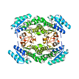
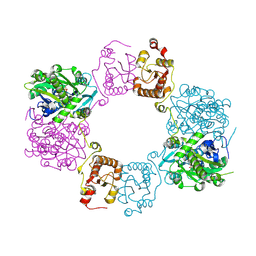
5BOE
 | | Crystal structure of Staphylococcus aureus enolase in complex with PEP | | 分子名称: | Enolase, GLYCEROL, MAGNESIUM ION, ... | | 著者 | Wang, C.L, Wu, Y.F, Han, L, Wu, M.H, Zhang, X, Zang, J.Y. | | 登録日 | 2015-05-27 | | 公開日 | 2015-12-09 | | 最終更新日 | 2023-11-08 | | 実験手法 | X-RAY DIFFRACTION (1.6 Å) | | 主引用文献 | Octameric structure of Staphylococcus aureus enolase in complex with phosphoenolpyruvate
Acta Crystallogr.,Sect.D, 71, 2015
|
|
8J8K
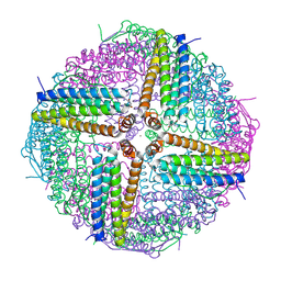
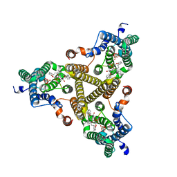
 | | Membrane bound PRTase, C3 symmetry, acceptor bound | | 分子名称: | Decaprenyl-phosphate phosphoribosyltransferase, MONO-TRANS, OCTA-CIS DECAPRENYL-PHOSPHATE | | 著者 | Wu, F.Y, Gao, S, Zhang, L, Rao, Z.H. | | 登録日 | 2023-05-01 | | 公開日 | 2024-02-07 | | 最終更新日 | 2024-04-17 | | 実験手法 | ELECTRON MICROSCOPY (3.36 Å) | | 主引用文献 | Structural analysis of phosphoribosyltransferase-mediated cell wall precursor synthesis in Mycobacterium tuberculosis.
Nat Microbiol, 9, 2024
|
|
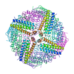
8J8J
 | | Membrane bound PRTase, C3 symmetry, donor bound | | 分子名称: | (1R)-2-{[(S)-{[(2S)-2,3-dihydroxypropyl]oxy}(hydroxy)phosphoryl]oxy}-1-[(hexadecanoyloxy)methyl]ethyl (9Z)-octadec-9-enoate, 1-O-pyrophosphono-5-O-phosphono-alpha-D-ribofuranose, Decaprenyl-phosphate phosphoribosyltransferase, ... | | 著者 | Wu, F.Y, Gao, S, Zhang, L, Rao, Z.H. | | 登録日 | 2023-05-01 | | 公開日 | 2024-02-07 | | 最終更新日 | 2024-04-17 | | 実験手法 | ELECTRON MICROSCOPY (2.76 Å) | | 主引用文献 | Structural analysis of phosphoribosyltransferase-mediated cell wall precursor synthesis in Mycobacterium tuberculosis.
Nat Microbiol, 9, 2024
|
|
8H4Q
 | |
8H4H
 | |
5II6
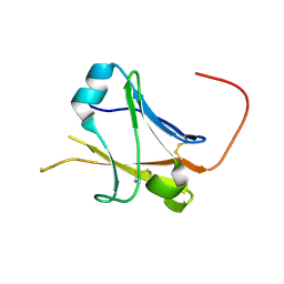 | | Crystal structure of the ZP-N1 domain of mouse sperm receptor ZP2 at 0.95 A resolution | | 分子名称: | Zona pellucida sperm-binding protein 2 | | 著者 | Dioguardi, E, Han, L, Nishimura, K, De Sanctis, D, Jovine, L. | | 登録日 | 2016-03-01 | | 公開日 | 2017-06-14 | | 最終更新日 | 2017-11-29 | | 実験手法 | X-RAY DIFFRACTION (0.95 Å) | | 主引用文献 | Structural Basis of Egg Coat-Sperm Recognition at Fertilization.
Cell, 169, 2017
|
|
7CUQ
 | | 2.55-Angstrom Cryo-EM structure of Cytochrome bo3 from Escherichia coli in Native Membrane | | 分子名称: | 1,2-Distearoyl-sn-glycerophosphoethanolamine, COPPER (II) ION, Cytochrome bo(3) ubiquinol oxidase subunit 1, ... | | 著者 | Li, J, Han, L, Gennis, R.B, Zhu, J.P, Zhang, K. | | 登録日 | 2020-08-24 | | 公開日 | 2021-08-25 | | 最終更新日 | 2024-05-29 | | 実験手法 | ELECTRON MICROSCOPY (2.64 Å) | | 主引用文献 | Cryo-EM structures of Escherichia coli cytochrome bo3 reveal bound phospholipids and ubiquinone-8 in a dynamic substrate binding site.
Proc.Natl.Acad.Sci.USA, 118, 2021
|
|
7CUW
 | | Ubiquinol Binding Site of Cytochrome bo3 from Escherichia coli | | 分子名称: | 1,2-Distearoyl-sn-glycerophosphoethanolamine, COPPER (II) ION, Cytochrome bo(3) ubiquinol oxidase subunit 1, ... | | 著者 | Li, J, Han, L, Gennis, R.B, Zhu, J.P, Zhang, K. | | 登録日 | 2020-08-25 | | 公開日 | 2021-08-25 | | 最終更新日 | 2024-05-29 | | 実験手法 | ELECTRON MICROSCOPY (2.63 Å) | | 主引用文献 | Cryo-EM structures of Escherichia coli cytochrome bo3 reveal bound phospholipids and ubiquinone-8 in a dynamic substrate binding site.
Proc.Natl.Acad.Sci.USA, 118, 2021
|
|
7CUB
 | | 2.55-Angstrom Cryo-EM structure of Cytochrome bo3 from Escherichia coli in Native Membrane | | 分子名称: | 1,2-Distearoyl-sn-glycerophosphoethanolamine, COPPER (II) ION, Cytochrome bo(3) ubiquinol oxidase subunit 1, ... | | 著者 | Li, J, Han, L, Gennis, R.B, Zhu, J.P, Zhang, K. | | 登録日 | 2020-08-22 | | 公開日 | 2021-08-25 | | 最終更新日 | 2024-05-29 | | 実験手法 | ELECTRON MICROSCOPY (2.55 Å) | | 主引用文献 | Cryo-EM structures of Escherichia coli cytochrome bo3 reveal bound phospholipids and ubiquinone-8 in a dynamic substrate binding site.
Proc.Natl.Acad.Sci.USA, 118, 2021
|
|


