7D6P
 
 | |
7D6T
 | |
7F5I
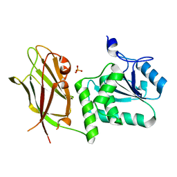
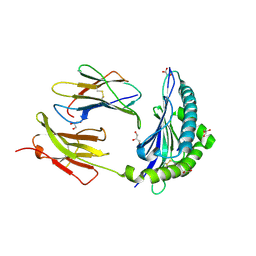
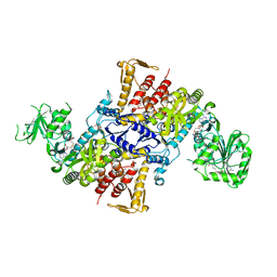
 | | X-ray structure of Clostridium perfringens-specific amidase endolysin | | 分子名称: | GLUTAMIC ACID, SODIUM ION, ZINC ION, ... | | 著者 | Kamitori, S, Tamai, E. | | 登録日 | 2021-06-22 | | 公開日 | 2022-05-04 | | 最終更新日 | 2023-11-29 | | 実験手法 | X-RAY DIFFRACTION (1.65 Å) | | 主引用文献 | Structural and biochemical characterization of the Clostridium perfringens-specific Zn 2+ -dependent amidase endolysin, Psa, catalytic domain.
Biochem.Biophys.Res.Commun., 576, 2021
|
|
8DB4
 | |
8HPK
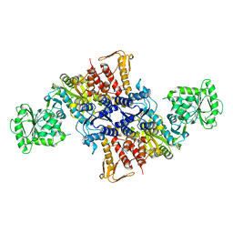
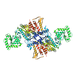
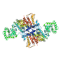
 | | Crystal structure of the bacterial oxalate transporter OxlT in an oxalate-bound occluded form | | 分子名称: | Fab fragment Heavy chein, Fab fragment Light chain, OXALATE ION, ... | | 著者 | Shimamura, T, Hirai, T, Yamashita, A. | | 登録日 | 2022-12-12 | | 公開日 | 2023-02-15 | | 最終更新日 | 2023-04-12 | | 実験手法 | X-RAY DIFFRACTION (3 Å) | | 主引用文献 | Structure and mechanism of oxalate transporter OxlT in an oxalate-degrading bacterium in the gut microbiota.
Nat Commun, 14, 2023
|
|
8HPJ
 | |
4KRU
 | |
4KRT
 | |
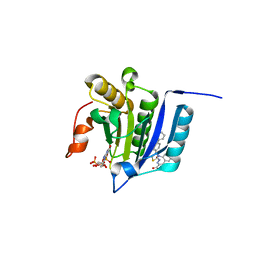
7JUK
 | | Crystal structure of PTEN with a tetra-phosphorylated tail (4p-crPTEN-13sp-T2, SDTTDSDPENEG) | | 分子名称: | PHOSPHATE ION, Phosphatidylinositol 3,4,5-trisphosphate 3-phosphatase and dual-specificity protein phosphatase PTEN | | 著者 | Dempsey, D, Phan, K, Cole, P, Gabelli, S.B. | | 登録日 | 2020-08-19 | | 公開日 | 2021-10-13 | | 最終更新日 | 2023-10-18 | | 実験手法 | X-RAY DIFFRACTION (3.15 Å) | | 主引用文献 | The structural basis of PTEN regulation by multi-site phosphorylation.
Nat.Struct.Mol.Biol., 28, 2021
|
|
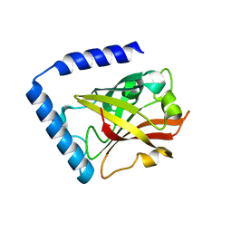
6MF8
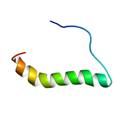 | | TCR alpha transmembrane domain | | 分子名称: | T-cell receptor alpha chain C region | | 著者 | Brazin, K.N, Reinherz, E.L. | | 登録日 | 2018-09-10 | | 公開日 | 2018-12-12 | | 最終更新日 | 2024-05-01 | | 実験手法 | SOLUTION NMR | | 主引用文献 | The T Cell Antigen Receptor alpha Transmembrane Domain Coordinates Triggering through Regulation of Bilayer Immersion and CD3 Subunit Associations.
Immunity, 49, 2018
|
|
7JVX
 | | Crystal structure of PTEN (aa 7-353 followed by spacer TGGGSGGTGGGSGGTGGGCY ligated to peptide pSDpTpTDpSDPENEPFDED) | | 分子名称: | PHOSPHATE ION, Phosphatidylinositol 3,4,5-trisphosphate 3-phosphatase and dual-specificity protein phosphatase PTEN | | 著者 | Dempsey, D, Phan, K, Cole, P, Gabelli, S.B. | | 登録日 | 2020-08-24 | | 公開日 | 2021-08-04 | | 最終更新日 | 2023-10-18 | | 実験手法 | X-RAY DIFFRACTION (3.2 Å) | | 主引用文献 | The structural basis of PTEN regulation by multi-site phosphorylation.
Nat.Struct.Mol.Biol., 28, 2021
|
|
7JUL
 | | Crystal structure of non phosphorylated PTEN (n-crPTEN-13sp-T1, SDTTDSDPENEG) | | 分子名称: | Phosphatidylinositol 3,4,5-trisphosphate 3-phosphatase and dual-specificity protein phosphatase PTEN | | 著者 | Dempsey, D, Phan, K, Cole, P, Gabelli, S.B. | | 登録日 | 2020-08-20 | | 公開日 | 2021-08-11 | | 最終更新日 | 2023-10-18 | | 実験手法 | X-RAY DIFFRACTION (2.53 Å) | | 主引用文献 | The structural basis of PTEN regulation by multi-site phosphorylation.
Nat.Struct.Mol.Biol., 28, 2021
|
|
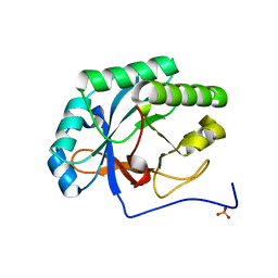
7JTX
 | |
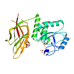
7JI2
 | | Crystal Structure of H2-Kb in complex with a OVA mutant peptide | | 分子名称: | Beta-2-microglobulin, GLYCEROL, H-2 class I histocompatibility antigen, ... | | 著者 | Li, X, Mallis, R.J, Mizsei, R, Tan, K, Reinherz, E.L, Wang, J. | | 登録日 | 2020-07-22 | | 公開日 | 2020-12-23 | | 最終更新日 | 2023-10-18 | | 実験手法 | X-RAY DIFFRACTION (1.95 Å) | | 主引用文献 | Pre-T cell receptors topologically sample self-ligands during thymocyte beta-selection.
Science, 371, 2021
|
|
7MEU
 | |
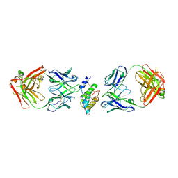
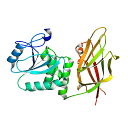
5XJM
 | | Complex structure of angiotensin II type 2 receptor with Fab | | 分子名称: | FabH, FabL, Sar1, ... | | 著者 | Asada, H, Horita, S, Shimamura, T, Iwata, S. | | 登録日 | 2017-05-02 | | 公開日 | 2018-07-11 | | 最終更新日 | 2023-11-22 | | 実験手法 | X-RAY DIFFRACTION (3.2 Å) | | 主引用文献 | Crystal structure of the human angiotensin II type 2 receptor bound to an angiotensin II analog
Nat. Struct. Mol. Biol., 25, 2018
|
|
5XCC
 | |
5XCB
 | |
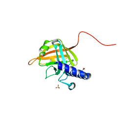
2RVH
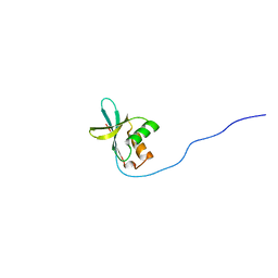 | | NMR structure of eIF1 | | 分子名称: | Eukaryotic translation initiation factor eIF-1 | | 著者 | Nagata, T, Obayashi, E, Asano, K. | | 登録日 | 2015-10-16 | | 公開日 | 2016-10-26 | | 最終更新日 | 2024-05-15 | | 実験手法 | SOLUTION NMR | | 主引用文献 | Molecular Landscape of the Ribosome Pre-initiation Complex during mRNA Scanning: Structural Role for eIF3c and Its Control by eIF5
Cell Rep, 18, 2017
|
|
5X70
 | |
5X6X
 | |
5X6Z
 | |
5X6Y
 | |
5X71
 | |
1GN7
 | |


