5YSH
 
 | |
1UCG
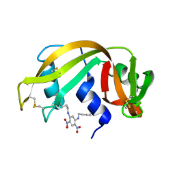
 | | Crystal structure of Ribonuclease MC1 N71T mutant | | 分子名称: | MANGANESE (II) ION, Ribonuclease MC | | 著者 | Suzuki, A, Numata, T, Yao, M, Tanaka, I, Kimura, M. | | 登録日 | 2003-04-14 | | 公開日 | 2003-04-29 | | 最終更新日 | 2023-10-25 | | 実験手法 | X-RAY DIFFRACTION (1.65 Å) | | 主引用文献 | Crystal structures of the ribonuclease MC1 mutants N71T and N71S in complex with 5'-GMP: structural basis for alterations in substrate specificity
Biochemistry, 42, 2003
|
|
7Y3J
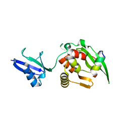
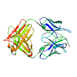 | | 24B3 antibody-peptide complex | | 分子名称: | 24B3 Heavy chain, 24B3 Light chain, ALA-LEU-VAL-PHE-PHE-ALA-PRO-ALA-VAL-GLY-SER | | 著者 | Irie, K, Irie, Y, Kita, A, Miki, K. | | 登録日 | 2022-06-11 | | 公開日 | 2022-08-17 | | 最終更新日 | 2023-11-29 | | 実験手法 | X-RAY DIFFRACTION (2.6 Å) | | 主引用文献 | Structural basis of the 24B3 antibody against the toxic conformer of amyloid beta with a turn at positions 22 and 23.
Biochem.Biophys.Res.Commun., 621, 2022
|
|
5X02
 | | Crystal structure of the FLT3 kinase domain bound to the inhibitor FF-10101 | | 分子名称: | N-[(2S)-1-[5-[2-[(4-cyanophenyl)amino]-4-(propylamino)pyrimidin-5-yl]pent-4-ynylamino]-1-oxidanylidene-propan-2-yl]-4-(dimethylamino)-N-methyl-but-2-enamide, Receptor-type tyrosine-protein kinase FLT3, SULFATE ION | | 著者 | Fujikawa, N, Hirano, D, Takasaki, M, Terada, D, Hagiwara, S, Park, S.-Y, Sugiyama, K. | | 登録日 | 2017-01-19 | | 公開日 | 2018-01-24 | | 最終更新日 | 2023-11-22 | | 実験手法 | X-RAY DIFFRACTION (2.401 Å) | | 主引用文献 | A novel irreversible FLT3 inhibitor, FF-10101, shows excellent efficacy against AML cells withFLT3mutations.
Blood, 131, 2018
|
|
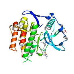
7Z74
 | | PI3KC2a core in complex with PITCOIN2 | | 分子名称: | 1,2-ETHANEDIOL, Phosphatidylinositol 4-phosphate 3-kinase C2 domain-containing subunit alpha, ~{N}-[4-(3-hydroxyphenyl)-1,3-thiazol-2-yl]-2-[4-oxidanylidene-3-(2-phenylethyl)pteridin-2-yl]sulfanyl-ethanamide | | 著者 | Lo, W.T, Roske, Y, Daumke, O, Haucke, V. | | 登録日 | 2022-03-15 | | 公開日 | 2022-08-31 | | 最終更新日 | 2024-01-31 | | 実験手法 | X-RAY DIFFRACTION (2.5 Å) | | 主引用文献 | Development of selective inhibitors of phosphatidylinositol 3-kinase C2 alpha.
Nat.Chem.Biol., 19, 2023
|
|
7Z75
 | | PI3KC2a core in complex with PITCOIN3 | | 分子名称: | 1,2-ETHANEDIOL, Phosphatidylinositol 4-phosphate 3-kinase C2 domain-containing subunit alpha, SULFATE ION, ... | | 著者 | Lo, W.T, Roske, Y, Daumke, O, Haucke, V. | | 登録日 | 2022-03-15 | | 公開日 | 2022-08-31 | | 最終更新日 | 2024-02-07 | | 実験手法 | X-RAY DIFFRACTION (2.59 Å) | | 主引用文献 | Development of selective inhibitors of phosphatidylinositol 3-kinase C2 alpha.
Nat.Chem.Biol., 19, 2023
|
|
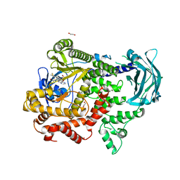
7VKF
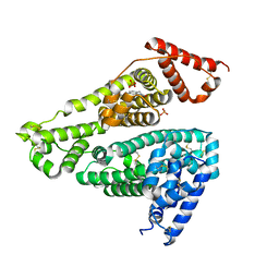
 | | Reduced enzyme of FAD-dpendent Glucose Dehydrogenase complex with D-glucono-1,5-lactone at pH8.5 | | 分子名称: | 2-[3-(2-HYDROXY-1,1-DIHYDROXYMETHYL-ETHYLAMINO)-PROPYLAMINO]-2-HYDROXYMETHYL-PROPANE-1,3-DIOL, D-glucono-1,5-lactone, DIHYDROFLAVINE-ADENINE DINUCLEOTIDE, ... | | 著者 | Nakajima, Y, Nishiya, Y, Ito, K. | | 登録日 | 2021-09-29 | | 公開日 | 2022-10-05 | | 最終更新日 | 2024-05-29 | | 実験手法 | X-RAY DIFFRACTION (1.6 Å) | | 主引用文献 | Conformational change of catalytic residue in reduced enzyme of FAD-dependent Glucose Dehydrogenase at pH6.5
To Be Published
|
|
1WE0
 | | Crystal structure of peroxiredoxin (AhpC) from Amphibacillus xylanus | | 分子名称: | AMMONIUM ION, alkyl hydroperoxide reductase C | | 著者 | Kitano, K, Kita, A, Hakoshima, T, Niimura, Y, Miki, K. | | 登録日 | 2004-05-21 | | 公開日 | 2005-03-29 | | 最終更新日 | 2018-02-07 | | 実験手法 | X-RAY DIFFRACTION (2.9 Å) | | 主引用文献 | Crystal structure of decameric peroxiredoxin (AhpC) from Amphibacillus xylanus
Proteins, 59, 2005
|
|
7VKD
 | |
5ZLQ
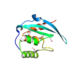
 | | Crystal Structure of C1orf123 | | 分子名称: | UPF0587 protein C1orf123, ZINC ION | | 著者 | Furukawa, Y, Lim, C.T, Tosha, T. | | 登録日 | 2018-03-29 | | 公開日 | 2018-10-10 | | 最終更新日 | 2024-03-27 | | 実験手法 | X-RAY DIFFRACTION (2 Å) | | 主引用文献 | Identification of a novel zinc-binding protein, C1orf123, as an interactor with a heavy metal-associated domain
PLoS ONE, 13, 2018
|
|
1RSM
 | | THE 2-ANGSTROMS RESOLUTION STRUCTURE OF A THERMOSTABLE RIBONUCLEASE A CHEMICALLY CROSS-LINKED BETWEEN LYSINE RESIDUES 7 AND 41 | | 分子名称: | DINITROPHENYLENE, RIBONUCLEASE A | | 著者 | Weber, P.C, Sheriff, S, Ohlendorf, D.H, Finzel, B.C, Salemme, F.R. | | 登録日 | 1985-08-27 | | 公開日 | 1986-01-21 | | 最終更新日 | 2024-06-05 | | 実験手法 | X-RAY DIFFRACTION (2 Å) | | 主引用文献 | The 2-A resolution structure of a thermostable ribonuclease A chemically cross-linked between lysine residues 7 and 41.
Proc.Natl.Acad.Sci.USA, 82, 1985
|
|
2ZYE
 | | Structure of HIV-1 Protease in Complex with Potent Inhibitor KNI-272 Determined by Neutron Crystallography | | 分子名称: | (4R)-N-tert-butyl-3-[(2S,3S)-2-hydroxy-3-({N-[(isoquinolin-5-yloxy)acetyl]-S-methyl-L-cysteinyl}amino)-4-phenylbutanoyl]-1,3-thiazolidine-4-carboxamide, protease | | 著者 | Adachi, M, Ohhara, T, Tamada, T, Okazaki, N, Kuroki, R. | | 登録日 | 2009-01-20 | | 公開日 | 2009-03-24 | | 最終更新日 | 2024-05-29 | | 実験手法 | NEUTRON DIFFRACTION (1.9 Å) | | 主引用文献 | Structure of HIV-1 protease in complex with potent inhibitor KNI-272 determined by high-resolution X-ray and neutron crystallography.
Proc.Natl.Acad.Sci.USA, 2009
|
|
2CW9
 | | Crystal structure of human Tim44 C-terminal domain | | 分子名称: | PENTAETHYLENE GLYCOL, translocase of inner mitochondrial membrane | | 著者 | Handa, N, Kishishita, S, Morita, S, Kinoshita, Y, Nagano, Y, Uda, H, Terada, T, Uchikubo, T, Takemoto, C, Jin, Z, Chrzas, J, Chen, L, Liu, Z.-J, Wang, B.-C, Shirouzu, M, Yokoyama, S, RIKEN Structural Genomics/Proteomics Initiative (RSGI) | | 登録日 | 2005-06-17 | | 公開日 | 2005-12-17 | | 最終更新日 | 2011-07-13 | | 実験手法 | X-RAY DIFFRACTION (1.9 Å) | | 主引用文献 | Structure of the human Tim44 C-terminal domain in complex with pentaethylene glycol: ligand-bound form.
Acta Crystallogr.,Sect.D, 63, 2007
|
|
5YC2
 | |
5YBX
 | |
7X7X
 | | Human serum albumin complex with deschloro-aripiprazole | | 分子名称: | 7-[4-(4-phenylpiperazin-1-yl)butoxy]-3,4-dihydro-1H-quinolin-2-one, PHOSPHATE ION, Serum albumin | | 著者 | Kawai, A, Otagiri, M. | | 登録日 | 2022-03-10 | | 公開日 | 2022-09-07 | | 最終更新日 | 2023-11-29 | | 実験手法 | X-RAY DIFFRACTION (2.1 Å) | | 主引用文献 | Chlorine Atoms of an Aripiprazole Molecule Control the Geometry and Motion of Aripiprazole and Deschloro-aripiprazole in Subdomain IIIA of Human Serum Albumin.
Acs Omega, 7, 2022
|
|
5YCA
 | | Crystal structure of inner membrane protein Bqt4 in complex with LEM2 | | 分子名称: | Lap-Emerin-Man domain protein 2, Ubiquitin-like protein SMT3,Bouquet formation protein 4 | | 著者 | Chen, Y, Hu, C. | | 登録日 | 2017-09-07 | | 公開日 | 2018-11-14 | | 最終更新日 | 2023-11-22 | | 実験手法 | X-RAY DIFFRACTION (1.57 Å) | | 主引用文献 | Structural insights into chromosome attachment to the nuclear envelope by an inner nuclear membrane protein Bqt4 in fission yeast.
Nucleic Acids Res., 47, 2019
|
|
5XJM
 | | Complex structure of angiotensin II type 2 receptor with Fab | | 分子名称: | FabH, FabL, Sar1, ... | | 著者 | Asada, H, Horita, S, Shimamura, T, Iwata, S. | | 登録日 | 2017-05-02 | | 公開日 | 2018-07-11 | | 最終更新日 | 2023-11-22 | | 実験手法 | X-RAY DIFFRACTION (3.2 Å) | | 主引用文献 | Crystal structure of the human angiotensin II type 2 receptor bound to an angiotensin II analog
Nat. Struct. Mol. Biol., 25, 2018
|
|
5XHG
 | | Crystal structure of Trastuzumab Fab fragment bearing Ne-(o-azidobenzyloxycarbonyl)-L-lysine | | 分子名称: | (2-azidophenyl)methyl hydrogen carbonate, 1,2-ETHANEDIOL, DI(HYDROXYETHYL)ETHER, ... | | 著者 | Kuratani, M, Yanagisawa, T, Sakamoto, K, Yokoyama, S. | | 登録日 | 2017-04-20 | | 公開日 | 2017-12-20 | | 最終更新日 | 2019-12-25 | | 実験手法 | X-RAY DIFFRACTION (1.76 Å) | | 主引用文献 | Extensive Survey of Antibody Invariant Positions for Efficient Chemical Conjugation Using Expanded Genetic Codes.
Bioconjug. Chem., 28, 2017
|
|
5X9P
 | | Crystal structure of the BCL6 BTB domain in complex with Compound 5 | | 分子名称: | 3-[[4-chloranyl-2-nitro-5-[(2-oxidanylidene-1,3-dihydrobenzimidazol-5-yl)amino]phenyl]amino]propanoic acid, B-cell lymphoma 6 protein | | 著者 | Sogabe, S, Ida, K, Lane, W, Snell, G. | | 登録日 | 2017-03-08 | | 公開日 | 2017-08-16 | | 最終更新日 | 2023-11-22 | | 実験手法 | X-RAY DIFFRACTION (1.86 Å) | | 主引用文献 | Discovery of a novel B-cell lymphoma 6 (BCL6)-corepressor interaction inhibitor by utilizing structure-based drug design
Bioorg. Med. Chem., 25, 2017
|
|
5X5M
 | |
6A6W
 | |
5X9O
 | | Crystal structure of the BCL6 BTB domain in complex with Compound 1a | | 分子名称: | 1,2-ETHANEDIOL, 5-[(2-chloranyl-4-nitro-phenyl)amino]-1,3-dihydrobenzimidazol-2-one, B-cell lymphoma 6 protein, ... | | 著者 | Sogabe, S, Ida, K, Lane, W, Snell, G. | | 登録日 | 2017-03-08 | | 公開日 | 2017-08-16 | | 最終更新日 | 2023-11-22 | | 実験手法 | X-RAY DIFFRACTION (1.58 Å) | | 主引用文献 | Discovery of a novel B-cell lymphoma 6 (BCL6)-corepressor interaction inhibitor by utilizing structure-based drug design
Bioorg. Med. Chem., 25, 2017
|
|
5XHF
 | | Crystal structure of Trastuzumab Fab fragment bearing p-azido-L-phenylalanine | | 分子名称: | polypeptide (H chain), polypeptide (L chain) | | 著者 | Kuratani, M, Yanagisawa, T, Sakamoto, K, Yokoyama, S. | | 登録日 | 2017-04-20 | | 公開日 | 2017-12-20 | | 最終更新日 | 2023-11-22 | | 実験手法 | X-RAY DIFFRACTION (3.205 Å) | | 主引用文献 | Extensive Survey of Antibody Invariant Positions for Efficient Chemical Conjugation Using Expanded Genetic Codes.
Bioconjug. Chem., 28, 2017
|
|
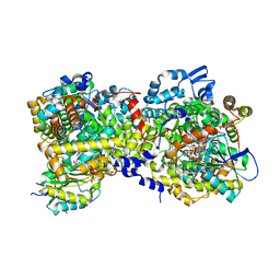
6A3J
 | | Levoglucosan dehydrogenase, complex with NADH and L-sorbose | | 分子名称: | 1,4-DIHYDRONICOTINAMIDE ADENINE DINUCLEOTIDE, 2-(N-MORPHOLINO)-ETHANESULFONIC ACID, Putative dehydrogenase, ... | | 著者 | Sugiura, M, Yamada, C, Arakawa, T, Fushinobu, S. | | 登録日 | 2018-06-15 | | 公開日 | 2018-09-26 | | 最終更新日 | 2023-11-22 | | 実験手法 | X-RAY DIFFRACTION (1.9 Å) | | 主引用文献 | Identification, functional characterization, and crystal structure determination of bacterial levoglucosan dehydrogenase.
J. Biol. Chem., 293, 2018
|
|


