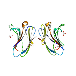
3HQF
 
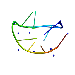
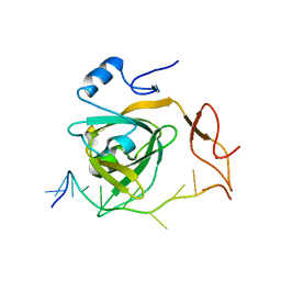
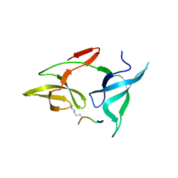
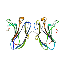
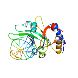 | | Crystal structure of restriction endonuclease EcoRII N-terminal effector-binding domain in complex with cognate DNA | | 分子名称: | 5'-D(*CP*GP*CP*CP*AP*GP*GP*GP*C)-3', 5'-D(*GP*CP*CP*CP*TP*GP*GP*CP*G)-3', Restriction endonuclease | | 著者 | Golovenko, D, Manakova, E, Grazulis, S, Tamulaitiene, G, Siksnys, V. | | 登録日 | 2009-06-06 | | 公開日 | 2009-09-22 | | 最終更新日 | 2023-09-06 | | 実験手法 | X-RAY DIFFRACTION (2.51 Å) | | 主引用文献 | Structural mechanisms for the 5'-CCWGG sequence recognition by the N- and C-terminal domains of EcoRII.
Nucleic Acids Res., 37, 2009
|
|
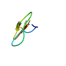
3HQG
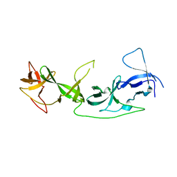
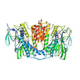
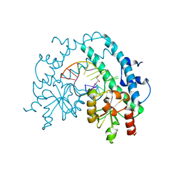 | | Crystal structure of restriction endonuclease EcoRII catalytic C-terminal domain in complex with cognate DNA | | 分子名称: | 5'-D(*TP*AP*GP*CP*CP*TP*GP*GP*TP*CP*GP*A)-3', 5'-D(*TP*CP*GP*AP*CP*CP*AP*GP*GP*CP*TP*A)-3', GLYCEROL, ... | | 著者 | Golovenko, D, Manakova, E, Grazulis, S, Tamulaitiene, G, Siksnys, V. | | 登録日 | 2009-06-06 | | 公開日 | 2009-09-22 | | 最終更新日 | 2023-09-06 | | 実験手法 | X-RAY DIFFRACTION (2.6 Å) | | 主引用文献 | Structural mechanisms for the 5'-CCWGG sequence recognition by the N- and C-terminal domains of EcoRII.
Nucleic Acids Res., 37, 2009
|
|
1YTS
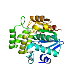
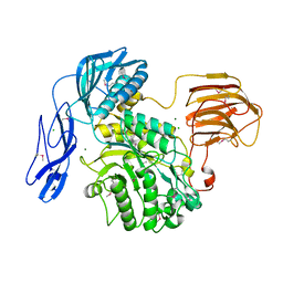
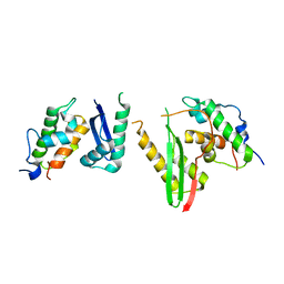
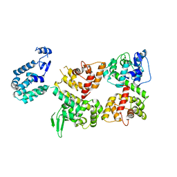
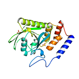 | | A LIGAND-INDUCED CONFORMATIONAL CHANGE IN THE YERSINIA PROTEIN TYROSINE PHOSPHATASE | | 分子名称: | SULFATE ION, YERSINIA PROTEIN TYROSINE PHOSPHATASE | | 著者 | Schubert, H.L, Stuckey, J.A, Fauman, E.B, Dixon, J.E, Saper, M.A. | | 登録日 | 1995-04-07 | | 公開日 | 1995-07-10 | | 最終更新日 | 2024-02-14 | | 実験手法 | X-RAY DIFFRACTION (2.5 Å) | | 主引用文献 | A ligand-induced conformational change in the Yersinia protein tyrosine phosphatase.
Protein Sci., 4, 1995
|
|
8JIO
 | |
7F5W
 | |
7YF7
 | |
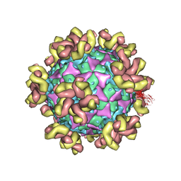
7WSW
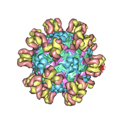
 | | Cryo-EM structure of the Potassium channel AKT1 from Arabidopsis thaliana | | 分子名称: | PHOSPHATIDYLETHANOLAMINE, POTASSIUM ION, Potassium channel AKT1 | | 著者 | Yang, G.H, Lu, Y.M, Zhang, Y.M, Jia, Y.T, Li, X.M, Lei, J.L. | | 登録日 | 2022-02-02 | | 公開日 | 2022-11-09 | | 実験手法 | ELECTRON MICROSCOPY (3.4 Å) | | 主引用文献 | Structural basis for the activity regulation of a potassium channel AKT1 from Arabidopsis.
Nat Commun, 13, 2022
|
|
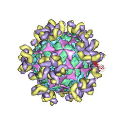
7XUF
 | | Cryo-EM structure of the AKT1-AtKC1 complex from Arabidopsis thaliana | | 分子名称: | POTASSIUM ION, Potassium channel AKT1, Potassium channel KAT3 | | 著者 | Yang, G.H, Lu, Y.M, Jia, Y.T, Yang, F, Zhang, Y.M, Xu, X, Li, X.M, Lei, J.L. | | 登録日 | 2022-05-18 | | 公開日 | 2022-11-09 | | 最終更新日 | 2024-07-03 | | 実験手法 | ELECTRON MICROSCOPY (3.3 Å) | | 主引用文献 | Structural basis for the activity regulation of a potassium channel AKT1 from Arabidopsis.
Nat Commun, 13, 2022
|
|
7Y01
 | |
7YT9
 | | crystal structure of AGD1-4 of Arabidopsis AGDP3 | | 分子名称: | AGD1-4 of Arabidopsis AGDP3 | | 著者 | Zhou, X, Du, J. | | 登録日 | 2022-08-13 | | 公開日 | 2022-10-12 | | 最終更新日 | 2024-05-29 | | 実験手法 | X-RAY DIFFRACTION (2.6 Å) | | 主引用文献 | The H3K9me2-binding protein AGDP3 limits DNA methylation and transcriptional gene silencing in Arabidopsis.
J Integr Plant Biol, 64, 2022
|
|
7YTA
 | |
6JQF
 | |
7Y7M
 | |
6KAG
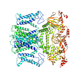
 | | Crystal structure of the SMARCB1/SMARCC2 subcomplex | | 分子名称: | SWI/SNF complex subunit SMARCC2, SWI/SNF-related matrix-associated actin-dependent regulator of chromatin subfamily B member 1 | | 著者 | Chen, G, Zhou, H, Giancotti, F.G, Long, J. | | 登録日 | 2019-06-22 | | 公開日 | 2020-09-23 | | 最終更新日 | 2024-03-27 | | 実験手法 | X-RAY DIFFRACTION (2.601 Å) | | 主引用文献 | A heterotrimeric SMARCB1-SMARCC2 subcomplex is required for the assembly and tumor suppression function of the BAF chromatin-remodeling complex.
Cell Discov, 6, 2020
|
|
7X1R
 | | Cryo-EM structure of human thioredoxin reductase bound by Au | | 分子名称: | FLAVIN-ADENINE DINUCLEOTIDE, GOLD ION, Thioredoxin reductase 1, ... | | 著者 | He, Z.S, Cao, P, Cao, S.H, He, B, Jiang, H.D, Gong, Y, Gao, X.Y. | | 登録日 | 2022-02-24 | | 公開日 | 2022-12-14 | | 実験手法 | ELECTRON MICROSCOPY (3.9 Å) | | 主引用文献 | Au4 cluster inhibits human thioredoxin reductase activity via specifically binding of Au to Cys189
Nano Today, 47, 2022
|
|
7XAC
 | |
7XBL
 | |
7YHH
 | |
7YHG
 | |
7YHF
 | |
7YHI
 | |
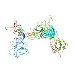
6LB8
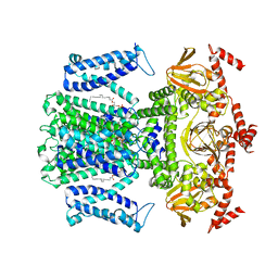
 | | Crystal structure of the Ca2+-free T4L-MICU1-MICU2 complex | | 分子名称: | Calcium uptake protein 2, mitochondrial, Endolysin,Calcium uptake protein 1 | | 著者 | Wu, W, Shen, Q, Zheng, J, Jia, Z. | | 登録日 | 2019-11-13 | | 公開日 | 2020-07-15 | | 最終更新日 | 2023-11-22 | | 実験手法 | X-RAY DIFFRACTION (3.283 Å) | | 主引用文献 | The structure of the MICU1-MICU2 complex unveils the regulation of the mitochondrial calcium uniporter.
Embo J., 39, 2020
|
|
7YMS
 | |
7YRH
 | |
7YRF
 | |


