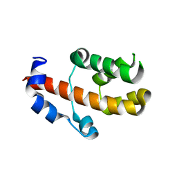
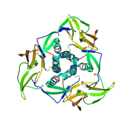
6JHY
 
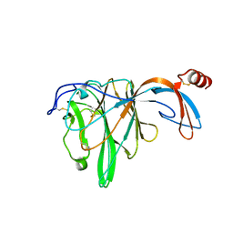 | |
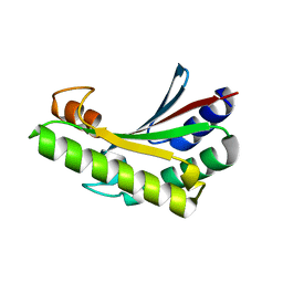
3V53
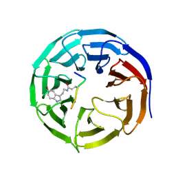
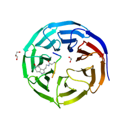
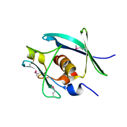
 | | Crystal structure of human RBM25 | | 分子名称: | RNA-binding protein 25 | | 著者 | Gong, D.S. | | 登録日 | 2011-12-16 | | 公開日 | 2012-12-12 | | 最終更新日 | 2024-03-20 | | 実験手法 | X-RAY DIFFRACTION (2.9 Å) | | 主引用文献 | Crystal structure and functional characterization of the human RBM25 PWI domain and its flanking basic region
Biochem.J., 450, 2013
|
|
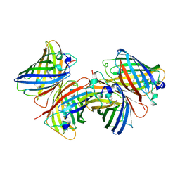
8WPZ
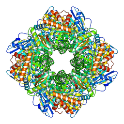 | |
1B9C
 | |
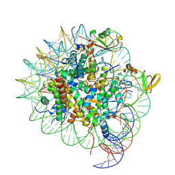
7CM3
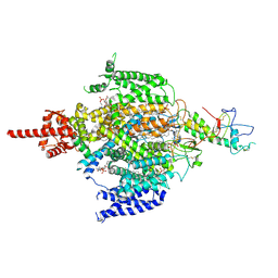 | | Cryo-EM structure of human NALCN in complex with FAM155A | | 分子名称: | 1,2-DIACYL-SN-GLYCERO-3-PHOSPHOCHOLINE, 2-acetamido-2-deoxy-beta-D-glucopyranose, 2-acetamido-2-deoxy-beta-D-glucopyranose-(1-4)-2-acetamido-2-deoxy-beta-D-glucopyranose, ... | | 著者 | Wu, J, Yan, Z, Ke, M. | | 登録日 | 2020-07-24 | | 公開日 | 2020-11-11 | | 最終更新日 | 2020-12-02 | | 実験手法 | ELECTRON MICROSCOPY (3.1 Å) | | 主引用文献 | Structure of the human sodium leak channel NALCN in complex with FAM155A.
Nat Commun, 11, 2020
|
|
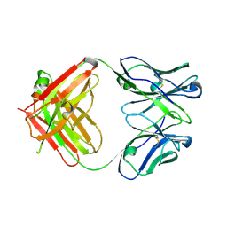
4HJR
 | | Crystal structure of F2YRS | | 分子名称: | Tyrosine-tRNA ligase | | 著者 | Wang, J, Tian, C, Gong, W, Li, F, Shi, P, Li, J, Ding, W. | | 登録日 | 2012-10-13 | | 公開日 | 2013-03-13 | | 最終更新日 | 2023-09-20 | | 実験手法 | X-RAY DIFFRACTION (2.5 Å) | | 主引用文献 | A genetically encoded 19F NMR probe for tyrosine phosphorylation.
Angew.Chem.Int.Ed.Engl., 52, 2013
|
|
7CFP
 | |
7CFQ
 | | Crystal structure of WDR5 in complex with H3K4me3Q5ser peptide | | 分子名称: | 1,2-ETHANEDIOL, GLYCEROL, H3K4me3Q5ser peptide, ... | | 著者 | Zhao, J, Zhang, X, Zang, J. | | 登録日 | 2020-06-27 | | 公開日 | 2021-07-07 | | 最終更新日 | 2023-11-29 | | 実験手法 | X-RAY DIFFRACTION (1.6 Å) | | 主引用文献 | Structural insights into the recognition of histone H3Q5 serotonylation by WDR5.
Sci Adv, 7, 2021
|
|
4HJX
 | | Crystal structure of F2YRS complexed with F2Y | | 分子名称: | 3,5-difluoro-L-tyrosine, Tyrosine-tRNA ligase | | 著者 | Wang, J, Tian, C, Gong, W, Li, F, Shi, P, Li, J, Ding, W. | | 登録日 | 2012-10-14 | | 公開日 | 2013-03-13 | | 最終更新日 | 2023-12-06 | | 実験手法 | X-RAY DIFFRACTION (2.91 Å) | | 主引用文献 | A genetically encoded 19F NMR probe for tyrosine phosphorylation.
Angew.Chem.Int.Ed.Engl., 52, 2013
|
|
4M3O
 | | Crystal structure of K.lactis Rtr1 NTD | | 分子名称: | KLLA0F12672p, ZINC ION | | 著者 | Hsu, P.L, Yang, W, Zheng, N, Varani, G. | | 登録日 | 2013-08-06 | | 公開日 | 2014-07-16 | | 最終更新日 | 2024-02-28 | | 実験手法 | X-RAY DIFFRACTION (2.1 Å) | | 主引用文献 | Rtr1 Is a Dual Specificity Phosphatase That Dephosphorylates Tyr1 and Ser5 on the RNA Polymerase II CTD.
J.Mol.Biol., 426, 2014
|
|
6JFW
 | | Crystal structure of PA0833 periplasmic domain from Pseudomonas aeruginosa reveals an unexpected enlarged peptidoglycan binding pocket | | 分子名称: | PA0833-PD protein | | 著者 | Lin, X, Ye, F, Lin, S, Yang, F.L, Chen, Z.M, Cao, Y, Chen, Z.J, Gu, J, Lu, G.W. | | 登録日 | 2019-02-12 | | 公開日 | 2019-03-20 | | 最終更新日 | 2023-11-22 | | 実験手法 | X-RAY DIFFRACTION (2.002 Å) | | 主引用文献 | Crystal structure of PA0833 periplasmic domain from Pseudomonas aeruginosa reveals an unexpected enlarged peptidoglycan binding pocket.
Biochem. Biophys. Res. Commun., 511, 2019
|
|
5YS2
 | |
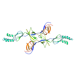
5YSL
 | | Crystal structure of antibody 1H1 Fab | | 分子名称: | 1H1 heavy chain, 1H1 light chain | | 著者 | Hu, X.L, Yang, F.L. | | 登録日 | 2017-11-14 | | 公開日 | 2017-12-27 | | 最終更新日 | 2023-11-22 | | 実験手法 | X-RAY DIFFRACTION (2.5 Å) | | 主引用文献 | Two classes of protective antibodies against Pseudorabies virus variant glycoprotein B: Implications for vaccine design.
PLoS Pathog., 13, 2017
|
|
6JKM
 | | Crystal structure of BubR1 kinase domain | | 分子名称: | ADENOSINE-5'-DIPHOSPHATE, GLYCEROL, MAGNESIUM ION, ... | | 著者 | Lin, L, Ye, S, Huang, Y, Liu, X, Zhang, R, Yao, X. | | 登録日 | 2019-03-01 | | 公開日 | 2019-06-26 | | 最終更新日 | 2023-11-22 | | 実験手法 | X-RAY DIFFRACTION (1.95 Å) | | 主引用文献 | BubR1 phosphorylates CENP-E as a switch enabling the transition from lateral association to end-on capture of spindle microtubules.
Cell Res., 29, 2019
|
|
6JKK
 | | Crystal structure of BubR1 kinase domain | | 分子名称: | DI(HYDROXYETHYL)ETHER, GLYCEROL, Mitotic checkpoint control protein kinase BUB1 | | 著者 | Lin, L, Ye, S, Huang, Y, Liu, X, Zhang, R, Yao, X. | | 登録日 | 2019-03-01 | | 公開日 | 2019-06-26 | | 最終更新日 | 2023-11-22 | | 実験手法 | X-RAY DIFFRACTION (1.85 Å) | | 主引用文献 | BubR1 phosphorylates CENP-E as a switch enabling the transition from lateral association to end-on capture of spindle microtubules.
Cell Res., 29, 2019
|
|
7YPX
 | | Cyanophage Pam3 fiber | | 分子名称: | Pam3 tail fiber proreins, tail fiber chaperone | | 著者 | Wei, Z.L, Jiang, Y.L, Zhou, C.Z. | | 登録日 | 2022-08-04 | | 公開日 | 2022-11-09 | | 最終更新日 | 2024-07-03 | | 実験手法 | ELECTRON MICROSCOPY (3.12 Å) | | 主引用文献 | Structural Insights into the Chaperone-Assisted Assembly of a Simplified Tail Fiber of the Myocyanophage Pam3.
Viruses, 14, 2022
|
|
7YQK
 | | cryo-EM structure of gammaH2AXK15ub-H4K20me2 nucleosome bound to 53BP1 | | 分子名称: | DNA (145-MER), Histone H2AX, Histone H2B, ... | | 著者 | Ai, H.S, GuoChao, C, Qingyue, G, Ze-Bin, T, Zhiheng, D, Xin, L, Fan, Y, Ziyu, X, Jia-Bin, L, Changlin, T, Liu, L. | | 登録日 | 2022-08-07 | | 公開日 | 2022-08-17 | | 最終更新日 | 2024-07-03 | | 実験手法 | ELECTRON MICROSCOPY (3.38 Å) | | 主引用文献 | Chemical Synthesis of Post-Translationally Modified H2AX Reveals Redundancy in Interplay between Histone Phosphorylation, Ubiquitination, and Methylation on the Binding of 53BP1 with Nucleosomes.
J.Am.Chem.Soc., 144, 2022
|
|
7YTU
 | |
7YTT
 | |
3EE5
 | | Crystal structure of human M340H-Beta1,4-Galactosyltransferase-I (M340H-B4GAL-T1) in complex with GLCNAC-Beta1,3-Gal-Beta-Naphthalenemethanol | | 分子名称: | 1,4-DIETHYLENE DIOXIDE, 2-(N-MORPHOLINO)-ETHANESULFONIC ACID, 2-acetamido-2-deoxy-beta-D-glucopyranose-(1-3)-beta-D-galactopyranose, ... | | 著者 | Ramakrishnan, B, Qasba, P.K. | | 登録日 | 2008-09-04 | | 公開日 | 2009-01-06 | | 最終更新日 | 2023-08-30 | | 実験手法 | X-RAY DIFFRACTION (2.2 Å) | | 主引用文献 | Deoxygenated Disaccharide Analogs as Specific Inhibitors of {beta}1-4-Galactosyltransferase 1 and Selectin-mediated Tumor Metastasis
J.Biol.Chem., 284, 2009
|
|
3OPO
 | |
3FLL
 | | Crystal structure of E55Q mutant of nitrophorin 4 | | 分子名称: | AMMONIA, Nitrophorin-4, PROTOPORPHYRIN IX CONTAINING FE | | 著者 | Montfort, W.R, Weichsel, A. | | 登録日 | 2008-12-18 | | 公開日 | 2009-02-10 | | 最終更新日 | 2023-09-06 | | 実験手法 | X-RAY DIFFRACTION (1.5 Å) | | 主引用文献 | Effect of mutation of carboxyl side-chain amino acids near the heme on the midpoint potentials and ligand binding constants of nitrophorin 2 and its NO, histamine, and imidazole complexes.
J.Am.Chem.Soc., 131, 2009
|
|
3OW7
 | |
6OVB
 | | Crystal structure of a Bacillus thuringiensis Cry1Da tryptic core variant | | 分子名称: | Active core crystal toxin protein 1D | | 著者 | Rydel, T.J, Halls, C, Evdokimov, A.G, Beishir, S.C. | | 登録日 | 2019-05-07 | | 公開日 | 2019-06-19 | | 最終更新日 | 2023-10-11 | | 実験手法 | X-RAY DIFFRACTION (2.611 Å) | | 主引用文献 | Bacillus thuringiensis Cry1Da_7 and Cry1B.868 Protein Interactions with Novel Receptors Allow Control of Resistant Fall Armyworms, Spodoptera frugiperda (J.E. Smith).
Appl.Environ.Microbiol., 85, 2019
|
|
7EFR
 | | Structure of SARS-CoV-2 spike receptor-binding domain in complex with high affinity ACE2 mutant (T27W,N330Y) | | 分子名称: | 2-acetamido-2-deoxy-beta-D-glucopyranose, Processed angiotensin-converting enzyme 2, Spike glycoprotein | | 著者 | Lu, G.W, Ye, F, Lin, X. | | 登録日 | 2021-03-23 | | 公開日 | 2021-11-24 | | 最終更新日 | 2023-11-29 | | 実験手法 | X-RAY DIFFRACTION (2.494 Å) | | 主引用文献 | S19W, T27W, and N330Y mutations in ACE2 enhance SARS-CoV-2 S-RBD binding toward both wild-type and antibody-resistant viruses and its molecular basis.
Signal Transduct Target Ther, 6, 2021
|
|


