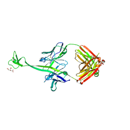
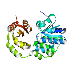
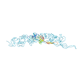
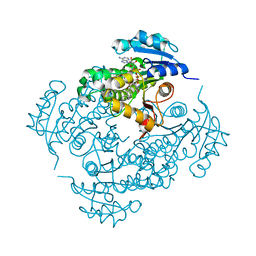
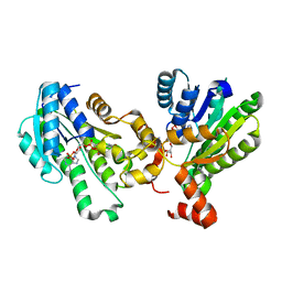
5D6Y
 
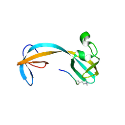 | | Crystal structure of double tudor domain of human lysine demethylase KDM4A complexed with histone H3K23me3 | | 分子名称: | Lysine-specific demethylase 4A, peptide H3K23me3 (19-28) | | 著者 | Wang, F, Su, Z, Miller, M.D, Denu, J.M, Phillips Jr, G.N, Enzyme Discovery for Natural Product Biosynthesis (NatPro) | | 登録日 | 2015-08-13 | | 公開日 | 2016-02-10 | | 最終更新日 | 2019-12-25 | | 実験手法 | X-RAY DIFFRACTION (2.287 Å) | | 主引用文献 | Reader domain specificity and lysine demethylase-4 family function.
Nat Commun, 7, 2016
|
|
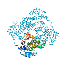
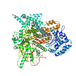
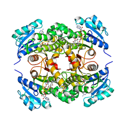
5D6W
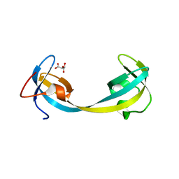 | | Crystal structure of double tudor domain of human lysine demethylase KDM4A | | 分子名称: | Lysine-specific demethylase 4A, S,R MESO-TARTARIC ACID | | 著者 | Wang, F, Su, Z, Denu, J.M, Phillips Jr, G.N, Enzyme Discovery for Natural Product Biosynthesis (NatPro) | | 登録日 | 2015-08-13 | | 公開日 | 2015-11-25 | | 最終更新日 | 2024-03-06 | | 実験手法 | X-RAY DIFFRACTION (1.992 Å) | | 主引用文献 | Reader domain specificity and lysine demethylase-4 family function.
Nat Commun, 7, 2016
|
|
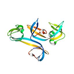
5D6X
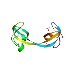 | | Crystal structure of double tudor domain of human lysine demethylase KDM4A | | 分子名称: | Lysine-specific demethylase 4A, SULFATE ION | | 著者 | Wang, F, Su, Z, Denu, J.M, Phillips Jr, G.N, Enzyme Discovery for Natural Product Biosynthesis (NatPro) | | 登録日 | 2015-08-13 | | 公開日 | 2015-11-25 | | 最終更新日 | 2024-03-06 | | 実験手法 | X-RAY DIFFRACTION (2.153 Å) | | 主引用文献 | Reader domain specificity and lysine demethylase-4 family function.
Nat Commun, 7, 2016
|
|
4K3E
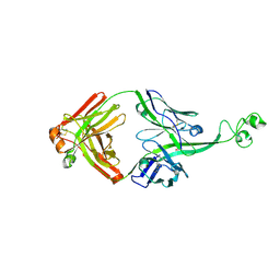 | | Crystal structure of bovine antibody BLV5B8 with ultralong CDR H3 | | 分子名称: | BOVINE ANTIBODY WITH ULTRALONG CDR H3, HEAVY CHAIN, LIGHT CHAIN, ... | | 著者 | Ekiert, D.C, Wang, F, Wilson, I.A. | | 登録日 | 2013-04-10 | | 公開日 | 2013-06-19 | | 最終更新日 | 2023-09-20 | | 実験手法 | X-RAY DIFFRACTION (2.2 Å) | | 主引用文献 | Reshaping antibody diversity.
Cell(Cambridge,Mass.), 153, 2013
|
|
4K3D
 | | Crystal structure of bovine antibody BLV1H12 with ultralong CDR H3 | | 分子名称: | BOVINE ANTIBODY WITH ULTRALONG CDR H3, HEAVY CHAIN, LIGHT CHAIN, ... | | 著者 | Ekiert, D.C, Wang, F, Wilson, I.A. | | 登録日 | 2013-04-10 | | 公開日 | 2013-06-19 | | 最終更新日 | 2023-09-20 | | 実験手法 | X-RAY DIFFRACTION (1.85 Å) | | 主引用文献 | Reshaping antibody diversity.
Cell(Cambridge,Mass.), 153, 2013
|
|
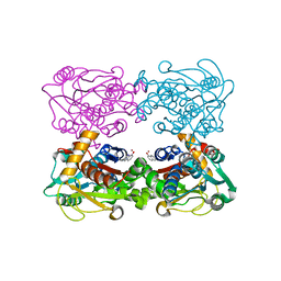
5JJU
 | | Crystal structure of Rv2837c complexed with 5'-pApA and 5'-AMP | | 分子名称: | ADENOSINE MONOPHOSPHATE, MANGANESE (II) ION, RNA (5'-R(P*AP*A)-3'), ... | | 著者 | Wang, F, He, Q, Liu, S, Gu, L. | | 登録日 | 2016-04-25 | | 公開日 | 2016-05-04 | | 最終更新日 | 2024-03-20 | | 実験手法 | X-RAY DIFFRACTION (2.312 Å) | | 主引用文献 | Structural and biochemical insight into the mechanism of Rv2837c from Mycobacterium tuberculosis as a c-di-NMP phosphodiesterase
J.Biol.Chem., 291, 2016
|
|
6X5I
 | |
4UC4
 | |
5CET
 | | Crystal structure of Rv2837c | | 分子名称: | Bifunctional oligoribonuclease and PAP phosphatase NrnA, MANGANESE (II) ION | | 著者 | Wang, F, He, Q, Zhu, D, Liu, S, Gu, L. | | 登録日 | 2015-07-07 | | 公開日 | 2015-12-23 | | 最終更新日 | 2024-03-20 | | 実験手法 | X-RAY DIFFRACTION (2 Å) | | 主引用文献 | Structural and Biochemical Insight into the Mechanism of Rv2837c from Mycobacterium tuberculosis as a c-di-NMP Phosphodiesterase
J.Biol.Chem., 291, 2016
|
|
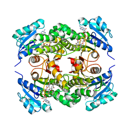
6XG6
 | | Full-length human mitochondrial Hsp90 (TRAP1) with ADP-BeF3 | | 分子名称: | ADENOSINE-5'-DIPHOSPHATE, BERYLLIUM TRIFLUORIDE ION, Heat shock protein 75 kDa, ... | | 著者 | Liu, Y.X, Wang, F, Agard, D.A. | | 登録日 | 2020-06-17 | | 公開日 | 2020-09-30 | | 最終更新日 | 2024-03-06 | | 実験手法 | ELECTRON MICROSCOPY (3.2 Å) | | 主引用文献 | General and robust covalently linked graphene oxide affinity grids for high-resolution cryo-EM.
Proc.Natl.Acad.Sci.USA, 117, 2020
|
|
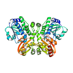
4WS9
 | | Crystal structure of sMAT N159G from Sulfolobus solfataricus | | 分子名称: | PHOSPHATE ION, S-adenosylmethionine synthase | | 著者 | Wang, F, Brady, E.L, Singh, S, Clinger, J.A, Huber, T.D, Thorson, J.S, Phillips Jr, G.N. | | 登録日 | 2014-10-26 | | 公開日 | 2014-11-05 | | 最終更新日 | 2023-09-27 | | 実験手法 | X-RAY DIFFRACTION (2.803 Å) | | 主引用文献 | Crystal structure of sMAT N159G from Sulfolobus solfataricus.
To Be Published
|
|
4XQ2
 | | Ensemble refinement of cystathione gamma lyase (CalE6) D7G from Micromonospora echinospora | | 分子名称: | 2-(N-MORPHOLINO)-ETHANESULFONIC ACID, CHLORIDE ION, CalE6, ... | | 著者 | Wang, F, Yennamalli, R.M, Singh, S, Tan, K, Thorson, J.S, Phillips Jr, G.N, Enzyme Discovery for Natural Product Biosynthesis (NatPro) | | 登録日 | 2015-01-18 | | 公開日 | 2015-04-15 | | 実験手法 | X-RAY DIFFRACTION (2.1 Å) | | 主引用文献 | The crystal structure of cystathione gamma lyase (CalE6) from Micromonospora echinospora
To Be Published
|
|
4XAU
 | | Crystal structure of AtS13 from Actinomadura melliaura | | 分子名称: | PYRIDOXAL-5'-PHOSPHATE, Putative aminotransferase | | 著者 | Wang, F, Singh, S, Xu, W, Thorson, J.S, Phillips Jr, G.N, Enzyme Discovery for Natural Product Biosynthesis (NatPro) | | 登録日 | 2014-12-15 | | 公開日 | 2014-12-24 | | 最終更新日 | 2023-09-27 | | 実験手法 | X-RAY DIFFRACTION (3.0012 Å) | | 主引用文献 | Structural characterization of AtmS13, a putative sugar aminotransferase involved in indolocarbazole AT2433 aminopentose biosynthesis.
Proteins, 83, 2015
|
|
8E5F
 | | Cryo-EM of P. calidifontis cytochrome filament | | 分子名称: | HEME C, c-type cytochrome | | 著者 | Wang, F, Cvirkaite-Krupovic, V, Krupovic, M, Egelman, E.H. | | 登録日 | 2022-08-22 | | 公開日 | 2023-05-10 | | 最終更新日 | 2023-07-26 | | 実験手法 | ELECTRON MICROSCOPY (3.8 Å) | | 主引用文献 | Extracellular cytochrome nanowires appear to be ubiquitous in prokaryotes.
Cell, 186, 2023
|
|
8E5G
 | | Cryo-EM of A. veneficus cytochrome filament | | 分子名称: | HEME C, c-type cytochrome | | 著者 | Wang, F, Baquero, D.P, Krupovic, M, Egelman, E.H. | | 登録日 | 2022-08-22 | | 公開日 | 2023-05-10 | | 最終更新日 | 2023-07-26 | | 実験手法 | ELECTRON MICROSCOPY (3.9 Å) | | 主引用文献 | Extracellular cytochrome nanowires appear to be ubiquitous in prokaryotes.
Cell, 186, 2023
|
|
3FNH
 | | Crystal structure of InhA bound to triclosan derivative | | 分子名称: | 2-(2,4-DICHLOROPHENOXY)-5-(2-PHENYLETHYL)PHENOL, Enoyl-[acyl-carrier-protein] reductase [NADH], NICOTINAMIDE-ADENINE-DINUCLEOTIDE | | 著者 | Wang, F. | | 登録日 | 2008-12-24 | | 公開日 | 2009-01-20 | | 最終更新日 | 2023-09-06 | | 実験手法 | X-RAY DIFFRACTION (2.8 Å) | | 主引用文献 | Triclosan Derivatives: Towards Potent Inhibitors of Drug-Sensitive and Drug-Resistant Mycobacterium tuberculosis.
Chemmedchem, 4, 2009
|
|
3FNF
 | | Crystal structure of InhA bound to triclosan derivative | | 分子名称: | 5-benzyl-2-(2,4-dichlorophenoxy)phenol, Enoyl-[acyl-carrier-protein] reductase [NADH], NICOTINAMIDE-ADENINE-DINUCLEOTIDE | | 著者 | Wang, F. | | 登録日 | 2008-12-24 | | 公開日 | 2009-01-20 | | 最終更新日 | 2023-09-06 | | 実験手法 | X-RAY DIFFRACTION (2.3 Å) | | 主引用文献 | Triclosan Derivatives: Towards Potent Inhibitors of Drug-Sensitive and Drug-Resistant Mycobacterium tuberculosis.
Chemmedchem, 4, 2009
|
|
8DST
 | |
3FNG
 | | Crystal structure of InhA bound to triclosan derivative | | 分子名称: | 5-(cyclohexylmethyl)-2-(2,4-dichlorophenoxy)phenol, Enoyl-[acyl-carrier-protein] reductase [NADH], NICOTINAMIDE-ADENINE-DINUCLEOTIDE | | 著者 | Wang, F. | | 登録日 | 2008-12-24 | | 公開日 | 2009-01-20 | | 最終更新日 | 2023-09-06 | | 実験手法 | X-RAY DIFFRACTION (1.97 Å) | | 主引用文献 | Triclosan Derivatives: Towards Potent Inhibitors of Drug-Sensitive and Drug-Resistant Mycobacterium tuberculosis.
Chemmedchem, 4, 2009
|
|
8EWG
 | | Cryo-EM structure of a riboendonclease | | 分子名称: | CRISPR-associated endonuclease Cas9, RNA (56-MER) | | 著者 | Li, Z, Wang, F. | | 登録日 | 2022-10-23 | | 公開日 | 2023-08-30 | | 最終更新日 | 2024-05-01 | | 実験手法 | ELECTRON MICROSCOPY (2.9 Å) | | 主引用文献 | Structural Basis for the Ribonuclease Activity of a Thermostable CRISPR-Cas13a from Thermoclostridium caenicola.
J.Mol.Biol., 435, 2023
|
|
8FJ5
 | | Structure of the Haloferax volcanii archaeal type IV pilus | | 分子名称: | Pilin_N domain-containing protein | | 著者 | Wang, F, Kreutzberger, M.A, Baquero, D.P, Krupovic, M, Egelman, E.H. | | 登録日 | 2022-12-19 | | 公開日 | 2023-06-28 | | 最終更新日 | 2024-06-19 | | 実験手法 | ELECTRON MICROSCOPY (2.9 Å) | | 主引用文献 | The evolution of archaeal flagellar filaments.
Proc.Natl.Acad.Sci.USA, 120, 2023
|
|
8FK7
 | | Structure of the Pyrobaculum calidifontis flagellar-like archaeal type IV pilus | | 分子名称: | Flagellin | | 著者 | Wang, F, Kreutzberger, M.A, Cvirkaite-Krupovic, V, Krupovic, M, Egelman, E.H. | | 登録日 | 2022-12-20 | | 公開日 | 2023-06-28 | | 最終更新日 | 2023-07-19 | | 実験手法 | ELECTRON MICROSCOPY (4.3 Å) | | 主引用文献 | The evolution of archaeal flagellar filaments.
Proc.Natl.Acad.Sci.USA, 120, 2023
|
|
5GT9
 | | The X-ray structure of 7beta-hydroxysteroid dehydrogenase | | 分子名称: | NADP NICOTINAMIDE-ADENINE-DINUCLEOTIDE PHOSPHATE, Oxidoreductase, short chain dehydrogenase/reductase family protein | | 著者 | Wang, F, Wang, R, Lv, Z, Chen, Q, Huo, X. | | 登録日 | 2016-08-19 | | 公開日 | 2017-05-17 | | 最終更新日 | 2023-11-08 | | 実験手法 | X-RAY DIFFRACTION (1.7 Å) | | 主引用文献 | Structure of NADP(+)-bound 7 beta-hydroxysteroid dehydrogenase reveals two cofactor-binding modes
Acta Crystallogr F Struct Biol Commun, 73, 2017
|
|
3FNE
 | | Crystal structure of InhA bound to triclosan derivative 17 | | 分子名称: | 2-(2,4-DICHLOROPHENOXY)-5-(PYRIDIN-2-YLMETHYL)PHENOL, Enoyl-[acyl-carrier-protein] reductase [NADH], NICOTINAMIDE-ADENINE-DINUCLEOTIDE | | 著者 | Wang, F. | | 登録日 | 2008-12-24 | | 公開日 | 2009-01-20 | | 最終更新日 | 2023-09-06 | | 実験手法 | X-RAY DIFFRACTION (1.98 Å) | | 主引用文献 | Triclosan Derivatives: Towards Potent Inhibitors of Drug-Sensitive and Drug-Resistant Mycobacterium tuberculosis.
Chemmedchem, 4, 2009
|
|
3FCA
 | | Genetic Incorporation of a Metal-ion Chelating Amino Acid into proteins as biophysical probe | | 分子名称: | Cysteine synthase, ZINC ION | | 著者 | Wang, F, Lee, H, Spraggon, G, Schultz, P.G. | | 登録日 | 2008-11-21 | | 公開日 | 2009-02-17 | | 最終更新日 | 2024-03-27 | | 実験手法 | X-RAY DIFFRACTION (2.149 Å) | | 主引用文献 | Genetic incorporation of a metal-ion chelating amino acid into proteins as a biophysical probe.
J.Am.Chem.Soc., 131, 2009
|
|


