Loading
PDBj
MenuPDBj@FacebookPDBj@TwitterPDBj@YouTubewwPDB FoundationwwPDB
RCSB PDBPDBeBMRBAdv. SearchSearch help
Search by PDB author
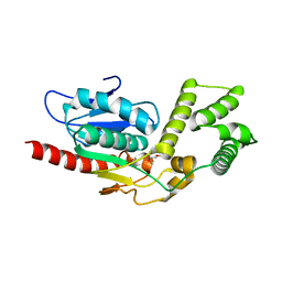
5V41
DownloadVisualize
BU of 5v41 by Molmil
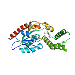
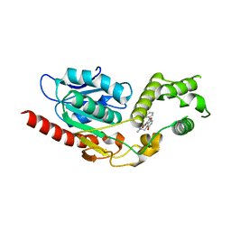
Crystal Structure of Mtb Pks13 Thioesterase domain in complex with inhibitor TAM5
Descriptor: Polyketide synthase Pks13 (Termination polyketide synthase), ethyl 5-hydroxy-4-(morpholin-4-ylmethyl)-2-phenyl-1-benzofuran-3-carboxylate
Authors:Aggarwal, A, Sacchettini, J.C.
Deposit date:2017-03-08
Release date:2017-07-05
Last modified:2023-10-04
Method:X-RAY DIFFRACTION (2.051 Å)
Cite:Development of a Novel Lead that Targets M. tuberculosis Polyketide Synthase 13.
Cell, 170, 2017
5V3W
DownloadVisualize
BU of 5v3w by Molmil
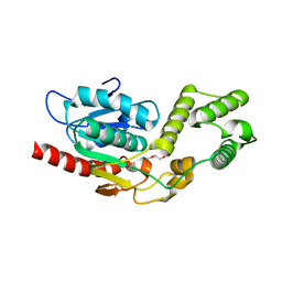
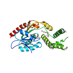
Crystal Structure of the Apo form of Thioesterase domain of Mtb Pks13
Descriptor: (2R)-2-{[(2R)-2-{[(2S)-2-{[(2R)-2-hydroxypropyl]oxy}propyl]oxy}propyl]oxy}propan-1-ol, Polyketide synthase Pks13 (Termination polyketide synthase)
Authors:Aggarwal, A, Sacchettini, J.C.
Deposit date:2017-03-08
Release date:2017-07-05
Last modified:2023-10-04
Method:X-RAY DIFFRACTION (1.723 Å)
Cite:Development of a Novel Lead that Targets M. tuberculosis Polyketide Synthase 13.
Cell, 170, 2017
5V42
DownloadVisualize
BU of 5v42 by Molmil
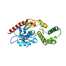
Crystal Structure of Mtb Pks13 Thioesterase domain in complex with inhibitor TAM3
Descriptor: Polyketide synthase Pks13 (Termination polyketide synthase), ethyl 5-hydroxy-2-phenyl-4-(pyrrolidin-1-ylmethyl)-1-benzofuran-3-carboxylate
Authors:Aggarwal, A, Sacchettini, J.C.
Deposit date:2017-03-08
Release date:2017-07-05
Last modified:2023-10-04
Method:X-RAY DIFFRACTION (1.987 Å)
Cite:Development of a Novel Lead that Targets M. tuberculosis Polyketide Synthase 13.
Cell, 170, 2017
5V3Y
DownloadVisualize
BU of 5v3y by Molmil
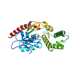
Crystal Structure of Mtb Pks13 Thioesterase domain in complex with inhibitor TAM16
Descriptor: 2-(4-hydroxyphenyl)-~{N}-methyl-5-oxidanyl-4-(piperidin-1-ylmethyl)-1-benzofuran-3-carboxamide, Polyketide synthase Pks13 (Termination polyketide synthase), SULFATE ION
Authors:Aggarwal, A, Sacchettini, J.C.
Deposit date:2017-03-08
Release date:2017-07-05
Last modified:2023-10-04
Method:X-RAY DIFFRACTION (1.98 Å)
Cite:Development of a Novel Lead that Targets M. tuberculosis Polyketide Synthase 13.
Cell, 170, 2017
5V3Z
DownloadVisualize
BU of 5v3z by Molmil
Crystal Structure of the D1607N mutant form of Thioesterase domain of Mtb Pks13
Descriptor: PENTAETHYLENE GLYCOL, Polyketide synthase Pks13 (Termination polyketide synthase), SULFATE ION
Authors:Aggarwal, A, Sacchettini, J.C.
Deposit date:2017-03-08
Release date:2017-07-05
Last modified:2023-10-04
Method:X-RAY DIFFRACTION (1.881 Å)
Cite:Development of a Novel Lead that Targets M. tuberculosis Polyketide Synthase 13.
Cell, 170, 2017
5V3X
DownloadVisualize
BU of 5v3x by Molmil
Crystal Structure of Mtb Pks13 Thioesterase domain in complex with inhibitor TAM1
Descriptor: Polyketide synthase Pks13 (Termination polyketide synthase), ethyl 5-hydroxy-4-[(4-methylpiperidin-1-yl)methyl]-2-phenyl-1-benzofuran-3-carboxylate
Authors:Aggarwal, A, Sacchettini, J.C.
Deposit date:2017-03-08
Release date:2017-07-05
Last modified:2023-10-04
Method:X-RAY DIFFRACTION (1.936 Å)
Cite:Development of a Novel Lead that Targets M. tuberculosis Polyketide Synthase 13.
Cell, 170, 2017
5V40
DownloadVisualize
BU of 5v40 by Molmil
Crystal Structure of Mtb Pks13 Thioesterase domain in complex with inhibitor TAM6
Descriptor: Polyketide synthase Pks13 (Termination polyketide synthase), ethyl 4-[(dimethylamino)methyl]-5-hydroxy-2-phenyl-1-benzofuran-3-carboxylate
Authors:Aggarwal, A, Sacchettini, J.C.
Deposit date:2017-03-08
Release date:2017-07-05
Last modified:2023-10-04
Method:X-RAY DIFFRACTION (1.991 Å)
Cite:Development of a Novel Lead that Targets M. tuberculosis Polyketide Synthase 13.
Cell, 170, 2017
7M7V
DownloadVisualize
BU of 7m7v by Molmil
Crystal Structure of Mtb Pks13 Thioesterase domain in complex with Compound 6
Descriptor: 5-hydroxy-2-(4-hydroxyphenyl)-N-methyl-4-[(2-oxa-6-azaspiro[3.4]octan-6-yl)methyl]-1-benzofuran-3-carboxamide, Polyketide synthase Pks13
Authors:Aggarwal, A, Sacchettini, J.C.
Deposit date:2021-03-29
Release date:2022-02-16
Last modified:2023-10-18
Method:X-RAY DIFFRACTION (2.29 Å)
Cite:Optimization of TAM16, a Benzofuran That Inhibits the Thioesterase Activity of Pks13; Evaluation toward a Preclinical Candidate for a Novel Antituberculosis Clinical Target.
J.Med.Chem., 65, 2022
7M5B
DownloadVisualize
BU of 7m5b by Molmil
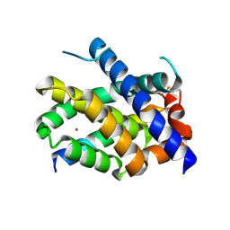
Crystal Structure of human BAK in complex with M3W5_BID
Descriptor: BH3-interacting domain death agonist p15, Bcl-2 homologous antagonist/killer, COPPER (II) ION
Authors:Singh, G, Aggarwal, A, Moldoveanu, T.
Deposit date:2021-03-23
Release date:2022-01-12
Last modified:2023-10-18
Method:X-RAY DIFFRACTION (1.85 Å)
Cite:Structural basis of BAK activation in mitochondrial apoptosis initiation.
Nat Commun, 13, 2022
7M5C
DownloadVisualize
BU of 7m5c by Molmil
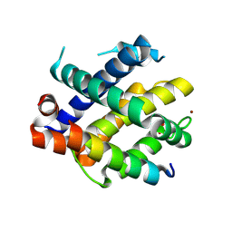
Crystal Structure of human BAK in complex with WT BAK BH3 peptide
Descriptor: Bcl-2 homologous antagonist/killer, COPPER (II) ION, SULFATE ION
Authors:Singh, G, Aggarwal, A, Moldoveanu, T.
Deposit date:2021-03-23
Release date:2022-01-12
Last modified:2023-10-18
Method:X-RAY DIFFRACTION (3.06 Å)
Cite:Structural basis of BAK activation in mitochondrial apoptosis initiation.
Nat Commun, 13, 2022
7M5A
DownloadVisualize
BU of 7m5a by Molmil
Crystal Structure of human BAK in complex with W3W5_BID
Descriptor: BH3-interacting domain death agonist p15, Bcl-2 homologous antagonist/killer
Authors:Singh, G, Aggarwal, A, Moldoveanu, T.
Deposit date:2021-03-23
Release date:2022-01-12
Last modified:2023-10-18
Method:X-RAY DIFFRACTION (1.5 Å)
Cite:Structural basis of BAK activation in mitochondrial apoptosis initiation.
Nat Commun, 13, 2022
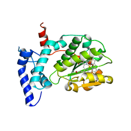
6W32
DownloadVisualize
BU of 6w32 by Molmil
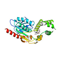
Crystal structure of Sfh5
Descriptor: PROTOPORPHYRIN IX CONTAINING FE, Phosphatidylinositol transfer protein SFH5
Authors:Gulten, G, Khan, D, Aggarwal, A, Krieger, I, Sacchettini, J.C, Bankaitis, V.A.
Deposit date:2020-03-08
Release date:2020-11-25
Last modified:2024-03-06
Method:X-RAY DIFFRACTION (2.9 Å)
Cite:A Sec14-like phosphatidylinositol transfer protein paralog defines a novel class of heme-binding proteins.
Elife, 9, 2020
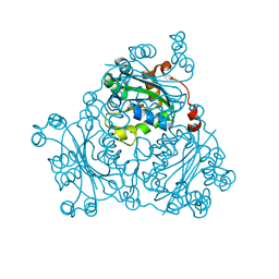
2VU5
DownloadVisualize
BU of 2vu5 by Molmil
Crystal structure of Pndk from Bacillus anthracis
Descriptor: NUCLEOSIDE DIPHOSPHATE KINASE
Authors:Misra, G, Aggarwal, A, Dube, D, Zaman, M.S, Singh, Y, Ramachandran, R.
Deposit date:2008-05-21
Release date:2009-03-10
Last modified:2024-05-08
Method:X-RAY DIFFRACTION (2 Å)
Cite:Crystal Structure of the Bacillus Anthracis Nucleoside Diphosphate Kinase and its Characterization Reveals an Enzyme Adapted to Perform Under Stress Conditions.
Proteins, 76, 2009
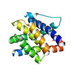
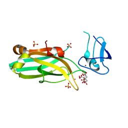
6UWA
DownloadVisualize
BU of 6uwa by Molmil
Mouse PKC C1B and C2 domains
Descriptor: CITRIC ACID, LEAD (II) ION, Protein kinase C, ...
Authors:Aggarwal, A, Mire, J, Sacchettini, J.C, Igumenova, T.
Deposit date:2019-11-04
Release date:2020-12-09
Last modified:2024-03-06
Method:X-RAY DIFFRACTION (1.2 Å)
Cite:Mouse PKC C1B and C2 domains
To Be Published
2LWE
DownloadVisualize
BU of 2lwe by Molmil
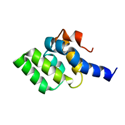
Solution structure of mutant (T170E) second CARD of human RIG-I
Descriptor: Probable ATP-dependent RNA helicase DDX58
Authors:Dutta, K, Ferrage, F, Aggarwal, A.
Deposit date:2012-07-27
Release date:2012-10-31
Last modified:2024-05-01
Method:SOLUTION NMR
Cite:Structure and Dynamics of the Second CARD of Human RIG-I Provide Mechanistic Insights into Regulation of RIG-I Activation.
Structure, 20, 2012
2LWD
DownloadVisualize
BU of 2lwd by Molmil
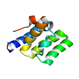
Solution structure of second CARD of human RIG-I
Descriptor: Probable ATP-dependent RNA helicase DDX58
Authors:Dutta, K, Ferrage, F, Aggarwal, A.
Deposit date:2012-07-27
Release date:2012-10-31
Last modified:2024-05-01
Method:SOLUTION NMR
Cite:Structure and Dynamics of the Second CARD of Human RIG-I Provide Mechanistic Insights into Regulation of RIG-I Activation.
Structure, 20, 2012
1IF1
DownloadVisualize
BU of 1if1 by Molmil
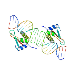
INTERFERON REGULATORY FACTOR 1 (IRF-1) COMPLEX WITH DNA
Descriptor: DNA (26-MER), PROTEIN (INTERFERON REGULATORY FACTOR 1)
Authors:Escalante, C.R, Yie, J, Thanos, D, Aggarwal, A.
Deposit date:1997-09-12
Release date:1998-02-25
Last modified:2024-02-07
Method:X-RAY DIFFRACTION (3 Å)
Cite:Structure of IRF-1 with bound DNA reveals determinants of interferon regulation.
Nature, 391, 1998
3SG7
DownloadVisualize
BU of 3sg7 by Molmil
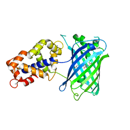
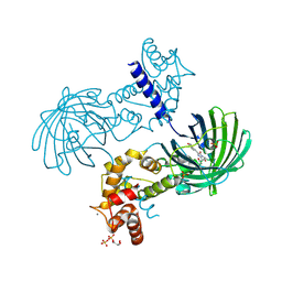
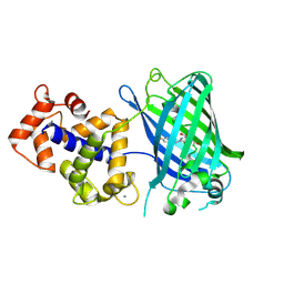
Crystal Structure of GCaMP3-KF(linker 1)
Descriptor: CALCIUM ION, Myosin light chain kinase, Green fluorescent protein, ...
Authors:Schreiter, E.R, Akerboom, J, Looger, L.L.
Deposit date:2011-06-14
Release date:2012-06-20
Last modified:2023-12-06
Method:X-RAY DIFFRACTION (1.9 Å)
Cite:Optimization of a GCaMP calcium indicator for neural activity imaging.
J.Neurosci., 32, 2012
8BON
DownloadVisualize
BU of 8bon by Molmil
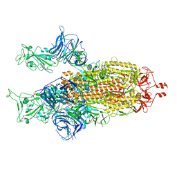
Structure of the SARS-CoV-2 spike glycoprotein in complex with the macrocyclic peptide S1B3inL1
Descriptor: 2-acetamido-2-deoxy-beta-D-glucopyranose, 2-acetamido-2-deoxy-beta-D-glucopyranose-(1-4)-2-acetamido-2-deoxy-beta-D-glucopyranose, Macrocyclic peptide S1B3inL1, ...
Authors:Hurdiss, D.L.
Deposit date:2022-11-15
Release date:2023-06-28
Last modified:2023-07-05
Method:ELECTRON MICROSCOPY (3.2 Å)
Cite:A broad-spectrum macrocyclic peptide inhibitor of the SARS-CoV-2 spike protein.
Proc.Natl.Acad.Sci.USA, 120, 2023
3SG5
DownloadVisualize
BU of 3sg5 by Molmil
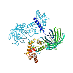
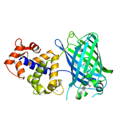
Crystal Structure of Dimeric GCaMP3-D380Y, QP(linker 1), LP(linker 2)
Descriptor: CALCIUM ION, GLYCEROL, Myosin light chain kinase, ...
Authors:Schreiter, E.R, Akerboom, J, Looger, L.L.
Deposit date:2011-06-14
Release date:2012-06-20
Last modified:2023-12-06
Method:X-RAY DIFFRACTION (1.9 Å)
Cite:Optimization of a GCaMP calcium indicator for neural activity imaging.
J.Neurosci., 32, 2012
3SG2
DownloadVisualize
BU of 3sg2 by Molmil
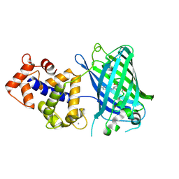
Crystal Structure of GCaMP2-T116V,D381Y
Descriptor: CALCIUM ION, Myosin light chain kinase, Green fluorescent protein, ...
Authors:Schreiter, E.R, Akerboom, J, Looger, L.L.
Deposit date:2011-06-14
Release date:2012-06-20
Last modified:2023-12-06
Method:X-RAY DIFFRACTION (2 Å)
Cite:Optimization of a GCaMP calcium indicator for neural activity imaging.
J.Neurosci., 32, 2012
3SG6
DownloadVisualize
BU of 3sg6 by Molmil
Crystal Structure of Dimeric GCaMP2-LIA(linker 1)
Descriptor: CALCIUM ION, Myosin light chain kinase, Green fluorescent protein, ...
Authors:Schreiter, E.R, Akerboom, J, Looger, L.L.
Deposit date:2011-06-14
Release date:2012-06-20
Last modified:2023-12-06
Method:X-RAY DIFFRACTION (1.7 Å)
Cite:Optimization of a GCaMP calcium indicator for neural activity imaging.
J.Neurosci., 32, 2012
3SG3
DownloadVisualize
BU of 3sg3 by Molmil
Crystal Structure of GCaMP3-D380Y
Descriptor: CALCIUM ION, Myosin light chain kinase, Green fluorescent protein, ...
Authors:Schreiter, E.R, Akerboom, J, Looger, L.L.
Deposit date:2011-06-14
Release date:2012-06-20
Last modified:2023-12-06
Method:X-RAY DIFFRACTION (2.1 Å)
Cite:Optimization of a GCaMP calcium indicator for neural activity imaging.
J.Neurosci., 32, 2012
3SG4
DownloadVisualize
BU of 3sg4 by Molmil
Crystal Structure of GCaMP3-D380Y, LP(linker 2)
Descriptor: CALCIUM ION, Myosin light chain kinase, Green fluorescent protein, ...
Authors:Schreiter, E.R, Akerboom, J, Looger, L.L.
Deposit date:2011-06-14
Release date:2012-06-20
Last modified:2023-12-06
Method:X-RAY DIFFRACTION (2.4 Å)
Cite:Optimization of a GCaMP calcium indicator for neural activity imaging.
J.Neurosci., 32, 2012
7E9Y
DownloadVisualize
BU of 7e9y by Molmil
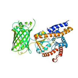
Crystal structure of eLACCO1
Descriptor: (2S)-2-HYDROXYPROPANOIC ACID, CALCIUM ION, Lactate-binding periplasmic protein TTHA0766,Lactate-binding periplasmic protein TTHA0766
Authors:Wen, Y, Campbell, R.E, Lemieux, M.J, Nasu, Y.
Deposit date:2021-03-05
Release date:2021-12-22
Last modified:2023-11-29
Method:X-RAY DIFFRACTION (2.25 Å)
Cite:A genetically encoded fluorescent biosensor for extracellular L-lactate.
Nat Commun, 12, 2021


12>

225681

PDB entries from 2024-10-02

PDB statisticsPDBj update infoContact PDBjnumon