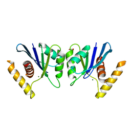
2G6H
 
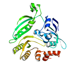
 | | Structure of rat nNOS heme domain (BH4 bound) in the reduced form | | Descriptor: | 5,6,7,8-TETRAHYDROBIOPTERIN, ACETATE ION, ARGININE, ... | | Authors: | Li, H, Igarashi, J, Jamal, J, Yang, W, Poulos, T.L. | | Deposit date: | 2006-02-24 | | Release date: | 2006-08-08 | | Last modified: | 2023-08-30 | | Method: | X-RAY DIFFRACTION (2 Å) | | Cite: | Structural studies of constitutive nitric oxide synthases with diatomic ligands bound.
J.Biol.Inorg.Chem., 11, 2006
|
|
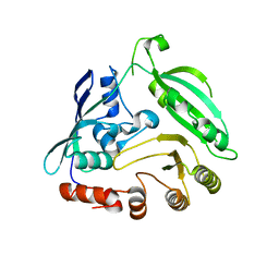
2COD
 | | Solution structure of the N-terminal PH domain of ARAP2 protein from human | | Descriptor: | Centaurin-delta 1 | | Authors: | Li, H, Tomizawa, T, Koshiba, S, Inoue, M, Kigawa, T, Yokoyama, S, RIKEN Structural Genomics/Proteomics Initiative (RSGI) | | Deposit date: | 2005-05-17 | | Release date: | 2005-11-17 | | Last modified: | 2024-05-29 | | Method: | SOLUTION NMR | | Cite: | Solution structure of the N-terminal PH domain of ARAP2 protein from human
To be Published
|
|
1V5P
 | | Solution Structure of the N-terminal Pleckstrin Homology Domain Of TAPP2 from Mouse | | Descriptor: | pleckstrin homology domain-containing, family A | | Authors: | Li, H, Hayashi, F, Koshiba, S, Inoue, M, Kigawa, T, Yokoyama, S, RIKEN Structural Genomics/Proteomics Initiative (RSGI) | | Deposit date: | 2003-11-25 | | Release date: | 2004-05-25 | | Last modified: | 2023-12-27 | | Method: | SOLUTION NMR | | Cite: | Solution Structure of the N-terminal Pleckstrin Homology Domain Of TAPP2 from Mouse
To be Published
|
|
1V88
 | | Solution Structure of the Pleckstrin Homology Domain of Oxysterol-Binding Protein-Related Protein 8 (KIAA1451 Protein) | | Descriptor: | Oxysterol binding protein-related protein 8 | | Authors: | Li, H, Tomizawa, T, Koshiba, S, Inoue, M, Kigawa, T, Yokoyama, S, RIKEN Structural Genomics/Proteomics Initiative (RSGI) | | Deposit date: | 2003-12-29 | | Release date: | 2004-06-29 | | Last modified: | 2023-12-27 | | Method: | SOLUTION NMR | | Cite: | Solution Structure of the Pleckstrin Homology Domain of Oxysterol-Binding Protein-Related Protein 8 (KIAA1451 Protein)
To be Published
|
|
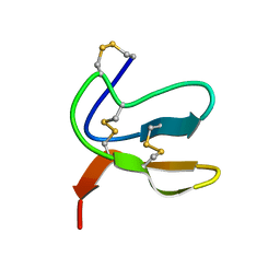
2CO9
 | | Solution structure of the HMG_box domain of thymus high mobility group box protein TOX from mouse | | Descriptor: | thymus high mobility group box protein TOX | | Authors: | Li, H, Saito, K, Koshiba, S, Inoue, M, Kigawa, T, Yokoyama, S, RIKEN Structural Genomics/Proteomics Initiative (RSGI) | | Deposit date: | 2005-05-17 | | Release date: | 2005-11-17 | | Last modified: | 2024-05-29 | | Method: | SOLUTION NMR | | Cite: | Solution structure of the HMG_box domain of thymus high mobility group box protein TOX from mouse
To be Published
|
|
2HAI
 | |
2COF
 | | Solution structure of the C-terminal PH domain of hypothetical protein KIAA1914 from human | | Descriptor: | Protein KIAA1914 | | Authors: | Li, H, Saito, K, Koshiba, S, Inoue, M, Kigawa, T, Yokoyama, S, RIKEN Structural Genomics/Proteomics Initiative (RSGI) | | Deposit date: | 2005-05-17 | | Release date: | 2005-11-17 | | Last modified: | 2024-05-29 | | Method: | SOLUTION NMR | | Cite: | Solution structure of the C-terminal PH domain of hypothetical protein KIAA1914 from human
To be Published
|
|
2COA
 | | Solution structure of the PH domain of protein kinase C, D2 type from human | | Descriptor: | Protein kinase C, D2 type | | Authors: | Li, H, Saito, K, Koshiba, S, Inoue, M, Kigawa, T, Yokoyama, S, RIKEN Structural Genomics/Proteomics Initiative (RSGI) | | Deposit date: | 2005-05-17 | | Release date: | 2005-11-17 | | Last modified: | 2024-05-29 | | Method: | SOLUTION NMR | | Cite: | Solution structure of the PH domain of protein kinase C, D2 type from human
To be Published
|
|
2COC
 | | Solution structure of the C-terminal PH domain of FYVE, RhoGEF and PH domain containing protein 3 (FGD3) from human | | Descriptor: | FYVE, RhoGEF and PH domain containing protein 3 | | Authors: | Li, H, Tomizawa, T, Tochio, N, Muto, Y, Koshiba, S, Inoue, M, Kigawa, T, Yokoyama, S, RIKEN Structural Genomics/Proteomics Initiative (RSGI) | | Deposit date: | 2005-05-17 | | Release date: | 2005-11-17 | | Last modified: | 2024-05-29 | | Method: | SOLUTION NMR | | Cite: | Solution structure of the C-terminal PH domain of FYVE, RhoGEF and PH domain containing protein 3 (FGD3) from human
To be Published
|
|
8J09
 | | Crystal structure of the Sld3 Cdc45-binding-domain, in complex with Cdc45 | | Descriptor: | Cell division control protein 45, DNA replication regulator SLD3 | | Authors: | Li, H, Ishizaka, I, Kato, K, Sun, X, Muramatsu, S, Itou, H, Ose, T, Araki, H, Yao, M. | | Deposit date: | 2023-04-10 | | Release date: | 2024-05-29 | | Last modified: | 2024-11-27 | | Method: | X-RAY DIFFRACTION (2.61 Å) | | Cite: | Structural and functional insights into Cdc45 recruitment by Sld7-Sld3 for CMG complex formation
Elife, 2024
|
|
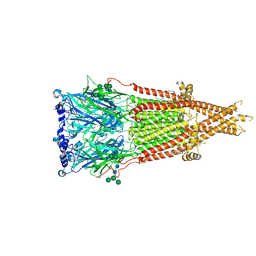
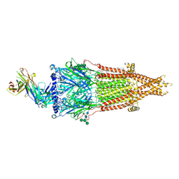
9DMQ
 | | Human muscle nAChR with fab3-bound | | Descriptor: | (2S)-3-(hexadecanoyloxy)-2-[(9Z)-octadec-9-enoyloxy]propyl 2-(trimethylammonio)ethyl phosphate, 2-acetamido-2-deoxy-beta-D-glucopyranose, 2-acetamido-2-deoxy-beta-D-glucopyranose-(1-4)-2-acetamido-2-deoxy-beta-D-glucopyranose, ... | | Authors: | Li, H, Hibbs, R.E. | | Deposit date: | 2024-09-14 | | Release date: | 2025-04-09 | | Last modified: | 2025-05-14 | | Method: | ELECTRON MICROSCOPY (2.06 Å) | | Cite: | Structure of human muscle AChR with fab3-bound
To Be Published
|
|
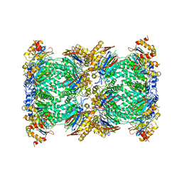
4G3T
 | | Mycobacterium smegmatis DprE1 - hexagonal crystal form | | Descriptor: | oxidoreductase DprE1 | | Authors: | Li, H, Jogl, G. | | Deposit date: | 2012-07-15 | | Release date: | 2012-12-05 | | Last modified: | 2024-02-28 | | Method: | X-RAY DIFFRACTION (2.346 Å) | | Cite: | Crystal structure of decaprenylphosphoryl-beta- D-ribose 2'-epimerase from Mycobacterium smegmatis.
Proteins, 81, 2013
|
|
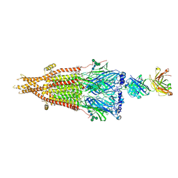
9DML
 | | Human muscle nAChR with fab2-bound | | Descriptor: | (2S)-3-(hexadecanoyloxy)-2-[(9Z)-octadec-9-enoyloxy]propyl 2-(trimethylammonio)ethyl phosphate, 2-acetamido-2-deoxy-beta-D-glucopyranose, Acetylcholine receptor subunit alpha, ... | | Authors: | Li, H, Hibbs, R.E. | | Deposit date: | 2024-09-13 | | Release date: | 2025-04-09 | | Last modified: | 2025-05-21 | | Method: | ELECTRON MICROSCOPY (2.24 Å) | | Cite: | Structure of human muscle AChR with fab2-bound
To Be Published
|
|
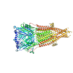
9DMJ
 | | Human muscle nAChR with two fab1b-bound | | Descriptor: | (2S)-3-(hexadecanoyloxy)-2-[(9Z)-octadec-9-enoyloxy]propyl 2-(trimethylammonio)ethyl phosphate, 2-acetamido-2-deoxy-beta-D-glucopyranose, 2-acetamido-2-deoxy-beta-D-glucopyranose-(1-4)-2-acetamido-2-deoxy-beta-D-glucopyranose, ... | | Authors: | Li, H, Hibbs, R.E. | | Deposit date: | 2024-09-13 | | Release date: | 2025-04-09 | | Last modified: | 2025-05-14 | | Method: | ELECTRON MICROSCOPY (2.19 Å) | | Cite: | Structure of human muscle AChR with two fab1b-bound
To Be Published
|
|
9DMH
 | | Human muscle nAChR ACh-bound state | | Descriptor: | (2S)-3-(hexadecanoyloxy)-2-[(9Z)-octadec-9-enoyloxy]propyl 2-(trimethylammonio)ethyl phosphate, 2-acetamido-2-deoxy-beta-D-glucopyranose-(1-4)-2-acetamido-2-deoxy-beta-D-glucopyranose, ACETYLCHOLINE, ... | | Authors: | Li, H, Hibbs, R.E. | | Deposit date: | 2024-09-13 | | Release date: | 2025-04-09 | | Last modified: | 2025-05-21 | | Method: | ELECTRON MICROSCOPY (2.06 Å) | | Cite: | Structure of human muscle AChR ACh-bound state
To Be Published
|
|
9DMK
 | | Human muscle nAChR with one fab1b-bound | | Descriptor: | (2S)-3-(hexadecanoyloxy)-2-[(9Z)-octadec-9-enoyloxy]propyl 2-(trimethylammonio)ethyl phosphate, 2-acetamido-2-deoxy-beta-D-glucopyranose, Acetylcholine receptor subunit alpha, ... | | Authors: | Li, H, Hibbs, R.E. | | Deposit date: | 2024-09-13 | | Release date: | 2025-04-09 | | Last modified: | 2025-05-21 | | Method: | ELECTRON MICROSCOPY (2.46 Å) | | Cite: | Structure of human muscle AChR with one fab1b-bound
To Be Published
|
|
9DMS
 | | Human muscle nAChR with fab6-bound | | Descriptor: | (2S)-3-(hexadecanoyloxy)-2-[(9Z)-octadec-9-enoyloxy]propyl 2-(trimethylammonio)ethyl phosphate, 2-acetamido-2-deoxy-beta-D-glucopyranose, Acetylcholine receptor subunit alpha, ... | | Authors: | Li, H, Hibbs, R.E. | | Deposit date: | 2024-09-14 | | Release date: | 2025-04-09 | | Last modified: | 2025-05-14 | | Method: | ELECTRON MICROSCOPY (1.92 Å) | | Cite: | Structure of human muscle AChR with fab6-bound
To Be Published
|
|
9DMT
 | | Human muscle nAChR with fab7-bound | | Descriptor: | (2S)-3-(hexadecanoyloxy)-2-[(9Z)-octadec-9-enoyloxy]propyl 2-(trimethylammonio)ethyl phosphate, 2-acetamido-2-deoxy-beta-D-glucopyranose, Acetylcholine receptor subunit alpha, ... | | Authors: | Li, H, Hibbs, R.E. | | Deposit date: | 2024-09-14 | | Release date: | 2025-04-09 | | Last modified: | 2025-05-28 | | Method: | ELECTRON MICROSCOPY (2.18 Å) | | Cite: | Structure of human muscle AChR with fab7-bound
To Be Published
|
|
9DMG
 | | Human muscle nAChR apo state | | Descriptor: | (2S)-3-(hexadecanoyloxy)-2-[(9Z)-octadec-9-enoyloxy]propyl 2-(trimethylammonio)ethyl phosphate, 2-acetamido-2-deoxy-beta-D-glucopyranose-(1-4)-2-acetamido-2-deoxy-beta-D-glucopyranose, Acetylcholine receptor subunit alpha, ... | | Authors: | Li, H, Hibbs, R.E. | | Deposit date: | 2024-09-13 | | Release date: | 2025-04-09 | | Last modified: | 2025-05-14 | | Method: | ELECTRON MICROSCOPY (2.05 Å) | | Cite: | Structure of human muscle AChR apo state
To Be Published
|
|
9DMV
 | | Human muscle nAChR with fab9-bound | | Descriptor: | (2S)-3-(hexadecanoyloxy)-2-[(9Z)-octadec-9-enoyloxy]propyl 2-(trimethylammonio)ethyl phosphate, 2-acetamido-2-deoxy-beta-D-glucopyranose, 2-acetamido-2-deoxy-beta-D-glucopyranose-(1-4)-2-acetamido-2-deoxy-beta-D-glucopyranose, ... | | Authors: | Li, H, Hibbs, R.E. | | Deposit date: | 2024-09-14 | | Release date: | 2025-04-09 | | Last modified: | 2025-05-14 | | Method: | ELECTRON MICROSCOPY (2.13 Å) | | Cite: | Structure of human muscle AChR with fab9-bound
To Be Published
|
|
5FMG
 | | Structure and function based design of Plasmodium-selective proteasome inhibitors | | Descriptor: | (2S)-N-[(E,2S)-1-(1H-indol-3-yl)-4-methylsulfonyl-but-3-en-2-yl]-2-[[(2S)-3-(1H-indol-3-yl)-2-(2-morpholin-4-ylethanoylamino)propanoyl]amino]-4-methyl-pentanamide, BETA3 PROTEASOME SUBUNIT, PUTATIVE, ... | | Authors: | Li, H, O'Donoghue, A.J, van der Linden, W.A, Xie, S.C, Yoo, E, Foe, I.T, Tilley, L, Craik, C.S, da Fonseca, P.C.A, Bogyo, M. | | Deposit date: | 2015-11-04 | | Release date: | 2016-03-02 | | Last modified: | 2024-11-13 | | Method: | ELECTRON MICROSCOPY (3.6 Å) | | Cite: | Structure and Function Based Design of Plasmodium-Selective Proteasome Inhibitors
Nature, 530, 2016
|
|
5E6F
 | | Canarypox virus resolvase | | Descriptor: | CNPV261 Holliday junction resolvase protein, D(-)-TARTARIC ACID, MAGNESIUM ION | | Authors: | Li, H, Hwang, Y, Perry, K, Bushman, F.D, Van Duyne, G.D. | | Deposit date: | 2015-10-09 | | Release date: | 2016-03-30 | | Last modified: | 2023-09-27 | | Method: | X-RAY DIFFRACTION (2.6 Å) | | Cite: | Structure and Metal Binding Properties of a Poxvirus Resolvase.
J.Biol.Chem., 291, 2016
|
|
4G3U
 | |
3NO9
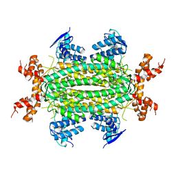 | | Crystal Structure of apo fumarate hydratase from Mycobacterium tuberculosis | | Descriptor: | Fumarate hydratase class II | | Authors: | Li, H, Swanson, S, Yu, M, Hung, L.-W, Sacchettini, J.C, TB Structural Genomics Consortium (TBSGC) | | Deposit date: | 2010-06-25 | | Release date: | 2010-07-14 | | Last modified: | 2023-09-06 | | Method: | X-RAY DIFFRACTION (2.48 Å) | | Cite: | Crystal Structure of apo fumarate hydratase from Mycobacterium tuberculosis
To be Published
|
|
2M3J
 | |


