Loading
PDBj
MenuPDBj@FacebookPDBj@TwitterPDBj@YouTubewwPDB FoundationwwPDB
RCSB PDBPDBeBMRBAdv. SearchSearch help
Search by PDB author
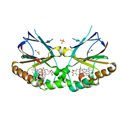
5K91
DownloadVisualize
BU of 5k91 by Molmil
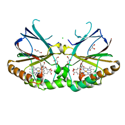
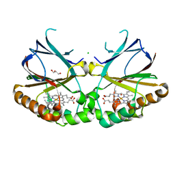
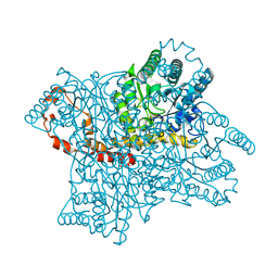
Crystal structure of dimeric chlorite dismutase from Cyanothece sp. PCC7425 in complex with fluoride
Descriptor: Chlorite dismutase, FLUORIDE ION, GLYCEROL, ...
Authors:Puehringer, D, Schaffner, I, Mlynek, G, Obinger, C, Djinovic-Carugo, K.
Deposit date:2016-05-31
Release date:2017-06-21
Last modified:2024-01-10
Method:X-RAY DIFFRACTION (1.18 Å)
Cite:Molecular Mechanism of Enzymatic Chlorite Detoxification: Insights from Structural and Kinetic Studies.
ACS Catal, 7, 2017
6AQ6
DownloadVisualize
BU of 6aq6 by Molmil
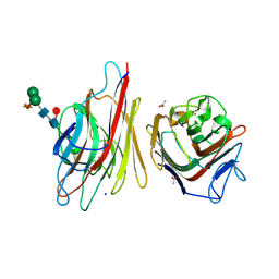
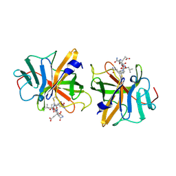
X-ray crystal structure of Erythrina crista-galli lectin in complex with N-acetyllactosamine
Descriptor: 2-acetamido-2-deoxy-beta-D-glucopyranose-(1-4)-2-acetamido-2-deoxy-beta-D-glucopyranose, ACETATE ION, CALCIUM ION, ...
Authors:Gerlits, O, Woods, R.J.
Deposit date:2017-08-18
Release date:2018-08-22
Last modified:2024-10-23
Method:X-RAY DIFFRACTION (1.903 Å)
Cite:Defining the Specificity of Carbohydrate-Protein Interactions by Quantifying Functional Group Contributions.
J Chem Inf Model, 58, 2018
5LZL
DownloadVisualize
BU of 5lzl by Molmil
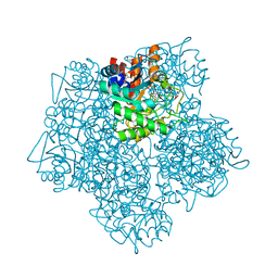
Pyrobaculum calidifontis 5-aminolaevulinic acid dehydratase
Descriptor: Delta-aminolevulinic acid dehydratase, ZINC ION
Authors:Azim, N, Erskine, P.T, Guo, J, Cooper, J.B.
Deposit date:2016-09-30
Release date:2016-10-12
Last modified:2024-01-17
Method:X-RAY DIFFRACTION (3.47 Å)
Cite:Structural studies of substrate and product complexes of 5-aminolaevulinic acid dehydratase from humans, Escherichia coli and the hyperthermophile Pyrobaculum calidifontis.
Acta Crystallogr D Struct Biol, 73, 2017
5MHB
DownloadVisualize
BU of 5mhb by Molmil
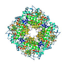
Product-Complex of E.coli 5-Amino Laevulinic Acid Dehydratase
Descriptor: 3-[5-(AMINOMETHYL)-4-(CARBOXYMETHYL)-1H-PYRROL-3-YL]PROPANOIC ACID, Delta-aminolevulinic acid dehydratase, GLYCEROL, ...
Authors:Norton, E, Erskine, P.T, Shoolingin-Jordan, P.M, Cooper, J.B.
Deposit date:2016-11-23
Release date:2016-12-07
Last modified:2024-01-17
Method:X-RAY DIFFRACTION (2.1 Å)
Cite:Structural studies of substrate and product complexes of 5-aminolaevulinic acid dehydratase from humans, Escherichia coli and the hyperthermophile Pyrobaculum calidifontis.
Acta Crystallogr D Struct Biol, 73, 2017
5NKV
DownloadVisualize
BU of 5nkv by Molmil
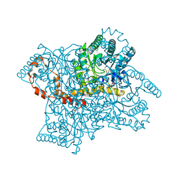
Crystal structure of dimeric chlorite dismutase from Cyanothece sp. PCC7425 at pH 9.0 and 293 K.
Descriptor: CHLORIDE ION, Chlorite Dismutase, GLYCEROL, ...
Authors:Puehringer, D, Schaffner, I, Mlynek, G, Obinger, C, Djinovic-Carugo, K.
Deposit date:2017-04-03
Release date:2018-01-31
Last modified:2024-05-08
Method:X-RAY DIFFRACTION (2 Å)
Cite:Molecular Mechanism of Enzymatic Chlorite Detoxification: Insights from Structural and Kinetic Studies.
ACS Catal, 7, 2017
5NKU
DownloadVisualize
BU of 5nku by Molmil
Joint neutron/X-ray structure of dimeric chlorite dismutase from Cyanothece sp. PCC7425
Descriptor: CHLORIDE ION, Chlorite Dismutase, GLYCEROL, ...
Authors:Puehringer, D, Schaffner, I, Mlynek, G, Obinger, C, Djinovic-Carugo, K.
Deposit date:2017-04-03
Release date:2018-02-28
Last modified:2024-05-01
Method:NEUTRON DIFFRACTION (2 Å), X-RAY DIFFRACTION
Cite:Molecular Mechanism of Enzymatic Chlorite Detoxification: Insights from Structural and Kinetic Studies.
ACS Catal, 7, 2017
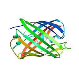
5EB7
DownloadVisualize
BU of 5eb7 by Molmil
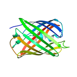
Crystal Structure of the Reversibly photoswitching chromoprotein Dathail, Metastable State
Descriptor: Reversibly photoswitching chromoprotein Dathail
Authors:Close, D.W, Langan, P.S, Bradbury, A.R.M.
Deposit date:2015-10-18
Release date:2016-04-06
Last modified:2023-11-15
Method:X-RAY DIFFRACTION (1.65 Å)
Cite:Evolution and characterization of a new reversibly photoswitching chromogenic protein, Dathail.
J.Mol.Biol., 428, 2016
5VG1
DownloadVisualize
BU of 5vg1 by Molmil
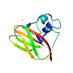
Neutron crystallographic structure of a Jonesia denitrificans lytic polysaccharide monooxygenase
Descriptor: COPPER (II) ION, Chitinase, PEROXIDE ION
Authors:Bacik, J.-P, Unkefer, C.J, Chen, J.C.H.
Deposit date:2017-04-10
Release date:2017-05-24
Last modified:2024-10-30
Method:NEUTRON DIFFRACTION (2.1 Å)
Cite:Neutron and Atomic Resolution X-ray Structures of a Lytic Polysaccharide Monooxygenase Reveal Copper-Mediated Dioxygen Binding and Evidence for N-Terminal Deprotonation.
Biochemistry, 56, 2017
5VG0
DownloadVisualize
BU of 5vg0 by Molmil
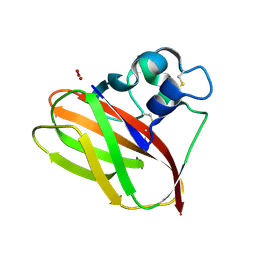
Room temperature X-ray crystallographic structure of a Jonesia denitrificans lytic polysaccharide monooxygenase at 1.1 angstrom resolution.
Descriptor: COPPER (II) ION, Chitinase, PEROXIDE ION
Authors:Bacik, J.-P, Unkefer, C.J, Chen, J.C.H.
Deposit date:2017-04-10
Release date:2017-05-24
Last modified:2023-10-04
Method:X-RAY DIFFRACTION (1.1 Å)
Cite:Neutron and Atomic Resolution X-ray Structures of a Lytic Polysaccharide Monooxygenase Reveal Copper-Mediated Dioxygen Binding and Evidence for N-Terminal Deprotonation.
Biochemistry, 56, 2017
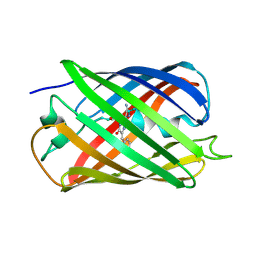
5EB6
DownloadVisualize
BU of 5eb6 by Molmil
Crystal Structure of the Reversibly photoswitching chromoprotein Dathail, Ground State
Descriptor: Reversible photoswitching chromoprotein Dathail
Authors:Close, D.W, Langan, P.S, Bradbury, A.R.M.
Deposit date:2015-10-18
Release date:2016-04-06
Last modified:2023-11-15
Method:X-RAY DIFFRACTION (1.65008247 Å)
Cite:Evolution and characterization of a new reversibly photoswitching chromogenic protein, Dathail.
J.Mol.Biol., 428, 2016
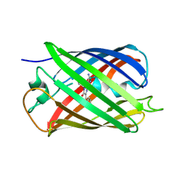
5EBJ
DownloadVisualize
BU of 5ebj by Molmil
Joint X-ray/neutron structure of reversibly photoswitching chromogenic protein, Dathail
Descriptor: photoswitching chromogenic protein
Authors:Kovalevsky, A.Y, Langan, P.S, Bradbury, A.R.M.
Deposit date:2015-10-19
Release date:2016-04-06
Last modified:2024-10-23
Method:NEUTRON DIFFRACTION (2.5 Å), X-RAY DIFFRACTION
Cite:Evolution and characterization of a new reversibly photoswitching chromogenic protein, Dathail.
J.Mol.Biol., 428, 2016
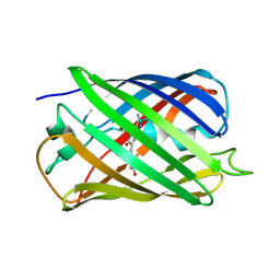
5EJU
DownloadVisualize
BU of 5eju by Molmil
Ensemble refinement of the Crystal Structure of the Reversibly photoswitching chromoprotein Dathail, Ground State
Descriptor: Reversibly photoswitching protein Dathail
Authors:Close, D.W, Langan, P.S, Bradbury, A.R.M.
Deposit date:2015-11-02
Release date:2016-04-06
Last modified:2024-10-30
Method:X-RAY DIFFRACTION (1.65 Å)
Cite:Evolution and characterization of a new reversibly photoswitching chromogenic protein, Dathail.
J.Mol.Biol., 428, 2016
5EXU
DownloadVisualize
BU of 5exu by Molmil
Reversibly photoswitching protein Dathail, Ensemble refinement
Descriptor: Reversibly photoswitching protein Dathail
Authors:Close, D.W, Langan, P.S, Bradbury, A.R.M.
Deposit date:2015-11-24
Release date:2016-04-06
Last modified:2024-10-16
Method:X-RAY DIFFRACTION (1.65 Å)
Cite:Evolution and characterization of a new reversibly photoswitching chromogenic protein, Dathail.
J.Mol.Biol., 428, 2016
2GUB
DownloadVisualize
BU of 2gub by Molmil
Crystal Structure of Metal Free D-Xylose Isomerase.
Descriptor: Xylose isomerase
Authors:Carrell, H.L, Katz, A.K, Glusker, J.P.
Deposit date:2006-04-29
Release date:2006-05-16
Last modified:2023-08-30
Method:X-RAY DIFFRACTION (1.8 Å)
Cite:Locating active-site hydrogen atoms in D-xylose isomerase: Time-of-flight neutron diffraction.
Proc.Natl.Acad.Sci.Usa, 103, 2006
2GLK
DownloadVisualize
BU of 2glk by Molmil
High-resolution study of D-Xylose isomerase, 0.94A resolution.
Descriptor: GLYCEROL, MANGANESE (II) ION, Xylose isomerase
Authors:Katz, A.K, Carrell, H.L, Hanson, B.L, Harp, J.M, Glusker, J.P, Bunick, G.J.
Deposit date:2006-04-05
Release date:2006-05-16
Last modified:2023-08-30
Method:X-RAY DIFFRACTION (0.94 Å)
Cite:Locating active-site hydrogen atoms in D-xylose isomerase: Time-of-flight neutron diffraction.
Proc.Natl.Acad.Sci.Usa, 103, 2006
4FYJ
DownloadVisualize
BU of 4fyj by Molmil
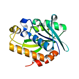
Crystal Structure of P. aeruginosa peptidyl-tRNA hydrolase
Descriptor: Peptidyl-tRNA hydrolase
Authors:Hughes, R.C, McFeeters, H, McFeeters, R.L.
Deposit date:2012-07-04
Release date:2012-12-12
Last modified:2023-09-13
Method:X-RAY DIFFRACTION (1.77 Å)
Cite:Recombinant production, crystallization and X-ray crystallographic structure determination of the peptidyl-tRNA hydrolase of Pseudomonas aeruginosa.
Acta Crystallogr.,Sect.F, 68, 2012
2IPH
DownloadVisualize
BU of 2iph by Molmil
X-ray Structure at 1.75 A Resolution of a Norovirus Protease Linked to an Active Site Directed Peptide Inhibitor
Descriptor: N-ACETYL-L-ALPHA-GLUTAMYL-L-PHENYLALANYL-L-GLUTAMINYL-N-[(1S)-4-AMINO-1-(2-CARBOXYETHYL)-4-OXOBUTYL]-L-LEUCINAMIDE, Thiol protease P3C
Authors:Hussey, R.J.
Deposit date:2006-10-12
Release date:2007-10-23
Last modified:2012-06-20
Method:X-RAY DIFFRACTION (1.75 Å)
Cite:A Structural Study of Norovirus 3C Protease Specificity: Binding of a Designed Active Site-Directed Peptide Inhibitor.
Biochemistry, 50, 2011
6K9R
DownloadVisualize
BU of 6k9r by Molmil
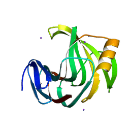
Crystal Structure Analysis of Endo-beta-1,4-xylanase II Complexed with Xylotriose
Descriptor: Endo-1,4-beta-xylanase 2, IODIDE ION, beta-D-xylopyranose-(1-4)-beta-D-xylopyranose-(1-4)-beta-D-xylopyranose
Authors:Li, C, Wan, Q.
Deposit date:2019-06-17
Release date:2020-07-08
Last modified:2023-11-22
Method:X-RAY DIFFRACTION (1.3 Å)
Cite:Studying the Role of a Single Mutation of a Family 11 Glycoside Hydrolase Using High-Resolution X-ray Crystallography.
Protein J., 39, 2020
6K9O
DownloadVisualize
BU of 6k9o by Molmil
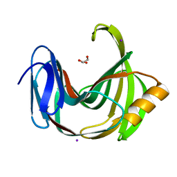
Crystal Structure Analysis of Protein
Descriptor: Endo-1,4-beta-xylanase 2, GLYCEROL, IODIDE ION
Authors:Li, C, Wan, Q.
Deposit date:2019-06-17
Release date:2020-06-17
Last modified:2023-11-22
Method:X-RAY DIFFRACTION (1.06 Å)
Cite:Studying the Role of a Single Mutation of a Family 11 Glycoside Hydrolase Using High-Resolution X-ray Crystallography.
Protein J., 39, 2020
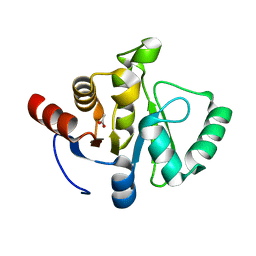
7TWR
DownloadVisualize
BU of 7twr by Molmil
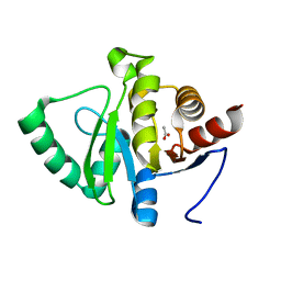
Crystal structure of SARS-CoV-2 NSP3 macrodomain at pH 8 (P43 crystal form)
Descriptor: ACETATE ION, Non-structural protein 3
Authors:Correy, G.J, Fraser, J.S.
Deposit date:2022-02-07
Release date:2022-02-23
Last modified:2023-10-18
Method:X-RAY DIFFRACTION (0.9 Å)
Cite:The mechanisms of catalysis and ligand binding for the SARS-CoV-2 NSP3 macrodomain from neutron and x-ray diffraction at room temperature.
Sci Adv, 8, 2022
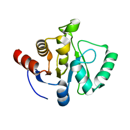
7TWF
DownloadVisualize
BU of 7twf by Molmil
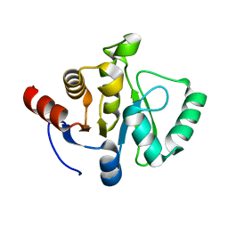
Crystal structure of SARS-CoV-2 NSP3 macrodomain at 293 K (P43 crystal form, 73 kGy)
Descriptor: Non-structural protein 3
Authors:Correy, G.J, Fraser, J.S.
Deposit date:2022-02-07
Release date:2022-02-23
Last modified:2023-10-18
Method:X-RAY DIFFRACTION (1.1 Å)
Cite:The mechanisms of catalysis and ligand binding for the SARS-CoV-2 NSP3 macrodomain from neutron and x-ray diffraction at room temperature.
Sci Adv, 8, 2022
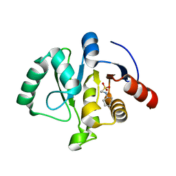
7TWP
DownloadVisualize
BU of 7twp by Molmil
Crystal structure of SARS-CoV-2 NSP3 macrodomain at pH 7 (P43 crystal form)
Descriptor: ACETATE ION, Non-structural protein 3
Authors:Correy, G.J, Fraser, J.S.
Deposit date:2022-02-07
Release date:2022-02-23
Last modified:2023-10-18
Method:X-RAY DIFFRACTION (0.9 Å)
Cite:The mechanisms of catalysis and ligand binding for the SARS-CoV-2 NSP3 macrodomain from neutron and x-ray diffraction at room temperature.
Sci Adv, 8, 2022
7TWH
DownloadVisualize
BU of 7twh by Molmil
Crystal structure of SARS-CoV-2 NSP3 macrodomain at 293 K (P43 crystal form, 290 kGy)
Descriptor: Non-structural protein 3
Authors:Correy, G.J, Fraser, J.S.
Deposit date:2022-02-07
Release date:2022-02-23
Last modified:2023-10-18
Method:X-RAY DIFFRACTION (1.1 Å)
Cite:The mechanisms of catalysis and ligand binding for the SARS-CoV-2 NSP3 macrodomain from neutron and x-ray diffraction at room temperature.
Sci Adv, 8, 2022
7TX3
DownloadVisualize
BU of 7tx3 by Molmil
Neutron crystal structure of SARS-CoV-2 NSP3 macrodomain at 293 K (P43 crystal form)
Descriptor: 2-[N-CYCLOHEXYLAMINO]ETHANE SULFONIC ACID, Non-structural protein 3
Authors:Correy, G.J, Fraser, J.S, Meilleur, F.
Deposit date:2022-02-07
Release date:2022-02-23
Last modified:2023-10-25
Method:NEUTRON DIFFRACTION (1.6 Å), X-RAY DIFFRACTION
Cite:The mechanisms of catalysis and ligand binding for the SARS-CoV-2 NSP3 macrodomain from neutron and x-ray diffraction at room temperature.
Sci Adv, 8, 2022
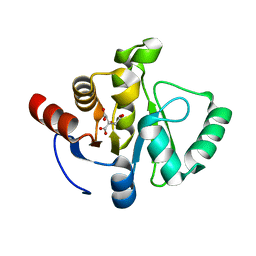
7TWN
DownloadVisualize
BU of 7twn by Molmil
Crystal structure of SARS-CoV-2 NSP3 macrodomain at pH 5 (P43 crystal form)
Descriptor: 2-[N-CYCLOHEXYLAMINO]ETHANE SULFONIC ACID, CITRIC ACID, Non-structural protein 3
Authors:Correy, G.J, Fraser, J.S.
Deposit date:2022-02-07
Release date:2022-02-23
Last modified:2023-10-18
Method:X-RAY DIFFRACTION (0.9 Å)
Cite:The mechanisms of catalysis and ligand binding for the SARS-CoV-2 NSP3 macrodomain from neutron and x-ray diffraction at room temperature.
Sci Adv, 8, 2022

226707

PDB entries from 2024-10-30

PDB statisticsPDBj update infoContact PDBjnumon