Loading
PDBj
MenuPDBj@FacebookPDBj@TwitterPDBj@YouTubewwPDB FoundationwwPDB
RCSB PDBPDBeBMRBAdv. SearchSearch help
Search by PDB author
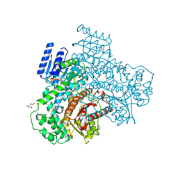
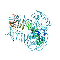
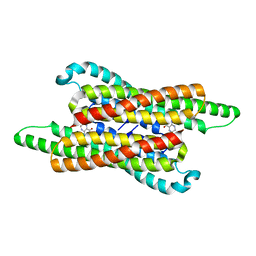
7VG5
DownloadVisualize
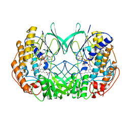
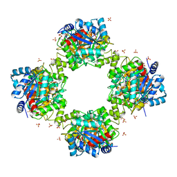
BU of 7vg5 by Molmil
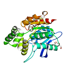
10,5-methenyltetrahydrofolate cyclohydrolase from Methylobacterium extorquens AM1 with tetrahydrofolate
Descriptor: (6S)-5,6,7,8-TETRAHYDROFOLATE, Methenyltetrahydrofolate cyclohydrolase
Authors:Kim, S, Lee, S, Kim, I.-K, Seo, H, Kim, K.-J.
Deposit date:2021-09-14
Release date:2022-07-06
Last modified:2023-11-29
Method:X-RAY DIFFRACTION (2.25 Å)
Cite:Structural insight into a molecular mechanism of methenyltetrahydrofolate cyclohydrolase from Methylobacterium extorquens AM1.
Int.J.Biol.Macromol., 202, 2022
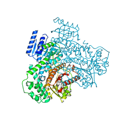
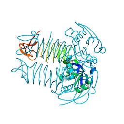
2QMQ
DownloadVisualize
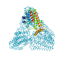
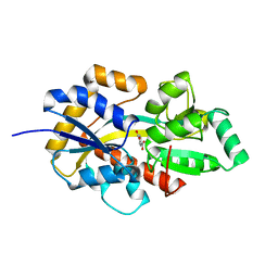
BU of 2qmq by Molmil
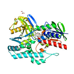
Crystal structure of a n-myc downstream regulated 2 protein (ndrg2, syld, ndr2, ai182517, au040374) from mus musculus at 1.70 A resolution
Descriptor: BENZOIC ACID, MAGNESIUM ION, NONAETHYLENE GLYCOL, ...
Authors:Joint Center for Structural Genomics (JCSG)
Deposit date:2007-07-16
Release date:2007-09-04
Last modified:2023-09-20
Method:X-RAY DIFFRACTION (1.7 Å)
Cite:Crystal structure of the human N-Myc downstream-regulated gene 2 protein provides insight into its role as a tumor suppressor.
J.Biol.Chem., 286, 2011
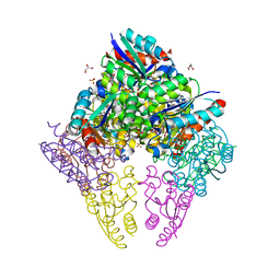
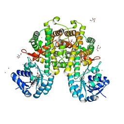
4REP
DownloadVisualize
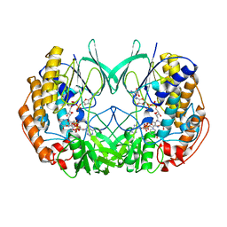
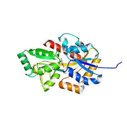
BU of 4rep by Molmil
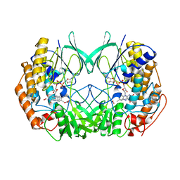
Crystal Structure of gamma-carotenoid desaturase
Descriptor: FLAVIN-ADENINE DINUCLEOTIDE, GLYCEROL, Gamma-carotene desaturase
Authors:Ahn, J.-W, Kim, E.-J, Kim, S, Kim, K.-J.
Deposit date:2014-09-23
Release date:2015-07-15
Last modified:2024-02-28
Method:X-RAY DIFFRACTION (1.97 Å)
Cite:Crystal structure of 1'-OH-carotenoid 3,4-desaturase from Nonlabens dokdonensis DSW-6.
Enzyme.Microb.Technol., 77, 2015
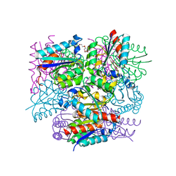
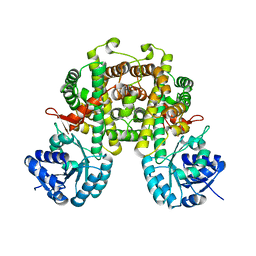
2XVJ
DownloadVisualize
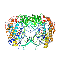
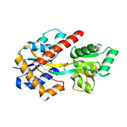
BU of 2xvj by Molmil
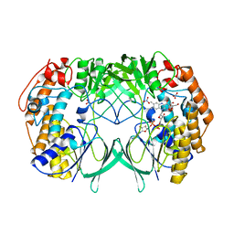
Crystal structure of the mutant bacterial flavin containing monooxygenase in complex with indole
Descriptor: 2-(N-MORPHOLINO)-ETHANESULFONIC ACID, FLAVIN-ADENINE DINUCLEOTIDE, FLAVIN-CONTAINING MONOOXYGENASE, ...
Authors:Cho, H.J, Kang, B.S.
Deposit date:2010-10-26
Release date:2011-05-04
Last modified:2024-05-08
Method:X-RAY DIFFRACTION (2.48 Å)
Cite:Structural and Functional Analysis of Bacterial Flavin-Containing Monooxygenase Reveals its Ping-Pong-Type Reaction Mechanism.
J.Struct.Biol., 175, 2011
2XVE
DownloadVisualize
BU of 2xve by Molmil
Crystal structure of bacterial flavin-containing monooxygenase
Descriptor: FLAVIN-ADENINE DINUCLEOTIDE, FLAVIN-CONTAINING MONOOXYGENASE, GLYCEROL, ...
Authors:Cho, H.J, Kang, B.S.
Deposit date:2010-10-26
Release date:2011-05-04
Last modified:2024-05-08
Method:X-RAY DIFFRACTION (1.99 Å)
Cite:Structural and Functional Analysis of Bacterial Flavin-Containing Monooxygenase Reveals its Ping-Pong-Type Reaction Mechanism.
J.Struct.Biol., 175, 2011
2XVI
DownloadVisualize
BU of 2xvi by Molmil
Crystal structure of the mutant bacterial flavin containing monooxygenase (Y207S)
Descriptor: 2-(N-MORPHOLINO)-ETHANESULFONIC ACID, FLAVIN-ADENINE DINUCLEOTIDE, FLAVIN-CONTAINING MONOOXYGENASE, ...
Authors:Cho, H.J, Kang, B.S.
Deposit date:2010-10-26
Release date:2011-05-04
Last modified:2024-05-08
Method:X-RAY DIFFRACTION (2.48 Å)
Cite:Structural and Functional Analysis of Bacterial Flavin-Containing Monooxygenase Reveals its Ping-Pong-Type Reaction Mechanism.
J.Struct.Biol., 175, 2011
6KD7
DownloadVisualize
BU of 6kd7 by Molmil
Crystal structure of geranylgeranyl pyrophosphate synthase
Descriptor: GLYCEROL, MAGNESIUM ION, PYROPHOSPHATE, ...
Authors:Kim, S, Kim, K.-J.
Deposit date:2019-07-01
Release date:2019-09-11
Last modified:2023-11-22
Method:X-RAY DIFFRACTION (2 Å)
Cite:Crystal structure of geranylgeranyl pyrophosphate synthase (crtE) from Nonlabens dokdonensis DSW-6.
Biochem.Biophys.Res.Commun., 518, 2019
2XVH
DownloadVisualize
BU of 2xvh by Molmil
Crystal structure of bacterial flavin containing monooxygenase in complex with NADP
Descriptor: 2-(N-MORPHOLINO)-ETHANESULFONIC ACID, FLAVIN-ADENINE DINUCLEOTIDE, FLAVIN-CONTAINING MONOOXYGENASE, ...
Authors:Cho, H.J, Kang, B.S.
Deposit date:2010-10-26
Release date:2011-05-04
Last modified:2024-05-08
Method:X-RAY DIFFRACTION (2.54 Å)
Cite:Structural and Functional Analysis of Bacterial Flavin-Containing Monooxygenase Reveals its Ping-Pong-Type Reaction Mechanism.
J.Struct.Biol., 175, 2011
2XVF
DownloadVisualize
BU of 2xvf by Molmil
Crystal structure of bacterial flavin-containing monooxygenase
Descriptor: FLAVIN-ADENINE DINUCLEOTIDE, FLAVIN-CONTAINING MONOOXYGENASE, GLYCEROL, ...
Authors:Cho, H.J, Kang, B.S.
Deposit date:2010-10-26
Release date:2011-05-04
Last modified:2024-10-23
Method:X-RAY DIFFRACTION (2.4 Å)
Cite:Structural and Functional Analysis of Bacterial Flavin-Containing Monooxygenase Reveals its Ping-Pong-Type Reaction Mechanism.
J.Struct.Biol., 175, 2011
8I70
DownloadVisualize
BU of 8i70 by Molmil
Crystal structure of NADP-binding form of malonyl-CoA reductase C-domain from Chloroflexus aurantiacus
Descriptor: GLYCEROL, L(+)-TARTARIC ACID, NADP NICOTINAMIDE-ADENINE-DINUCLEOTIDE PHOSPHATE, ...
Authors:Kim, S, Kim, K.-J.
Deposit date:2023-01-30
Release date:2023-06-14
Last modified:2024-05-29
Method:X-RAY DIFFRACTION (1.9 Å)
Cite:Cryo-EM structure of bifunctional malonyl-CoA reductase from Chloroflexus aurantiacus reveals a dynamic domain movement for high enzymatic activity.
Int.J.Biol.Macromol., 242, 2023
8I6Z
DownloadVisualize
BU of 8i6z by Molmil
Crystal structure of apo-form of malonyl-CoA reductase C-domain from Chloroflexus aurantiacus
Descriptor: GLYCEROL, L(+)-TARTARIC ACID, Short-chain dehydrogenase/reductase SDR
Authors:Kim, S, Kim, K.-J.
Deposit date:2023-01-30
Release date:2023-06-14
Last modified:2024-05-29
Method:X-RAY DIFFRACTION (1.95 Å)
Cite:Cryo-EM structure of bifunctional malonyl-CoA reductase from Chloroflexus aurantiacus reveals a dynamic domain movement for high enzymatic activity.
Int.J.Biol.Macromol., 242, 2023
8IDU
DownloadVisualize
BU of 8idu by Molmil
Crystal structure of substrate bound-form dehydroquinate dehydratase from Corynebacterium glutamicum
Descriptor: 1,3,4-TRIHYDROXY-5-OXO-CYCLOHEXANECARBOXYLIC ACID, 3-dehydroquinate dehydratase, GLYCEROL, ...
Authors:Lee, C.H, Kim, S, Kim, K.-J.
Deposit date:2023-02-14
Release date:2023-12-27
Last modified:2024-01-10
Method:X-RAY DIFFRACTION (2 Å)
Cite:Structural and Biochemical Analysis of 3-Dehydroquinate Dehydratase from Corynebacterium glutamicum .
J Microbiol Biotechnol., 33, 2023
8IDR
DownloadVisualize
BU of 8idr by Molmil
Crystal structure of apo-form of dehydroquinate dehydratase from Corynebacterium glutamicum
Descriptor: 3-dehydroquinate dehydratase, CITRATE ANION, TETRAETHYLENE GLYCOL
Authors:Lee, C.H, Kim, S, Kim, K.-J.
Deposit date:2023-02-14
Release date:2023-12-27
Last modified:2024-01-10
Method:X-RAY DIFFRACTION (1.8 Å)
Cite:Structural and Biochemical Analysis of 3-Dehydroquinate Dehydratase from Corynebacterium glutamicum .
J Microbiol Biotechnol., 33, 2023
7VG9
DownloadVisualize
BU of 7vg9 by Molmil
Crystal structure of phosphotransbutyrylase from Clostridium acetobutylicum
Descriptor: Phosphate butyryltransferase, SULFATE ION
Authors:Kim, S, Kim, K.-J.
Deposit date:2021-09-15
Release date:2021-10-27
Last modified:2023-11-29
Method:X-RAY DIFFRACTION (2.91 Å)
Cite:Crystal structure and molecular mechanism of phosphotransbutyrylase from Clostridium acetobutylicum .
J Microbiol Biotechnol., 31, 2021
8HKB
DownloadVisualize
BU of 8hkb by Molmil
TPA bound-form of Periplasmic terephthalate binding protein (TBP) from Ideonella sakaiensis mutant K184D
Descriptor: Periplasmic terephthalate binding protein (TBP), terephthalic acid
Authors:Lee, S.H, Seo, H, Kim, K.-J.
Deposit date:2022-11-25
Release date:2023-06-21
Last modified:2024-05-22
Method:X-RAY DIFFRACTION (1.4 Å)
Cite:Molecular mechanism underlying high-affinity terephthalate binding and conformational change of TBP from Ideonella sakaiensis.
Int.J.Biol.Macromol., 243, 2023
8HKA
DownloadVisualize
BU of 8hka by Molmil
TPA bound-form of Periplasmic terephthalate binding protein (TBP) from Ideonella sakaiensis
Descriptor: Periplasmic terephthalate binding protein (TBP), terephthalic acid
Authors:Lee, S.H, Seo, H, Kim, K.-J.
Deposit date:2022-11-25
Release date:2023-06-21
Last modified:2024-05-22
Method:X-RAY DIFFRACTION (1.02 Å)
Cite:Molecular mechanism underlying high-affinity terephthalate binding and conformational change of TBP from Ideonella sakaiensis.
Int.J.Biol.Macromol., 243, 2023
8HK9
DownloadVisualize
BU of 8hk9 by Molmil
Apo-form of Periplasmic terephthalate binding protein (TBP) from Ideonella sakaiensis
Descriptor: GLYCEROL, Periplasmic terephthalate binding protein (TBP)
Authors:Lee, S.H, Seo, H, Kim, K.-J.
Deposit date:2022-11-25
Release date:2023-06-21
Last modified:2024-05-22
Method:X-RAY DIFFRACTION (2 Å)
Cite:Molecular mechanism underlying high-affinity terephthalate binding and conformational change of TBP from Ideonella sakaiensis.
Int.J.Biol.Macromol., 243, 2023
5E3Q
DownloadVisualize
BU of 5e3q by Molmil
Crystal structure of DapD in complex with succinyl-CoA from Corynebacterium glutamicum
Descriptor: 2,3,4,5-tetrahydropyridine-2,6-dicarboxylate N-succinyltransferase, SUCCINYL-COENZYME A
Authors:Sagong, H.-Y, Kim, K.-J.
Deposit date:2015-10-03
Release date:2015-11-04
Last modified:2023-11-08
Method:X-RAY DIFFRACTION (1.8 Å)
Cite:Crystal Structure and Biochemical Characterization of Tetrahydrodipicolinate N-Succinyltransferase from Corynebacterium glutamicum.
J.Agric.Food Chem., 63, 2015
5E3R
DownloadVisualize
BU of 5e3r by Molmil
Crystal structure of DapD in complex with 2-aminopimelate from Corynebacterium glutamicum
Descriptor: (2S)-2-aminoheptanedioic acid, 2,3,4,5-tetrahydropyridine-2,6-dicarboxylate N-succinyltransferase
Authors:Sagong, H.-Y, Kim, K.-J.
Deposit date:2015-10-03
Release date:2015-11-04
Last modified:2023-11-08
Method:X-RAY DIFFRACTION (1.85 Å)
Cite:Crystal Structure and Biochemical Characterization of Tetrahydrodipicolinate N-Succinyltransferase from Corynebacterium glutamicum.
J.Agric.Food Chem., 63, 2015
8I4Q
DownloadVisualize
BU of 8i4q by Molmil
Crystal structure of 6-phosphogluconate dehydrogenase from Corynebacterium glutamicum
Descriptor: 2-AMINO-2-HYDROXYMETHYL-PROPANE-1,3-DIOL, 6-phosphogluconate dehydrogenase, decarboxylating, ...
Authors:Yu, H, Kim, K.-J.
Deposit date:2023-01-20
Release date:2023-07-26
Last modified:2024-05-29
Method:X-RAY DIFFRACTION (1.9 Å)
Cite:Crystal Structures of 6-Phosphogluconate Dehydrogenase from Corynebacterium glutamicum.
J Microbiol Biotechnol., 33, 2023
8I4N
DownloadVisualize
BU of 8i4n by Molmil
Crystal strcuture of 6-phosphogluconate dehydrogenase from Corynebacterium glutamicum
Descriptor: 6-phosphogluconate dehydrogenase, decarboxylating
Authors:Yu, H, Kim, K.-J.
Deposit date:2023-01-20
Release date:2023-07-26
Last modified:2024-05-29
Method:X-RAY DIFFRACTION (2.41 Å)
Cite:Crystal Structures of 6-Phosphogluconate Dehydrogenase from Corynebacterium glutamicum.
J Microbiol Biotechnol., 33, 2023
7D02
DownloadVisualize
BU of 7d02 by Molmil
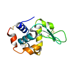
Lysozyme structure SASE2 from SASE mode
Descriptor: Lysozyme C
Authors:Kang, H.S, Lee, S.J.
Deposit date:2020-09-09
Release date:2021-03-31
Last modified:2023-11-29
Method:X-RAY DIFFRACTION (1.65 Å)
Cite:High-brightness self-seeded X-ray free-electron laser covering the 3.5 keV to 14.6 keV range
Nat Photonics, 2021
7D04
DownloadVisualize
BU of 7d04 by Molmil
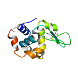
Lysozyme structure SS3 from SS mode
Descriptor: Lysozyme C
Authors:Kang, H.S, Lee, S.J.
Deposit date:2020-09-09
Release date:2021-03-31
Last modified:2024-10-16
Method:X-RAY DIFFRACTION (1.7 Å)
Cite:High-brightness self-seeded X-ray free-electron laser covering the 3.5 keV to 14.6 keV range
Nat Photonics, 2021
7D05
DownloadVisualize
BU of 7d05 by Molmil
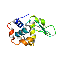
Lysozyme structure SASE3 from SASE mode
Descriptor: Lysozyme C
Authors:Kang, H.S, Lee, S.J.
Deposit date:2020-09-09
Release date:2021-03-31
Last modified:2024-10-16
Method:X-RAY DIFFRACTION (1.7 Å)
Cite:High-brightness self-seeded X-ray free-electron laser covering the 3.5 keV to 14.6 keV range
Nat Photonics, 2021
5E3P
DownloadVisualize
BU of 5e3p by Molmil
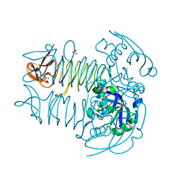
Crystal structure of DapD from Corynebacterium glutamicum
Descriptor: 2,3,4,5-tetrahydropyridine-2,6-dicarboxylate N-succinyltransferase, SULFATE ION, TETRAETHYLENE GLYCOL
Authors:Sagong, H.-Y, Kim, K.-J.
Deposit date:2015-10-03
Release date:2015-11-04
Last modified:2023-11-08
Method:X-RAY DIFFRACTION (2.01 Å)
Cite:Crystal Structure and Biochemical Characterization of Tetrahydrodipicolinate N-Succinyltransferase from Corynebacterium glutamicum.
J.Agric.Food Chem., 63, 2015

227111

PDB entries from 2024-11-06

PDB statisticsPDBj update infoContact PDBjnumon