Loading
PDBj
MenuPDBj@FacebookPDBj@X(formerly Twitter)PDBj@BlueSkyPDBj@YouTubewwPDB FoundationwwPDB
RCSB PDBPDBeBMRBAdv. SearchSearch help
Search by PDB author
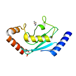
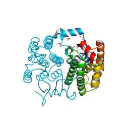
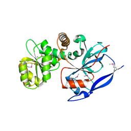
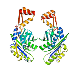
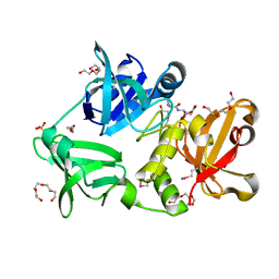
3DCX
DownloadVisualize
BU of 3dcx by Molmil
Crystal structure of a duf1696 family protein with a pleckstrin-homology domain (shew_0819) from shewanella loihica pv-4 at 2.00 A resolution
Descriptor: (4S)-2-METHYL-2,4-PENTANEDIOL, CHLORIDE ION, Protein of Unknown Function (DUF1696) with Pleckstrin-homology Domains
Authors:Joint Center for Structural Genomics (JCSG)
Deposit date:2008-06-04
Release date:2008-07-29
Last modified:2024-10-16
Method:X-RAY DIFFRACTION (2 Å)
Cite:Bacterial pleckstrin homology domains: a prokaryotic origin for the PH domain.
J.Mol.Biol., 396, 2010
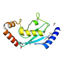
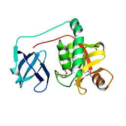
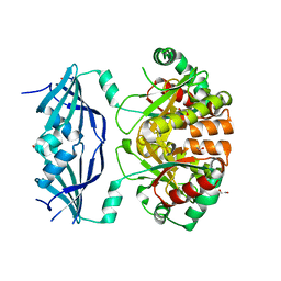
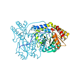
3H41
DownloadVisualize
BU of 3h41 by Molmil
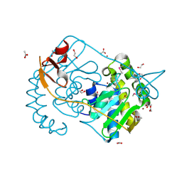
CRYSTAL STRUCTURE OF A NLPC/P60 FAMILY PROTEIN (BCE_2878) FROM BACILLUS CEREUS ATCC 10987 AT 1.79 A RESOLUTION
Descriptor: ALANINE, D-GLUTAMIC ACID, NLP/P60 family protein, ...
Authors:Joint Center for Structural Genomics (JCSG)
Deposit date:2009-04-17
Release date:2009-05-05
Last modified:2023-02-01
Method:X-RAY DIFFRACTION (1.79 Å)
Cite:Structure of the gamma-D-glutamyl-L-diamino acid endopeptidase YkfC from Bacillus cereus in complex with L-Ala-[gamma]-D-Glu: insights into substrate recognition by NlpC/P60 cysteine peptidases
Acta Crystallogr.,Sect.F, 66, 2010
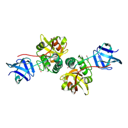
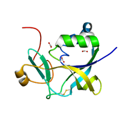
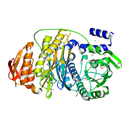
7AMF
DownloadVisualize
BU of 7amf by Molmil
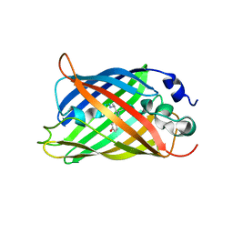
Crystal structure of rsFolder2 in its non-fluorescent off-state
Descriptor: GLYCEROL, Green fluorescent protein
Authors:Moreno-Chicano, T, El Khatib, M, Colletier, J.-P.
Deposit date:2020-10-08
Release date:2022-08-24
Last modified:2024-01-31
Method:X-RAY DIFFRACTION (1.63 Å)
Cite:Rational Control of Off-State Heterogeneity in a Photoswitchable Fluorescent Protein Provides Switching Contrast Enhancement.
Chemphyschem, 23, 2022
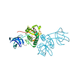
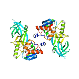
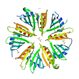
7AMB
DownloadVisualize
BU of 7amb by Molmil
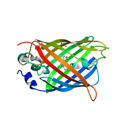
Crystal structure of rsFolder2 in its fluorescent on-state
Descriptor: GLYCEROL, Green fluorescent protein
Authors:Moreno-Chicano, T, El Khatib, M, Colletier, J.-P.
Deposit date:2020-10-08
Release date:2022-08-24
Last modified:2024-01-31
Method:X-RAY DIFFRACTION (1.63 Å)
Cite:Rational Control of Off-State Heterogeneity in a Photoswitchable Fluorescent Protein Provides Switching Contrast Enhancement.
Chemphyschem, 23, 2022
2GVI
DownloadVisualize
BU of 2gvi by Molmil
Crystal structure of a putative formylmethanofuran dehydrogenase subunit e (ta1109) from thermoplasma acidophilum at 1.87 A resolution
Descriptor: 1,2-ETHANEDIOL, ACETIC ACID, UNKNOWN LIGAND, ...
Authors:Joint Center for Structural Genomics (JCSG)
Deposit date:2006-05-02
Release date:2006-06-13
Last modified:2023-01-25
Method:X-RAY DIFFRACTION (1.87 Å)
Cite:Structures of three members of Pfam PF02663 (FmdE) implicated in microbial methanogenesis reveal a conserved alpha+beta core domain and an auxiliary C-terminal treble-clef zinc finger.
Acta Crystallogr.,Sect.F, 66, 2010
7MCK
DownloadVisualize
BU of 7mck by Molmil
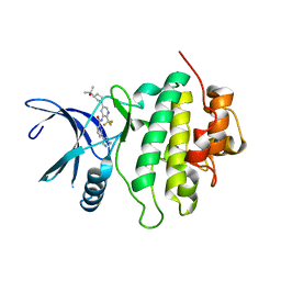
Structure of CHK1 10-pt. mutant complex with LRRK2 inhibitor 18
Descriptor: N-{5-[(3S)-3-(2-hydroxypropan-2-yl)pyrrolidin-1-yl]-2-(trifluoromethyl)pyridin-3-yl}-6-(1-methyl-1H-pyrazol-4-yl)pyridine-2-carboxamide, Serine/threonine-protein kinase Chk1
Authors:Palte, R.
Deposit date:2021-04-02
Release date:2021-06-16
Last modified:2023-10-18
Method:X-RAY DIFFRACTION (1.65 Å)
Cite:Optimization of brain-penetrant picolinamide derived leucine-rich repeat kinase 2 (LRRK2) inhibitors.
Rsc Med Chem, 12, 2021
7PJM
DownloadVisualize
BU of 7pjm by Molmil
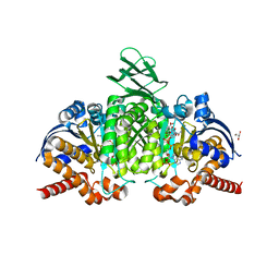
Crystal Structure of Ivosidenib-resistant IDH1 variant R132C S280F in complex with NADPH and Ca2+/2-Oxoglutarate
Descriptor: 2-OXOGLUTARIC ACID, CALCIUM ION, CHLORIDE ION, ...
Authors:Reinbold, R, Rabe, P, Abboud, M.I, Schofield, C.J.
Deposit date:2021-08-24
Release date:2022-07-13
Last modified:2024-11-13
Method:X-RAY DIFFRACTION (2.1 Å)
Cite:Resistance to the isocitrate dehydrogenase 1 mutant inhibitor ivosidenib can be overcome by alternative dimer-interface binding inhibitors.
Nat Commun, 13, 2022
7PJN
DownloadVisualize
BU of 7pjn by Molmil
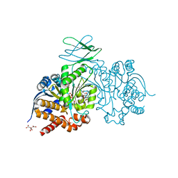
Crystal Structure of Ivosidenib-resistant IDH1 variant R132C S280F in complex with NADPH and inhibitor DS-1001B
Descriptor: (E)-3-(1-(5-(2-fluoropropan-2-yl)-3-(2,4,6-trichlorophenyl)isoxazole-4-carbonyl)-3-methyl-1H-indol-4-yl)acrylic acid, CITRIC ACID, GLYCEROL, ...
Authors:Reinbold, R, Rabe, P, Abboud, M.I, Schofield, C.J, Clifton, I.J.
Deposit date:2021-08-24
Release date:2022-07-13
Last modified:2024-10-09
Method:X-RAY DIFFRACTION (2.45 Å)
Cite:Resistance to the isocitrate dehydrogenase 1 mutant inhibitor ivosidenib can be overcome by alternative dimer-interface binding inhibitors.
Nat Commun, 13, 2022
8JVL
DownloadVisualize
BU of 8jvl by Molmil
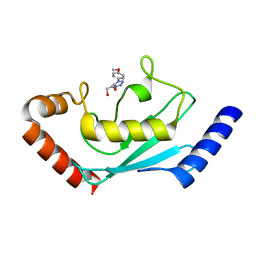
Identification and characterization of inhibitors covalently modifying catalytic cysteine of UBE2T and blocking ubiquitin transfer
Descriptor: 1,2-ETHANEDIOL, 1-(4-methoxyphenyl)-1,2,3,4-tetrazole, Ubiquitin-conjugating enzyme E2 T
Authors:Anantharajan, J, Baburajendran, N.
Deposit date:2023-06-28
Release date:2023-11-29
Method:X-RAY DIFFRACTION (2.06 Å)
Cite:Identification and characterization of inhibitors covalently modifying catalytic cysteine of UBE2T and blocking ubiquitin transfer.
Biochem.Biophys.Res.Commun., 689, 2023
8JVE
DownloadVisualize
BU of 8jve by Molmil
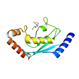
Identification and characterization of inhibitors covalently modifying catalytic cysteine of UBE2T and blocking ubiquitin transfer
Descriptor: 1,2-ETHANEDIOL, 1-(3-methoxyphenyl)-1,2,3,4-tetrazole, Ubiquitin-conjugating enzyme E2 T
Authors:Anantharajan, J, Baburajendran, N.
Deposit date:2023-06-28
Release date:2023-11-29
Method:X-RAY DIFFRACTION (1.76 Å)
Cite:Identification and characterization of inhibitors covalently modifying catalytic cysteine of UBE2T and blocking ubiquitin transfer.
Biochem.Biophys.Res.Commun., 689, 2023
8JVD
DownloadVisualize
BU of 8jvd by Molmil
Identification of small-molecule binding sites of a ubiquitin-conjugating enzyme-UBE2T through fragment-based screening
Descriptor: 5-fluoranyl-1,3-benzoxazol-2-amine, Ubiquitin-conjugating enzyme E2 T
Authors:Anantharajan, J, Baburajendran, N.
Deposit date:2023-06-28
Release date:2024-02-28
Method:X-RAY DIFFRACTION (1.696 Å)
Cite:Identification of small-molecule binding sites of a ubiquitin-conjugating enzyme-UBE2T through fragment-based screening.
Protein Sci., 33, 2024
8JUC
DownloadVisualize
BU of 8juc by Molmil
Identification of small-molecule binding sites of a ubiquitin-conjugating enzyme-UBE2T through fragment-based screening
Descriptor: 1,2-ETHANEDIOL, 7-methyl-2-(trifluoromethyl)-3~{H}-[1,2,4]triazolo[1,5-a]pyridin-5-one, Ubiquitin-conjugating enzyme E2 T
Authors:Anantharajan, J, Baburajendran, N.
Deposit date:2023-06-26
Release date:2024-02-28
Method:X-RAY DIFFRACTION (1.54 Å)
Cite:Identification of small-molecule binding sites of a ubiquitin-conjugating enzyme-UBE2T through fragment-based screening.
Protein Sci., 33, 2024
2FG0
DownloadVisualize
BU of 2fg0 by Molmil
Crystal structure of a putative gamma-d-glutamyl-l-diamino acid endopeptidase (npun_r0659) from nostoc punctiforme pcc 73102 at 1.79 A resolution
Descriptor: COG0791: Cell wall-associated hydrolases (invasion-associated proteins), GLYCEROL
Authors:Joint Center for Structural Genomics (JCSG)
Deposit date:2005-12-20
Release date:2006-01-10
Last modified:2024-11-13
Method:X-RAY DIFFRACTION (1.79 Å)
Cite:Structural Basis of Murein Peptide Specificity of a gamma-D-Glutamyl-L-Diamino Acid Endopeptidase.
Structure, 17, 2009
2EVR
DownloadVisualize
BU of 2evr by Molmil
CRYSTAL STRUCTURE OF A PUTATIVE GAMMA-D-GLUTAMYL-L-DIAMINO ACID ENDOPEPTIDASE (NPUN_R0659) FROM NOSTOC PUNCTIFORME PCC 73102 AT 1.60 A RESOLUTION
Descriptor: 1,2-ETHANEDIOL, CHLORIDE ION, COG0791: Cell wall-associated hydrolases (invasion-associated proteins), ...
Authors:Joint Center for Structural Genomics (JCSG)
Deposit date:2005-10-31
Release date:2005-11-22
Last modified:2024-10-16
Method:X-RAY DIFFRACTION (1.6 Å)
Cite:Structural Basis of Murein Peptide Specificity of a gamma-D-Glutamyl-L-Diamino Acid Endopeptidase.
Structure, 17, 2009
2FNO
DownloadVisualize
BU of 2fno by Molmil
Crystal structure of a glutathione s-transferase (atu5508) from agrobacterium tumefaciens str. c58 at 2.00 A resolution
Descriptor: AGR_pAT_752p, THIOCYANATE ION
Authors:Joint Center for Structural Genomics (JCSG)
Deposit date:2006-01-11
Release date:2006-02-14
Last modified:2023-01-25
Method:X-RAY DIFFRACTION (2 Å)
Cite:Comparative structural analysis of a novel glutathioneS-transferase (ATU5508) from Agrobacterium tumefaciens at 2.0 A resolution.
Proteins, 65, 2006
2HBW
DownloadVisualize
BU of 2hbw by Molmil
Crystal structure of a putative endopeptidase (ava_3396) from anabaena variabilis atcc 29413 at 1.05 A resolution
Descriptor: ACETATE ION, NLP/P60 protein, UNKNOWN LIGAND
Authors:Joint Center for Structural Genomics (JCSG)
Deposit date:2006-06-14
Release date:2006-08-08
Last modified:2023-01-25
Method:X-RAY DIFFRACTION (1.05 Å)
Cite:Structural Basis of Murein Peptide Specificity of a gamma-D-Glutamyl-L-Diamino Acid Endopeptidase.
Structure, 17, 2009
1VKB
DownloadVisualize
BU of 1vkb by Molmil
Crystal structure of an aig2-like protein (a2ld1, ggact, mgc7867) from mus musculus at 1.90 A resolution
Descriptor: FORMIC ACID, hypothetical protein
Authors:Joint Center for Structural Genomics (JCSG)
Deposit date:2004-05-07
Release date:2004-05-18
Last modified:2024-10-30
Method:X-RAY DIFFRACTION (1.9 Å)
Cite:Crystal structure of a conserved hypothetical protein (gi: 13879369) from Mouse at 1.90 A resolution reveals a new fold.
Proteins, 61, 2005
1VKY
DownloadVisualize
BU of 1vky by Molmil
Crystal structure of S-adenosylmethionine tRNA ribosyltransferase (TM0574) from Thermotoga maritima at 2.00 A resolution
Descriptor: S-adenosylmethionine:tRNA ribosyltransferase-isomerase, UNKNOWN LIGAND
Authors:Joint Center for Structural Genomics (JCSG)
Deposit date:2004-06-28
Release date:2004-08-17
Last modified:2024-05-22
Method:X-RAY DIFFRACTION (2 Å)
Cite:Crystal structure of S-adenosylmethionine:tRNA ribosyltransferase-isomerase (QueA) from Thermotoga maritima at 2.0 A resolution reveals a new fold.
Proteins, 59, 2005
5JHL
DownloadVisualize
BU of 5jhl by Molmil
Crystal structure of zika virus envelope protein in complex with a flavivirus broadly-protective antibody
Descriptor: Antibody Heavy chain, antibody Light chain, envelope protein
Authors:Dai, L, Shi, Y, Qi, J, Gao, G.F.
Deposit date:2016-04-21
Release date:2016-05-11
Last modified:2024-11-06
Method:X-RAY DIFFRACTION (3 Å)
Cite:Structures of the Zika Virus Envelope Protein and Its Complex with a Flavivirus Broadly Protective Antibody.
Cell Host Microbe, 19, 2016
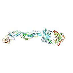
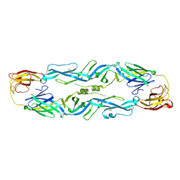
5JHM
DownloadVisualize
BU of 5jhm by Molmil
Crystal structure of Zika virus Envelope protein
Descriptor: Envelope protein
Authors:Dai, L, Shi, Y, Qi, J, Gao, G.F.
Deposit date:2016-04-21
Release date:2016-05-11
Last modified:2024-11-06
Method:X-RAY DIFFRACTION (2 Å)
Cite:Structures of the Zika Virus Envelope Protein and Its Complex with a Flavivirus Broadly Protective Antibody.
Cell Host Microbe, 19, 2016
1VLR
DownloadVisualize
BU of 1vlr by Molmil
Crystal structure of mRNA decapping enzyme (DcpS) from Mus musculus at 1.83 A resolution
Descriptor: 1,2-ETHANEDIOL, mRNA decapping enzyme
Authors:Joint Center for Structural Genomics (JCSG)
Deposit date:2004-08-10
Release date:2004-08-24
Last modified:2023-09-20
Method:X-RAY DIFFRACTION (1.83 Å)
Cite:Crystal structure of an Apo mRNA decapping enzyme (DcpS) from Mouse at 1.83 A resolution.
Proteins, 60, 2005
1VJ1
DownloadVisualize
BU of 1vj1 by Molmil
Crystal structure of putative NADPH-dependent oxidoreductase from Mus musculus at 2.10 A resolution
Descriptor: putative NADPH-dependent oxidoreductase
Authors:Joint Center for Structural Genomics (JCSG)
Deposit date:2003-12-03
Release date:2003-12-09
Last modified:2023-01-25
Method:X-RAY DIFFRACTION (2.1 Å)
Cite:Crystal structure of a putative NADPH-dependent oxidoreductase (GI: 18204011) from mouse at 2.10 A resolution
Proteins, 56, 2004
1VQ0
DownloadVisualize
BU of 1vq0 by Molmil
Crystal structure of 33 kDa chaperonin (Heat shock protein 33 homolog) (HSP33) (TM1394) from Thermotoga maritima at 2.20 A resolution
Descriptor: 1,2-ETHANEDIOL, 33 kDa chaperonin, CHLORIDE ION, ...
Authors:Joint Center for Structural Genomics (JCSG)
Deposit date:2004-11-30
Release date:2004-12-14
Last modified:2023-09-20
Method:X-RAY DIFFRACTION (2.2 Å)
Cite:Crystal structure of Hsp33 chaperone (TM1394) from Thermotoga maritima at 2.20 A resolution.
Proteins, 61, 2005
1VJO
DownloadVisualize
BU of 1vjo by Molmil
Crystal structure of Alanine--glyoxylate aminotransferase (ALR1004) from Nostoc sp. at 1.70 A resolution
Descriptor: PYRIDOXAL-5'-PHOSPHATE, alanine--glyoxylate aminotransferase
Authors:Joint Center for Structural Genomics (JCSG)
Deposit date:2004-03-16
Release date:2004-03-23
Last modified:2023-01-25
Method:X-RAY DIFFRACTION (1.7 Å)
Cite:Crystal structure of an alanine-glyoxylate aminotransferase from Anabaena sp. at 1.70 A resolution reveals a noncovalently linked PLP cofactor
Proteins, 58, 2005
1VK3
DownloadVisualize
BU of 1vk3 by Molmil
Crystal structure of Phosphoribosylformylglycinamidine synthase II (TM1246) from Thermotoga maritima at 2.15 A resolution
Descriptor: CHLORIDE ION, Phosphoribosylformylglycinamidine synthase II
Authors:Joint Center for Structural Genomics (JCSG)
Deposit date:2004-04-23
Release date:2004-05-11
Last modified:2023-01-25
Method:X-RAY DIFFRACTION (2.15 Å)
Cite:Crystal structure of phosphoribosylformylglycinamidine synthase II (smPurL) from Thermotoga maritima at 2.15 A resolution.
Proteins, 63, 2006

227344

PDB entries from 2024-11-13

PDB statisticsPDBj update infoContact PDBjnumon