Loading
PDBj
MenuPDBj@FacebookPDBj@TwitterPDBj@YouTubewwPDB FoundationwwPDB
RCSB PDBPDBeBMRBAdv. SearchSearch help
Search by PDB author
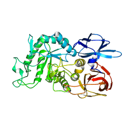
3CGH
DownloadVisualize
BU of 3cgh by Molmil
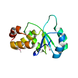
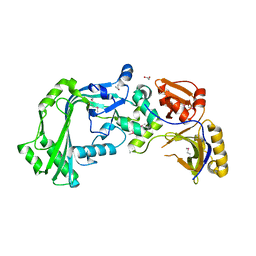
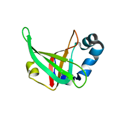
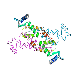
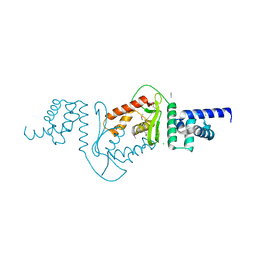
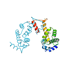
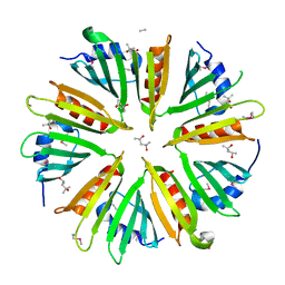
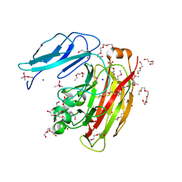
Crystal structure of a susd homolog (bt_3984) from bacteroides thetaiotaomicron vpi-5482 at 1.70 A resolution
Descriptor: ACETATE ION, SusD homolog, ZINC ION
Authors:Joint Center for Structural Genomics (JCSG)
Deposit date:2008-03-05
Release date:2008-03-18
Last modified:2023-02-01
Method:X-RAY DIFFRACTION (1.7 Å)
Cite:Structure of BT_3984, a member of the SusD/RagB family of nutrient-binding molecules.
Acta Crystallogr.,Sect.F, 66, 2010
8IUA
DownloadVisualize
BU of 8iua by Molmil
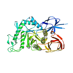
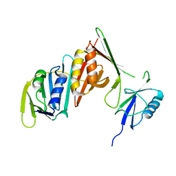
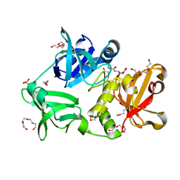
Crystal structure of GH66 endodextranase from Flavobacterium johnsoniae in complex with isomaltose
Descriptor: Candidate dextranase Glycoside hydrolase family 66, SODIUM ION, alpha-D-glucopyranose-(1-6)-alpha-D-glucopyranose, ...
Authors:Nakamura, S, Miyazaki, T.
Deposit date:2023-03-24
Release date:2023-06-14
Last modified:2024-05-29
Method:X-RAY DIFFRACTION (1.8 Å)
Cite:Bacteroidota polysaccharide utilization system for branched dextran exopolysaccharides from lactic acid bacteria.
J.Biol.Chem., 299, 2023
8IU8
DownloadVisualize
BU of 8iu8 by Molmil
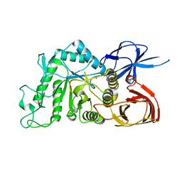
Crystal structure of GH66 endodextranase from Flavobacterium johnsoniae
Descriptor: Candidate dextranase Glycoside hydrolase family 66, GLYCEROL
Authors:Nakamura, S, Miyazaki, T.
Deposit date:2023-03-24
Release date:2023-06-14
Last modified:2024-05-29
Method:X-RAY DIFFRACTION (1.85 Å)
Cite:Bacteroidota polysaccharide utilization system for branched dextran exopolysaccharides from lactic acid bacteria.
J.Biol.Chem., 299, 2023
8IU9
DownloadVisualize
BU of 8iu9 by Molmil
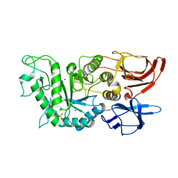
Crystal structure of GH66 endodextranase from Flavobacterium johnsoniae in complex with glucose
Descriptor: Candidate dextranase Glycoside hydrolase family 66, SODIUM ION, alpha-D-glucopyranose, ...
Authors:Nakamura, S, Miyazaki, T.
Deposit date:2023-03-24
Release date:2023-06-14
Last modified:2024-05-29
Method:X-RAY DIFFRACTION (1.8 Å)
Cite:Bacteroidota polysaccharide utilization system for branched dextran exopolysaccharides from lactic acid bacteria.
J.Biol.Chem., 299, 2023
8IUB
DownloadVisualize
BU of 8iub by Molmil
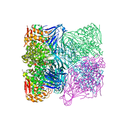
Crystal structure of GH66 endodextranase from Flavobacterium johnsoniae in complex with isomaltotriose
Descriptor: Candidate dextranase Glycoside hydrolase family 66, SODIUM ION, SULFATE ION, ...
Authors:Nakamura, S, Miyazaki, T.
Deposit date:2023-03-24
Release date:2023-06-14
Last modified:2024-05-29
Method:X-RAY DIFFRACTION (1.18 Å)
Cite:Bacteroidota polysaccharide utilization system for branched dextran exopolysaccharides from lactic acid bacteria.
J.Biol.Chem., 299, 2023
8IUC
DownloadVisualize
BU of 8iuc by Molmil
Crystal structure of GH65 alpha-1,2-glucosidase from Flavobacterium johnsoniae in complex with isomaltose
Descriptor: 1,2-ETHANEDIOL, Candidate alpha glycoside phosphorylase Glycoside hydrolase family 65, alpha-D-glucopyranose-(1-6)-beta-D-glucopyranose
Authors:Nakamura, S, Miyazaki, T.
Deposit date:2023-03-24
Release date:2023-06-14
Last modified:2024-05-29
Method:X-RAY DIFFRACTION (1.56 Å)
Cite:Bacteroidota polysaccharide utilization system for branched dextran exopolysaccharides from lactic acid bacteria.
J.Biol.Chem., 299, 2023
2F46
DownloadVisualize
BU of 2f46 by Molmil
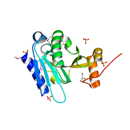
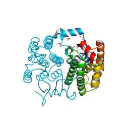
Crystal structure of a putative phosphatase (nma1982) from neisseria meningitidis z2491 at 1.41 A resolution
Descriptor: CHLORIDE ION, UNKNOWN LIGAND, hypothetical protein
Authors:Joint Center for Structural Genomics (JCSG)
Deposit date:2005-11-22
Release date:2006-02-07
Last modified:2023-01-25
Method:X-RAY DIFFRACTION (1.41 Å)
Cite:Crystal structure of NMA1982 from Neisseria meningitidis at 1.5 A resolution provides a structural scaffold for nonclassical, eukaryotic-like phosphatases.
Proteins, 69, 2007
2JVA
DownloadVisualize
BU of 2jva by Molmil
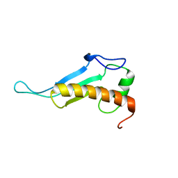
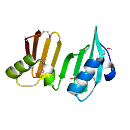
NMR solution structure of peptidyl-tRNA hydrolase domain protein from Pseudomonas syringae pv. tomato. Northeast Structural Genomics Consortium target PsR211
Descriptor: Peptidyl-tRNA hydrolase domain protein
Authors:Singarapu, K.K, Sukumaran, D, Parish, D, Eletsky, A, Zhang, Q, Zhao, L, Jiang, M, Maglaqui, M, Xiao, R, Liu, J, Baran, M.C, Swapna, G.V.T, Huang, Y.J, Acton, T.B, Rost, B, Montelione, G.T, Szyperski, T, Northeast Structural Genomics Consortium (NESG)
Deposit date:2007-09-14
Release date:2007-10-02
Last modified:2024-05-08
Method:SOLUTION NMR
Cite:NMR structure of the peptidyl-tRNA hydrolase domain from Pseudomonas syringae expands the structural coverage of the hydrolysis domains of class 1 peptide chain release factors.
Proteins, 71, 2008
3K5J
DownloadVisualize
BU of 3k5j by Molmil
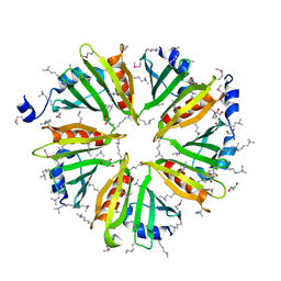
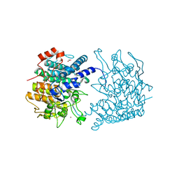
Crystal structure of Putative SUFU (suppressor of fused protein) homolog (YP_208451.1) from Neisseria gonorrhoeae FA 1090 at 1.40 A resolution
Descriptor: GLYCEROL, SULFATE ION, Suppressor of fused family protein
Authors:Joint Center for Structural Genomics (JCSG)
Deposit date:2009-10-07
Release date:2010-01-26
Last modified:2019-07-17
Method:X-RAY DIFFRACTION (1.4 Å)
Cite:The crystal structure of a bacterial Sufu-like protein defines a novel group of bacterial proteins that are similar to the N-terminal domain of human Sufu.
Protein Sci., 19, 2010
3HSA
DownloadVisualize
BU of 3hsa by Molmil
Crystal structure of pleckstrin homology domain (YP_926556.1) from SHEWANELLA AMAZONENSIS SB2B at 1.99 A resolution
Descriptor: DI(HYDROXYETHYL)ETHER, GLYCEROL, pleckstrin homology domain
Authors:Joint Center for Structural Genomics (JCSG)
Deposit date:2009-06-10
Release date:2009-06-23
Last modified:2023-02-01
Method:X-RAY DIFFRACTION (1.99 Å)
Cite:Bacterial pleckstrin homology domains: a prokaryotic origin for the PH domain.
J.Mol.Biol., 396, 2010
3KK7
DownloadVisualize
BU of 3kk7 by Molmil
Crystal structure of Putative cell invasion protein with MAC/Perforin domain (NP_812351.1) from BACTERIODES THETAIOTAOMICRON VPI-5482 at 2.46 A resolution
Descriptor: (4S)-2-METHYL-2,4-PENTANEDIOL, 1,2-ETHANEDIOL, CHLORIDE ION, ...
Authors:Joint Center for Structural Genomics (JCSG)
Deposit date:2009-11-04
Release date:2009-11-24
Last modified:2023-02-01
Method:X-RAY DIFFRACTION (2.46 Å)
Cite:Structure of a membrane-attack complex/perforin (MACPF) family protein from the human gut symbiont Bacteroides thetaiotaomicron.
Acta Crystallogr.,Sect.F, 66, 2010
3F1Z
DownloadVisualize
BU of 3f1z by Molmil
Crystal structure of putative nucleic acid-binding lipoprotein (YP_001337197.1) from Klebsiella pneumoniae subsp. pneumoniae MGH 78578 at 2.46 A resolution
Descriptor: DI(HYDROXYETHYL)ETHER, putative nucleic acid-binding lipoprotein
Authors:Joint Center for Structural Genomics (JCSG)
Deposit date:2008-10-28
Release date:2008-11-18
Last modified:2023-02-01
Method:X-RAY DIFFRACTION (2.46 Å)
Cite:The structure of KPN03535 (gi|152972051), a novel putative lipoprotein from Klebsiella pneumoniae, reveals an OB-fold.
Acta Crystallogr.,Sect.F, 66, 2010
3NNQ
DownloadVisualize
BU of 3nnq by Molmil
Crystal Structure of the N-terminal domain of Moloney murine leukemia virus integrase, Northeast Structural Genomics Consortium Target OR3
Descriptor: ACETATE ION, N-terminal domain of Moloney murine leukemia virus integrase, ZINC ION
Authors:Guan, R, Xiao, R, Acton, T, Jiang, M, Roth, M, Montelione, G.T, Northeast Structural Genomics Consortium (NESG)
Deposit date:2010-06-24
Release date:2010-07-14
Last modified:2023-07-26
Method:X-RAY DIFFRACTION (2.693 Å)
Cite:X-ray crystal structure of the N-terminal region of Moloney murine leukemia virus integrase and its implications for viral DNA recognition.
Proteins, 85, 2017
3DEE
DownloadVisualize
BU of 3dee by Molmil
CRYSTAL STRUCTURE OF A PUTATIVE REGULATORY PROTEIN INVOLVED IN TRANSCRIPTION (NGO1945) FROM NEISSERIA GONORRHOEAE FA 1090 AT 2.25 A RESOLUTION
Descriptor: CHLORIDE ION, IMIDAZOLE, Putative regulatory protein
Authors:Joint Center for Structural Genomics (JCSG)
Deposit date:2008-06-09
Release date:2008-08-05
Last modified:2023-02-01
Method:X-RAY DIFFRACTION (2.1 Å)
Cite:The structure of the first representative of Pfam family PF09836 reveals a two-domain organization and suggests involvement in transcriptional regulation.
Acta Crystallogr.,Sect.F, 66, 2010
5JJG
DownloadVisualize
BU of 5jjg by Molmil
Structure of magnesium-loaded ALG-2
Descriptor: ISOPROPYL ALCOHOL, MAGNESIUM ION, Pcalcium-binding protein ALG-2, ...
Authors:Tanner, J.J.
Deposit date:2016-04-23
Release date:2016-09-07
Last modified:2023-09-27
Method:X-RAY DIFFRACTION (1.72 Å)
Cite:EF5 Is the High-Affinity Mg(2+) Site in ALG-2.
Biochemistry, 55, 2016
3OGI
DownloadVisualize
BU of 3ogi by Molmil
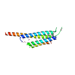
Crystal structure of the Mycobacterium tuberculosis H37Rv EsxOP complex (Rv2346c-Rv2347c)
Descriptor: Putative ESAT-6-like protein 6, Putative ESAT-6-like protein 7
Authors:Arbing, M.A, Chan, S, Zhou, T.T, Ahn, C, Harris, L, Kuo, E, Sawaya, M.R, Cascio, D, Eisenberg, D, Integrated Center for Structure and Function Innovation (ISFI), TB Structural Genomics Consortium (TBSGC)
Deposit date:2010-08-16
Release date:2010-08-25
Last modified:2014-05-21
Method:X-RAY DIFFRACTION (2.549 Å)
Cite:Heterologous expression of mycobacterial Esx complexes in Escherichia coli for structural studies is facilitated by the use of maltose binding protein fusions.
Plos One, 8, 2013
3GF8
DownloadVisualize
BU of 3gf8 by Molmil
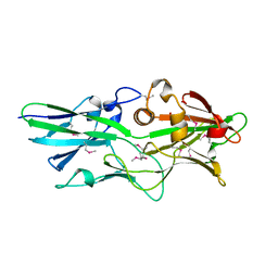
Crystal structure of putative polysaccharide binding proteins (DUF1812) (NP_809975.1) from BACTEROIDES THETAIOTAOMICRON VPI-5482 at 2.20 A resolution
Descriptor: 1,2-ETHANEDIOL, putative polysaccharide binding proteins (DUF1812)
Authors:Joint Center for Structural Genomics (JCSG)
Deposit date:2009-02-26
Release date:2009-03-24
Last modified:2023-02-01
Method:X-RAY DIFFRACTION (2.2 Å)
Cite:A conserved fold for fimbrial components revealed by the crystal structure of a putative fimbrial assembly protein (BT1062) from Bacteroides thetaiotaomicron at 2.2 A resolution
Acta Crystallogr.,Sect.F, 66, 2010
3DUE
DownloadVisualize
BU of 3due by Molmil
CRYSTAL STRUCTURE OF A PUTATIVE PERIPLASMIC PROTEIN FROM DUF2874 FAMILY (BVU_2987) FROM BACTEROIDES VULGATUS ATCC 8482 AT 1.85 A RESOLUTION
Descriptor: CACODYLATE ION, Putative periplasmic protein
Authors:Joint Center for Structural Genomics (JCSG)
Deposit date:2008-07-17
Release date:2008-08-05
Last modified:2023-02-01
Method:X-RAY DIFFRACTION (1.85 Å)
Cite:The structure of BVU2987 from Bacteroides vulgatus reveals a superfamily of bacterial periplasmic proteins with possible inhibitory function.
Acta Crystallogr.,Sect.F, 66, 2010
3DCX
DownloadVisualize
BU of 3dcx by Molmil
Crystal structure of a duf1696 family protein with a pleckstrin-homology domain (shew_0819) from shewanella loihica pv-4 at 2.00 A resolution
Descriptor: (4S)-2-METHYL-2,4-PENTANEDIOL, CHLORIDE ION, Protein of Unknown Function (DUF1696) with Pleckstrin-homology Domains
Authors:Joint Center for Structural Genomics (JCSG)
Deposit date:2008-06-04
Release date:2008-07-29
Last modified:2023-02-01
Method:X-RAY DIFFRACTION (2 Å)
Cite:Bacterial pleckstrin homology domains: a prokaryotic origin for the PH domain.
J.Mol.Biol., 396, 2010
3PGE
DownloadVisualize
BU of 3pge by Molmil
Structure of sumoylated PCNA
Descriptor: Proliferating cell nuclear antigen, SUMO-modified proliferating cell nuclear antigen
Authors:Freudenthal, B.D, Brogie, J.E, Gakhar, L, Washington, T.
Deposit date:2010-11-01
Release date:2010-12-29
Last modified:2023-09-06
Method:X-RAY DIFFRACTION (2.8 Å)
Cite:Crystal Structure of SUMO-Modified Proliferating Cell Nuclear Antigen.
J.Mol.Biol., 406, 2011
2FEA
DownloadVisualize
BU of 2fea by Molmil
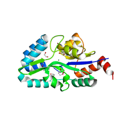
Crystal structure of MtnX phosphatase from Bacillus Subtilis at 2.00 A resolution
Descriptor: 1,2-ETHANEDIOL, 2-hydroxy-3-keto-5-methylthiopentenyl-1-phosphate phosphatase, MAGNESIUM ION, ...
Authors:Joint Center for Structural Genomics (JCSG)
Deposit date:2005-12-15
Release date:2005-12-27
Last modified:2023-01-25
Method:X-RAY DIFFRACTION (2 Å)
Cite:Crystal structure of MtnX phosphatase from Bacillus subtilis at 2.0 A resolution provides a structural basis for bipartite phosphomonoester hydrolysis of 2-hydroxy-3-keto-5-methylthiopentenyl-1-phosphate.
Proteins, 69, 2007
7M42
DownloadVisualize
BU of 7m42 by Molmil
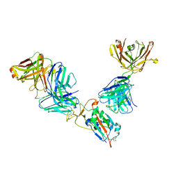
Complex of SARS-CoV-2 receptor binding domain with the Fab fragments of neutralizing antibodies REGN10985 and REGN10989
Descriptor: REGN10985 antibody Fab fragment heavy chain, REGN10985 antibody Fab fragment light chain, REGN10989 antibody Fab fragment heavy chain, ...
Authors:Zhou, Y, Romero Hernandez, A, Saotome, K, Franklin, M.C.
Deposit date:2021-03-19
Release date:2021-07-28
Last modified:2021-08-25
Method:ELECTRON MICROSCOPY (3.3 Å)
Cite:The monoclonal antibody combination REGEN-COV protects against SARS-CoV-2 mutational escape in preclinical and human studies.
Cell, 184, 2021
3HBZ
DownloadVisualize
BU of 3hbz by Molmil
Crystal structure of a putative glycoside hydrolase (bt_2081) from bacteroides thetaiotaomicron vpi-5482 at 2.05 A resolution
Descriptor: ACETATE ION, CACODYLATE ION, CALCIUM ION, ...
Authors:Joint Center for Structural Genomics (JCSG)
Deposit date:2009-05-05
Release date:2009-05-26
Last modified:2023-02-01
Method:X-RAY DIFFRACTION (2.05 Å)
Cite:Structure of Bacteroides thetaiotaomicron BT2081 at 2.05 A resolution: the first structural representative of a new protein family that may play a role in carbohydrate metabolism.
Acta Crystallogr.,Sect.F, 66, 2010
3H41
DownloadVisualize
BU of 3h41 by Molmil
CRYSTAL STRUCTURE OF A NLPC/P60 FAMILY PROTEIN (BCE_2878) FROM BACILLUS CEREUS ATCC 10987 AT 1.79 A RESOLUTION
Descriptor: ALANINE, D-GLUTAMIC ACID, NLP/P60 family protein, ...
Authors:Joint Center for Structural Genomics (JCSG)
Deposit date:2009-04-17
Release date:2009-05-05
Last modified:2023-02-01
Method:X-RAY DIFFRACTION (1.79 Å)
Cite:Structure of the gamma-D-glutamyl-L-diamino acid endopeptidase YkfC from Bacillus cereus in complex with L-Ala-[gamma]-D-Glu: insights into substrate recognition by NlpC/P60 cysteine peptidases
Acta Crystallogr.,Sect.F, 66, 2010
2FNO
DownloadVisualize
BU of 2fno by Molmil
Crystal structure of a glutathione s-transferase (atu5508) from agrobacterium tumefaciens str. c58 at 2.00 A resolution
Descriptor: AGR_pAT_752p, THIOCYANATE ION
Authors:Joint Center for Structural Genomics (JCSG)
Deposit date:2006-01-11
Release date:2006-02-14
Last modified:2023-01-25
Method:X-RAY DIFFRACTION (2 Å)
Cite:Comparative structural analysis of a novel glutathioneS-transferase (ATU5508) from Agrobacterium tumefaciens at 2.0 A resolution.
Proteins, 65, 2006

223790

PDB entries from 2024-08-14

PDB statisticsPDBj update infoContact PDBjnumon