Loading
PDBj
MenuPDBj@FacebookPDBj@TwitterPDBj@YouTubewwPDB FoundationwwPDB
RCSB PDBPDBeBMRBAdv. SearchSearch help
Search by PDB author
6UDE
DownloadVisualize
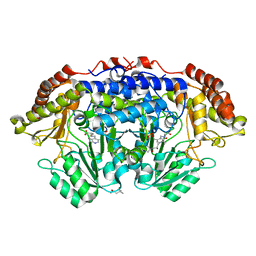
BU of 6ude by Molmil
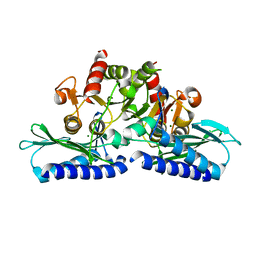
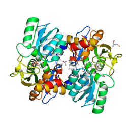
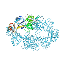
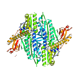
Crystal structure of Glycerol kinase from Elizabethkingia anophelis NUHP1 in complex with ADP and glycerol
Descriptor: ADENOSINE-5'-DIPHOSPHATE, GLYCEROL, Glycerol kinase, ...
Authors:Seattle Structural Genomics Center for Infectious Disease (SSGCID)
Deposit date:2019-09-19
Release date:2019-10-16
Last modified:2023-10-11
Method:X-RAY DIFFRACTION (1.95 Å)
Cite:Crystal structure of Glycerol kinase from Elizabethkingia anophelis NUHP1 in complex with ADP and glycerol
TO BE PUBLISHED
6ULD
DownloadVisualize
BU of 6uld by Molmil
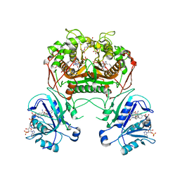
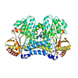
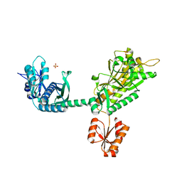
Crystal structure of serine hydroxymethyltransferase from Mycobacterium tuberculosis with bound PLP forming a Schiff base with substrate Serine in one monomer and PLP forming a Schiff base with product Glycine in the other monomer
Descriptor: (4S)-2-METHYL-2,4-PENTANEDIOL, GLYCINE, PYRIDOXAL-5'-PHOSPHATE, ...
Authors:Seattle Structural Genomics Center for Infectious Disease (SSGCID)
Deposit date:2019-10-07
Release date:2019-10-16
Last modified:2023-10-11
Method:X-RAY DIFFRACTION (1.5 Å)
Cite:Crystal structure of serine hydroxymethyltransferase from Mycobacterium tuberculosis with bound PLP forming a Schiff base with substrate Serine in one monomer and PLP forming a Schiff base with product Glycine in the other monomer
to be published
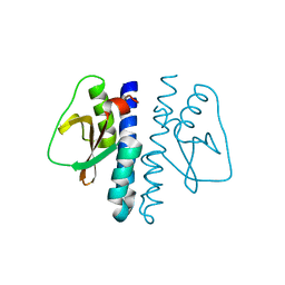
3LV0
DownloadVisualize
BU of 3lv0 by Molmil
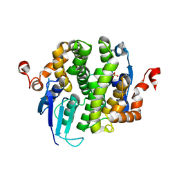
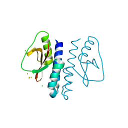
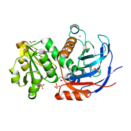
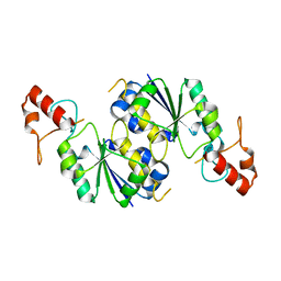
Crystal structure of extragenic suppressor protein suhB from Bartonella henselae, native
Descriptor: Extragenic suppressor protein suhB, MAGNESIUM ION
Authors:Seattle Structural Genomics Center for Infectious Disease (SSGCID)
Deposit date:2010-02-18
Release date:2010-03-02
Last modified:2023-09-06
Method:X-RAY DIFFRACTION (1.85 Å)
Cite:Crystal structure of extragenic suppressor protein suhB from Bartonella henselae
To be Published
3DNG
DownloadVisualize
BU of 3dng by Molmil
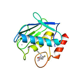
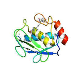
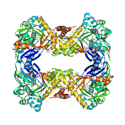
Crystal structure of the complex between MMP-8 and a non-zinc chelating inhibitor
Descriptor: (5S)-5-(2-amino-2-oxoethyl)-4-oxo-N-[(3-oxo-3,4-dihydro-2H-1,4-benzoxazin-6-yl)methyl]-3,4,5,6,7,8-hexahydro[1]benzothieno[2,3-d]pyrimidine-2-carboxamide, CALCIUM ION, Neutrophil collagenase, ...
Authors:Pochetti, G, Montanari, R, Mazza, F.
Deposit date:2008-07-02
Release date:2009-03-03
Last modified:2023-11-01
Method:X-RAY DIFFRACTION (2 Å)
Cite:Extra Binding Region Induced by Non-Zinc Chelating Inhibitors into the S(1)' Subsite of Matrix Metalloproteinase 8 (MMP-8)
J.Med.Chem., 52, 2009
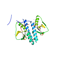
3LGJ
DownloadVisualize
BU of 3lgj by Molmil
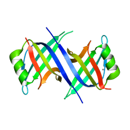
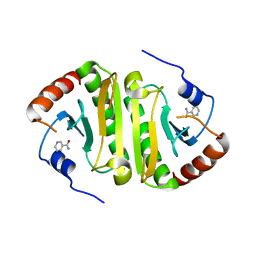
Crystal structure of single-stranded binding protein (ssb) from Bartonella henselae
Descriptor: CALCIUM ION, Single-stranded DNA-binding protein
Authors:Seattle Structural Genomics Center for Infectious Disease (SSGCID)
Deposit date:2010-01-20
Release date:2010-02-16
Last modified:2023-09-06
Method:X-RAY DIFFRACTION (2.5 Å)
Cite:Crystal structure of single-stranded binding protein (ssb) from Bartonella henselae
To be Published
3LAA
DownloadVisualize
BU of 3laa by Molmil
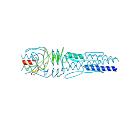
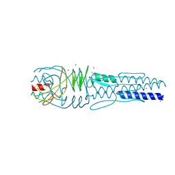
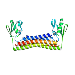
Crystal structure of the trimeric autotransporter adhesin head domain BpaA from Burkholderia pseudomallei
Descriptor: Haemagglutinin family protein
Authors:Seattle Structural Genomics Center for Infectious Disease (SSGCID)
Deposit date:2010-01-06
Release date:2010-05-12
Last modified:2023-09-06
Method:X-RAY DIFFRACTION (1.35 Å)
Cite:Structure of a Burkholderia pseudomallei trimeric autotransporter adhesin head.
Plos One, 5, 2010
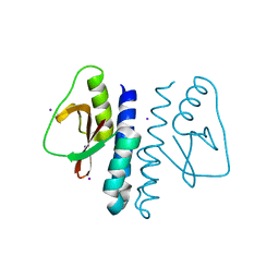
3LB5
DownloadVisualize
BU of 3lb5 by Molmil
Crystal structure of Hit-like protein involved in cell-cycle regulation from Bartonella henselae with unknown ligand
Descriptor: Hit-like protein involved in cell-cycle regulation, UNKNOWN LIGAND
Authors:Seattle Structural Genomics Center for Infectious Disease (SSGCID)
Deposit date:2010-01-07
Release date:2010-01-19
Last modified:2023-09-06
Method:X-RAY DIFFRACTION (1.9 Å)
Cite:Crystal structure of Hit-like protein involved in cell-cycle regulation from Bartonella henselae with unknown ligand
To be Published
6UYN
DownloadVisualize
BU of 6uyn by Molmil
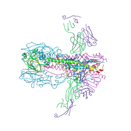
Crystal structure of influenza A virus hemagglutinin from A/Ohio/09/2015 bound to the stalk-binding CR6261 antibody Fab
Descriptor: 2-acetamido-2-deoxy-beta-D-glucopyranose, 2-acetamido-2-deoxy-beta-D-glucopyranose-(1-4)-2-acetamido-2-deoxy-beta-D-glucopyranose, CHLORIDE ION, ...
Authors:Seattle Structural Genomics Center for Infectious Disease, Seattle Structural Genomics Center for Infectious Disease (SSGCID)
Deposit date:2019-11-13
Release date:2020-09-02
Last modified:2023-10-11
Method:X-RAY DIFFRACTION (2.85 Å)
Cite:Crystal structure of influenza A virus hemagglutinin from A/Ohio/09/2015 bound to the stalk-binding CR6261 antibody Fab
To Be Published
3LG6
DownloadVisualize
BU of 3lg6 by Molmil
Crystal structure of putative glutathione transferase from Coccidioides immitis
Descriptor: SULFATE ION, putative glutathione transferase
Authors:Seattle Structural Genomics Center for Infectious Disease (SSGCID)
Deposit date:2010-01-19
Release date:2010-01-26
Last modified:2023-09-06
Method:X-RAY DIFFRACTION (2.2 Å)
Cite:Structures of a putative zeta-class glutathione S-transferase from the pathogenic fungus Coccidioides immitis.
Acta Crystallogr.,Sect.F, 67, 2011
3LA9
DownloadVisualize
BU of 3la9 by Molmil
Crystal structure of the trimeric autotransporter adhesin head domain BpaA from Burkholderia pseudomallei, iodide phased
Descriptor: Haemagglutinin family protein, IODIDE ION
Authors:Seattle Structural Genomics Center for Infectious Disease (SSGCID)
Deposit date:2010-01-06
Release date:2010-05-12
Last modified:2024-02-21
Method:X-RAY DIFFRACTION (2.05 Å)
Cite:Structure of a Burkholderia pseudomallei trimeric autotransporter adhesin head.
Plos One, 5, 2010
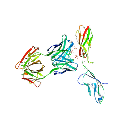
6VEL
DownloadVisualize
BU of 6vel by Molmil
Crystal Structure of Human E-cadherin bound by mouse monoclonal antibody 66E8Fab
Descriptor: 66E8 Fab Heavy Chain, 66E8 Fab Light Chain, CALCIUM ION, ...
Authors:Seattle Structural Genomics Center for Infectious Disease, Seattle Structural Genomics Center for Infectious Disease (SSGCID)
Deposit date:2020-01-02
Release date:2020-01-29
Last modified:2024-10-09
Method:X-RAY DIFFRACTION (2.65 Å)
Cite:Regulation of multiple dimeric states of E-cadherin by adhesion activating antibodies revealed through Cryo-EM and X-ray crystallography.
Pnas Nexus, 1, 2022
3LR3
DownloadVisualize
BU of 3lr3 by Molmil
Periplasmic domain of the risS sensor protein from Burkholderia pesuromallei, low pH native structure
Descriptor: Sensor protein
Authors:Seattle Structural Genomics Center for Infectious Disease (SSGCID)
Deposit date:2010-02-10
Release date:2010-06-16
Last modified:2023-09-06
Method:X-RAY DIFFRACTION (2.1 Å)
Cite:Structural basis for pH sensing by the periplasmic domain of the risS histidine kinase from Burkholderia pseudomallei
To be Published
3LR0
DownloadVisualize
BU of 3lr0 by Molmil
Periplasmic domain of the risS sensor protein from Burkholderia pseudomallei, iodide phased at low pH
Descriptor: IODIDE ION, Sensor protein
Authors:Seattle Structural Genomics Center for Infectious Disease (SSGCID)
Deposit date:2010-02-10
Release date:2010-06-16
Last modified:2024-02-21
Method:X-RAY DIFFRACTION (1.9 Å)
Cite:Structural basis for pH sensing by the periplasmic domain of the risS histidine kinase from Bukholderia pseudomallei
To be Published
3LR5
DownloadVisualize
BU of 3lr5 by Molmil
Periplasmic domain of the risS sensor protein from Burkholderia pseudomallei, iodide phased at neutral pH
Descriptor: IODIDE ION, Sensor protein
Authors:Seattle Structural Genomics Center for Infectious Disease (SSGCID)
Deposit date:2010-02-10
Release date:2010-06-16
Last modified:2024-02-21
Method:X-RAY DIFFRACTION (2.3 Å)
Cite:Structural basis for pH sensing by the periplsamic domain of the risS histidine kinase from Burkholderia pseudomallei
To be Published
3LR4
DownloadVisualize
BU of 3lr4 by Molmil
Periplasmic domain of the risS sensor protein from Burkholderia pseudomallei, barium phased at low pH
Descriptor: BARIUM ION, CHLORIDE ION, Sensor protein
Authors:Seattle Structural Genomics Center for Infectious Disease (SSGCID)
Deposit date:2010-02-10
Release date:2010-06-16
Last modified:2024-02-21
Method:X-RAY DIFFRACTION (1.9 Å)
Cite:Structural basis for pH sensing by the periplasmic domain of the risS histidine kinase from Burkholderia pseudomallei
To be Published
6VUD
DownloadVisualize
BU of 6vud by Molmil
Crystal structure of a ribosome recycling factor from Ehrlichia chaffeensis
Descriptor: CHLORIDE ION, Ribosome-recycling factor
Authors:Seattle Structural Genomics Center for Infectious Disease (SSGCID)
Deposit date:2020-02-15
Release date:2020-03-18
Last modified:2024-04-03
Method:X-RAY DIFFRACTION (2.35 Å)
Cite:Crystal structure of a ribosome recycling factor from Ehrlichia chaffeensis
to be published
6VJU
DownloadVisualize
BU of 6vju by Molmil
Crystal Structure of Cystathionine beta synthase from Legionella pneumophila with LLP, PLP, and homocysteine
Descriptor: 1,2-ETHANEDIOL, 2-AMINO-4-MERCAPTO-BUTYRIC ACID, ACETATE ION, ...
Authors:Seattle Structural Genomics Center for Infectious Disease (SSGCID)
Deposit date:2020-01-17
Release date:2020-02-05
Last modified:2024-04-03
Method:X-RAY DIFFRACTION (1.3 Å)
Cite:Crystal Structure of Cystathionine beta synthase from Legionella pneumophila with LLP, PLP, and homocysteine
to be published
3K2H
DownloadVisualize
BU of 3k2h by Molmil
Co-crystal structure of dihydrofolate reductase/thymidylate synthase from Babesia bovis with dUMP, Pemetrexed and NADP
Descriptor: 1,2-ETHANEDIOL, 2'-DEOXYURIDINE 5'-MONOPHOSPHATE, 2-{4-[2-(2-AMINO-4-OXO-4,7-DIHYDRO-3H-PYRROLO[2,3-D]PYRIMIDIN-5-YL)-ETHYL]-BENZOYLAMINO}-PENTANEDIOIC ACID, ...
Authors:Seattle Structural Genomics Center for Infectious Disease (SSGCID)
Deposit date:2009-09-30
Release date:2009-10-13
Last modified:2023-09-06
Method:X-RAY DIFFRACTION (2.2 Å)
Cite:Inhibitor-bound complexes of dihydrofolate reductase-thymidylate synthase from Babesia bovis.
Acta Crystallogr.,Sect.F, 67, 2011
3DPF
DownloadVisualize
BU of 3dpf by Molmil
Crystal structure of the complex between MMP-8 and a non-zinc chelating inhibitor
Descriptor: ACETOHYDROXAMIC ACID, CALCIUM ION, N-{[2-(2-amino-3,4-dioxocyclobut-1-en-1-yl)-1,2,3,4-tetrahydroisoquinolin-7-yl]methyl}-4-oxo-3,5,6,8-tetrahydro-4H-thiopyrano[4',3':4,5]thieno[2,3-d]pyrimidine-2-carboxamide 7,7-dioxide, ...
Authors:Pochetti, G, Montanari, R, Mazza, F.
Deposit date:2008-07-08
Release date:2009-03-03
Last modified:2023-11-01
Method:X-RAY DIFFRACTION (2.1 Å)
Cite:Extra Binding Region Induced by Non-Zinc Chelating Inhibitors into the S(1)' Subsite of Matrix Metalloproteinase 8 (MMP-8)
J.Med.Chem., 52, 2009
3K5P
DownloadVisualize
BU of 3k5p by Molmil
Crystal structure of amino acid-binding ACT: D-isomer specific 2-hydroxyacid dehydrogenase catalytic domain from Brucella melitensis
Descriptor: 1,2-ETHANEDIOL, D-3-phosphoglycerate dehydrogenase, GLYCEROL
Authors:Seattle Structural Genomics Center for Infectious Disease (SSGCID)
Deposit date:2009-10-07
Release date:2009-11-03
Last modified:2023-09-06
Method:X-RAY DIFFRACTION (2.15 Å)
Cite:Crystal structure of amino acid-binding ACT: D-isomer specific 2-hydroxyacid dehydrogenase catalytic domain from Brucella melitensis
To be Published
6C49
DownloadVisualize
BU of 6c49 by Molmil
Crystal Structure of Alcohol Dehydrogenase from Acinetobacter baumannii
Descriptor: 1,2-ETHANEDIOL, Alcohol dehydrogenase, DI(HYDROXYETHYL)ETHER, ...
Authors:Seattle Structural Genomics Center for Infectious Disease (SSGCID)
Deposit date:2018-01-11
Release date:2018-01-24
Last modified:2024-03-13
Method:X-RAY DIFFRACTION (1.85 Å)
Cite:Crystal Structure of Alcohol Dehydrogenase from Acinetobacter baumannii
To be Published
6WCI
DownloadVisualize
BU of 6wci by Molmil
Crystal structure of a cysteine desulfurase SufS from Stenotrophomonas maltophilia K279a
Descriptor: 1,2-ETHANEDIOL, Cysteine desulfurase, SULFATE ION
Authors:Seattle Structural Genomics Center for Infectious Disease (SSGCID)
Deposit date:2020-03-30
Release date:2020-04-08
Last modified:2023-11-15
Method:X-RAY DIFFRACTION (2.05 Å)
Cite:Crystal structure of a cysteine desulfurase SufS from Stenotrophomonas maltophilia K279a
To Be Published
6UYH
DownloadVisualize
BU of 6uyh by Molmil
Crystal structure of prolyl-tRNA synthetase from Naegleria fowleri in complex with halofuginone and AMPPNP
Descriptor: 1,2-ETHANEDIOL, 7-bromo-6-chloro-3-{3-[(2R,3S)-3-hydroxypiperidin-2-yl]-2-oxopropyl}quinazolin-4(3H)-one, MAGNESIUM ION, ...
Authors:Seattle Structural Genomics Center for Infectious Disease (SSGCID)
Deposit date:2019-11-13
Release date:2019-11-27
Last modified:2023-10-11
Method:X-RAY DIFFRACTION (1.75 Å)
Cite:Crystal structure of prolyl-tRNA synthetase from Naegleria fowleri in complex with halofuginone and AMPPNP
TO BE PUBLISHED
6VU9
DownloadVisualize
BU of 6vu9 by Molmil
Crystal structure of threonyl-tRNA synthetase (ThrRS) from Stenotrophomonas maltophilia K279a
Descriptor: SULFATE ION, Threonine--tRNA ligase, ZINC ION
Authors:Seattle Structural Genomics Center for Infectious Disease (SSGCID)
Deposit date:2020-02-14
Release date:2020-03-04
Last modified:2023-10-11
Method:X-RAY DIFFRACTION (2.2 Å)
Cite:Crystal structure of threonyl-tRNA synthetase (ThrRS) from Stenotrophomonas maltophilia K279a
To Be Published
3KNU
DownloadVisualize
BU of 3knu by Molmil
Crystal structure of tRNA (guanine-N1)-methyltransferase from Anaplasma phagocytophilum
Descriptor: tRNA (guanine-N(1)-)-methyltransferase
Authors:Seattle Structural Genomics Center for Infectious Disease (SSGCID)
Deposit date:2009-11-12
Release date:2009-11-24
Last modified:2023-09-06
Method:X-RAY DIFFRACTION (2.25 Å)
Cite:Crystal structure of tRNA (guanine-N1)-methyltransferase from Anaplasma phagocytophilum
To be Published

227111

PDB entries from 2024-11-06

PDB statisticsPDBj update infoContact PDBjnumon