Loading
PDBj
MenuPDBj@FacebookPDBj@X(formerly Twitter)PDBj@BlueSkyPDBj@YouTubewwPDB FoundationwwPDB
RCSB PDBPDBeBMRBAdv. SearchSearch help
Search by PDB author
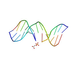
2HSL
DownloadVisualize
BU of 2hsl by Molmil
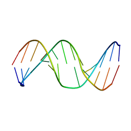
NMR structure of 13mer duplex DNA containing an abasic site, averaged structure (alpha anomer)
Descriptor: 5'-D(*CP*CP*AP*AP*AP*GP*(D1P)P*AP*CP*CP*GP*GP*G)-3', 5'-D(*CP*CP*CP*GP*GP*TP*AP*CP*TP*TP*TP*GP*G)-3'
Authors:Chen, J, Dupradeau, F.Y, Case, D.A, Turner, C.J, Stubbe, J.
Deposit date:2006-07-22
Release date:2007-05-29
Last modified:2024-05-01
Method:SOLUTION NMR
Cite:Nuclear magnetic resonance structural studies and molecular modeling of duplex DNA containing normal and 4'-oxidized abasic sites.
Biochemistry, 46, 2007
2HSS
DownloadVisualize
BU of 2hss by Molmil
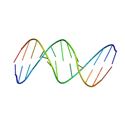
13mer duplex DNA containg an abasic site with beta anomer, averaged structure
Descriptor: 5'-D(*CP*CP*AP*AP*AP*GP*(AAB)P*AP*CP*CP*GP*GP*G)-3', 5'-D(*CP*CP*CP*GP*GP*TP*AP*CP*TP*TP*TP*GP*G)-3'
Authors:Chen, J, Dupradeau, F.Y, Case, D.A, Turner, C.J, Stubbe, J.
Deposit date:2006-07-22
Release date:2007-05-29
Last modified:2024-05-01
Method:SOLUTION NMR
Cite:Nuclear magnetic resonance structural studies and molecular modeling of duplex DNA containing normal and 4'-oxidized abasic sites.
Biochemistry, 46, 2007
1G5K
DownloadVisualize
BU of 1g5k by Molmil
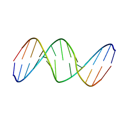
NMR Structrure of d(CCAAAGXACTGGG), X is a 3'-phosphoglycolate, 5'-phosphate gapped lesion, 10 structures
Descriptor: 2-PHOSPHOGLYCOLIC ACID, 5'-D(*CP*CP*AP*AP*AP*G)-3', 5'-D(*CP*CP*CP*AP*GP*TP*AP*CP*TP*TP*TP*GP*G)-3', ...
Authors:Junker, H.-D, Hoehn, S.T, Bunt, R.C, Marathius, V, Chen, J, Turner, C.J, Stubbe, J.
Deposit date:2002-10-14
Release date:2003-01-07
Last modified:2024-05-22
Method:SOLUTION NMR
Cite:Synthesis, Characterization and Solution Structure of Tethered Oligonucleotides Containing an Internal 3'-Phosphoglycolate, 5'-Phosphate Gapped Lesion
Nucleic Acids Res., 30, 2002
1GJ1
DownloadVisualize
BU of 1gj1 by Molmil
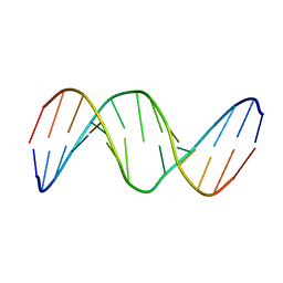
NMR structure of d(CCAAAGXACTGGG), X is a 3'phosphoglycolate, 5'phosphate gapped lesion
Descriptor: 2-PHOSPHOGLYCOLIC ACID, 5'-D(*CP*CP*AP*AP*AP*G)-3', 5'-D(*CP*CP*CP*AP*GP*TP*AP*CP*TP*TP*TP*GP*G)-3', ...
Authors:Junker, H.-D, Hoehn, S.T, Bunt, R.C, Marathius, V, Chen, J, Turner, C.J, Stubbe, J.
Deposit date:2002-10-14
Release date:2003-01-07
Last modified:2024-05-22
Method:SOLUTION NMR
Cite:Synthesis, Characterization and Solution Structure of Tethered Oligonucleotides Containing an Internal 3'-Phosphoglycolate, 5'-Phosphate Gapped Lesion
Nucleic Acids Res., 30, 2002
1G5E
DownloadVisualize
BU of 1g5e by Molmil
NMR STRUCTURE OF AN OLIGONUCLEOTIDE CONTAINING AN ABASIC SITE: BETA ANOMER
Descriptor: 5'-D(*CP*CP*AP*AP*AP*GP*(AAB)P*AP*CP*TP*GP*GP*G)-3', 5'-D(*CP*CP*CP*AP*GP*TP*AP*CP*TP*TP*TP*GP*G)-3'
Authors:Hoehn, S.T, Turner, C.J, Stubbe, J.
Deposit date:2000-10-31
Release date:2001-08-22
Last modified:2024-05-22
Method:SOLUTION NMR
Cite:Solution structure of an oligonucleotide containing an abasic site: evidence for an unusual deoxyribose conformation.
Nucleic Acids Res., 29, 2001
1GJ0
DownloadVisualize
BU of 1gj0 by Molmil
NMR STRUCTURE OF AN OLIGONUCLEOTIDE CONTAINING AN ABASIC SITE: BETA ANOMER
Descriptor: 5'-D(*CP*CP*AP*AP*AP*GP*(AAB)P*AP*CP*TP*GP*GP*G)-3', 5'-D(*CP*CP*CP*AP*GP*TP*AP*CP*TP*TP*TP*GP*G)-3'
Authors:Hoehn, S.T, Turner, C.J, Stubbe, J.
Deposit date:2000-10-31
Release date:2001-08-22
Last modified:2023-12-27
Method:SOLUTION NMR
Cite:Solution structure of an oligonucleotide containing an abasic site: evidence for an unusual deoxyribose conformation.
Nucleic Acids Res., 29, 2001
1G5D
DownloadVisualize
BU of 1g5d by Molmil
NMR STRUCTURE OF AN OLIGONUCLEOTIDE CONTAINING AN ABASIC SITE: ALPHA ANOMER
Descriptor: 5'-D(*CP*CP*AP*AP*AP*GP*(D1P)P*AP*CP*TP*GP*GP*G)-3', 5'-D(*CP*CP*CP*AP*GP*TP*AP*CP*TP*TP*TP*GP*G)-3'
Authors:Hoehn, S.T, Turner, C.J, Stubbe, J.
Deposit date:2000-10-31
Release date:2001-08-22
Last modified:2024-05-22
Method:SOLUTION NMR
Cite:Solution structure of an oligonucleotide containing an abasic site: evidence for an unusual deoxyribose conformation.
Nucleic Acids Res., 29, 2001
1GIZ
DownloadVisualize
BU of 1giz by Molmil
NMR STRUCTURE OF AN OLIGONUCLEOTIDE CONTAINING AN ABASIC SITE: ALPHA ANOMER
Descriptor: 5'-D(*CP*CP*AP*AP*AP*GP*(D1P)P*AP*CP*TP*GP*GP*G)-3', 5'-D(*CP*CP*CP*AP*GP*TP*AP*CP*TP*TP*TP*GP*G)-3'
Authors:Hoehn, S.T, Turner, C.J, Stubbe, J.
Deposit date:2000-10-31
Release date:2001-08-22
Last modified:2023-12-27
Method:SOLUTION NMR
Cite:Solution structure of an oligonucleotide containing an abasic site: evidence for an unusual deoxyribose conformation.
Nucleic Acids Res., 29, 2001
1GSO
DownloadVisualize
BU of 1gso by Molmil
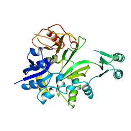
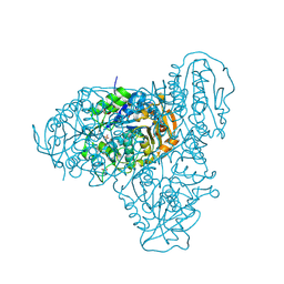
GLYCINAMIDE RIBONUCLEOTIDE SYNTHETASE (GAR-SYN) FROM E. COLI.
Descriptor: PROTEIN (GLYCINAMIDE RIBONUCLEOTIDE SYNTHETASE)
Authors:Wang, W, Kappock, T.J, Stubbe, J, Ealick, S.E.
Deposit date:1998-09-08
Release date:1998-12-09
Last modified:2024-02-07
Method:X-RAY DIFFRACTION (1.6 Å)
Cite:X-ray crystal structure of glycinamide ribonucleotide synthetase from Escherichia coli.
Biochemistry, 37, 1998
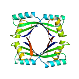
1QCZ
DownloadVisualize
BU of 1qcz by Molmil
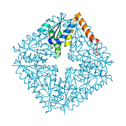
CRYSTAL STRUCTURE OF E. COLI PURE, AN UNUSUAL MUTASE THAT CATALYZES THE CONVERSION OF N5-CARBOXYAMINOIMIDAZOLE RIBONUCLEOTIDE (N5-CAIR) TO 4-CARBOXYAMINOIMIDAZOLE RIBONUCLEOTIDE (CAIR) IN THE PURINE BIOSYNTHETIC PATHWAY
Descriptor: N5-CARBOXYAMINOIMIDAZOLE RIBONUCLEOTIDE MUTASE
Authors:Ealick, S.E, Mathews, I.I.
Deposit date:1999-05-10
Release date:1999-11-10
Last modified:2024-10-30
Method:X-RAY DIFFRACTION (1.5 Å)
Cite:Crystal structure of Escherichia coli PurE, an unusual mutase in the purine biosynthetic pathway.
Structure Fold.Des., 7, 1999
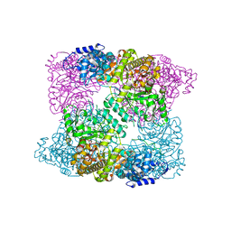
2AV8
DownloadVisualize
BU of 2av8 by Molmil
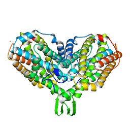
Y122F MUTANT OF RIBONUCLEOTIDE REDUCTASE FROM ESCHERICHIA COLI
Descriptor: FE (II) ION, MU-OXO-DIIRON, RIBONUCLEOTIDE REDUCTASE R2
Authors:Han, S, Arvai, A, Tainer, J.A.
Deposit date:1997-09-30
Release date:1998-10-28
Last modified:2024-05-22
Method:X-RAY DIFFRACTION (2.46 Å)
Cite:Characterization of Y122F R2 of Escherichia coli ribonucleotide reductase by time-resolved physical biochemical methods and X-ray crystallography.
Biochemistry, 37, 1998
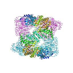
4U3E
DownloadVisualize
BU of 4u3e by Molmil
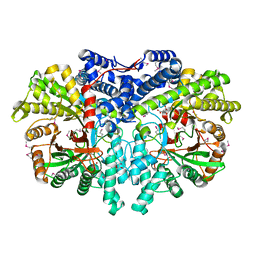
Anaerobic ribonucleotide reductase
Descriptor: ACETATE ION, CITRIC ACID, GLYCEROL, ...
Authors:Funk, M.A, Drennan, C.L.
Deposit date:2014-07-20
Release date:2014-09-03
Last modified:2024-10-23
Method:X-RAY DIFFRACTION (1.64 Å)
Cite:The class III ribonucleotide reductase from Neisseria bacilliformis can utilize thioredoxin as a reductant.
Proc.Natl.Acad.Sci.USA, 111, 2014
6AUI
DownloadVisualize
BU of 6aui by Molmil
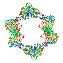
Human ribonucleotide reductase large subunit (alpha) with dATP and CDP
Descriptor: 2'-DEOXYADENOSINE 5'-TRIPHOSPHATE, CYTIDINE-5'-DIPHOSPHATE, MAGNESIUM ION, ...
Authors:Brignole, E.J, Drennan, C.L, Asturias, F.J, Tsai, K.L, Penczek, P.A.
Deposit date:2017-09-01
Release date:2018-04-18
Last modified:2024-03-13
Method:ELECTRON MICROSCOPY (3.3 Å)
Cite:3.3- angstrom resolution cryo-EM structure of human ribonucleotide reductase with substrate and allosteric regulators bound.
Elife, 7, 2018
4ERP
DownloadVisualize
BU of 4erp by Molmil
Crystal structure of a gemcitabine-diphosphate inhibited E. coli class Ia ribonucleotide reductase complex
Descriptor: ADENOSINE-5'-TRIPHOSPHATE, MU-OXO-DIIRON, Ribonucleoside-diphosphate reductase 1 subunit alpha, ...
Authors:Zimanyi, C.M, Drennan, C.L.
Deposit date:2012-04-20
Release date:2012-07-04
Last modified:2023-09-13
Method:X-RAY DIFFRACTION (4.45 Å)
Cite:Tangled up in knots: structures of inactivated forms of E. coli class Ia ribonucleotide reductase.
Structure, 20, 2012
5T6O
DownloadVisualize
BU of 5t6o by Molmil
Structure of the catalytic domain of the class I polyhydroxybutyrate synthase from Cupriavidus necator
Descriptor: Poly-beta-hydroxybuterate polymerase, SULFATE ION
Authors:Wittenborn, E.C, Jost, M, Drennan, C.L.
Deposit date:2016-09-01
Release date:2016-10-26
Last modified:2024-11-13
Method:X-RAY DIFFRACTION (1.8 Å)
Cite:Structure of the Catalytic Domain of the Class I Polyhydroxybutyrate Synthase from Cupriavidus necator.
J.Biol.Chem., 291, 2016
4N82
DownloadVisualize
BU of 4n82 by Molmil
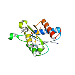
X-ray crystal structure of Streptococcus sanguinis NrdIox
Descriptor: FLAVIN MONONUCLEOTIDE, Ribonucleotide reductase, SULFATE ION
Authors:Boal, A.K, Rosenzweig, A.C.
Deposit date:2013-10-16
Release date:2014-01-08
Last modified:2023-09-20
Method:X-RAY DIFFRACTION (1.88 Å)
Cite:Streptococcus sanguinis Class Ib Ribonucleotide Reductase: HIGH ACTIVITY WITH BOTH IRON AND MANGANESE COFACTORS AND STRUCTURAL INSIGHTS.
J.Biol.Chem., 289, 2014
4N83
DownloadVisualize
BU of 4n83 by Molmil
X-ray crystal structure of Streptococcus sanguinis dimanganese(II)-NrdF
Descriptor: MANGANESE (II) ION, Ribonucleoside-diphosphate reductase subunit beta
Authors:Boal, A.K, Rosenzweig, A.C.
Deposit date:2013-10-16
Release date:2014-01-08
Last modified:2023-09-20
Method:X-RAY DIFFRACTION (2.65 Å)
Cite:Streptococcus sanguinis Class Ib Ribonucleotide Reductase: HIGH ACTIVITY WITH BOTH IRON AND MANGANESE COFACTORS AND STRUCTURAL INSIGHTS.
J.Biol.Chem., 289, 2014
3HJZ
DownloadVisualize
BU of 3hjz by Molmil
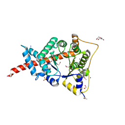
The structure of an aldolase from Prochlorococcus marinus
Descriptor: 1,2-ETHANEDIOL, CHLORIDE ION, DI(HYDROXYETHYL)ETHER, ...
Authors:Singer, A.U, Xu, X, Cui, H, Joachimiak, A, Edwards, A.M, Savchenko, A, Midwest Center for Structural Genomics (MCSG)
Deposit date:2009-05-22
Release date:2009-06-09
Last modified:2024-11-20
Method:X-RAY DIFFRACTION (1.9 Å)
Cite:Phage auxiliary metabolic genes and the redirection of cyanobacterial host carbon metabolism.
Proc.Natl.Acad.Sci.USA, 108, 2011
5CI4
DownloadVisualize
BU of 5ci4 by Molmil
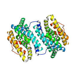
Ribonucleotide reductase beta subunit
Descriptor: MU-OXO-DIIRON, Ribonucleoside-diphosphate reductase 1, beta subunit, ...
Authors:Funk, M.A, Drennan, C.L.
Deposit date:2015-07-10
Release date:2016-06-22
Last modified:2023-09-27
Method:X-RAY DIFFRACTION (2.05 Å)
Cite:Biophysical Characterization of Fluorotyrosine Probes Site-Specifically Incorporated into Enzymes: E. coli Ribonucleotide Reductase As an Example.
J.Am.Chem.Soc., 138, 2016
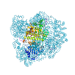
9DB2
DownloadVisualize
BU of 9db2 by Molmil
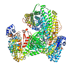
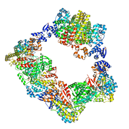
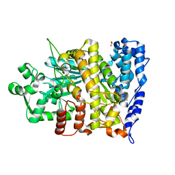
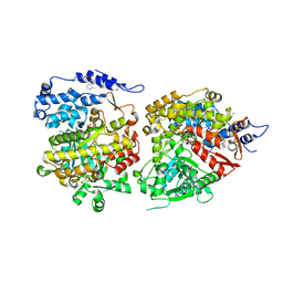
Class Ia ribonucleotide reductase with mechanism-based inhibitor N3CDP
Descriptor: 2'-DEOXYADENOSINE 5'-TRIPHOSPHATE, 4-amino-1-{(3xi)-3-C-amino-2-deoxy-5-O-[(S)-hydroxy(phosphonooxy)phosphoryl]-beta-D-threo-pentofuranosyl}pyrimidin-2(1H)-one, ADENOSINE-5'-TRIPHOSPHATE, ...
Authors:Westmoreland, D.E, Drennan, C.L.
Deposit date:2024-08-23
Release date:2024-11-06
Method:ELECTRON MICROSCOPY (2.6 Å)
Cite:2.6- angstrom resolution cryo-EM structure of a class Ia ribonucleotide reductase trapped with mechanism-based inhibitor N 3 CDP.
Proc.Natl.Acad.Sci.USA, 121, 2024
3D54
DownloadVisualize
BU of 3d54 by Molmil
Structure of PurLQS from Thermotoga maritima
Descriptor: ADENOSINE-5'-DIPHOSPHATE, Formylglycinamide ribonucleotide amidotransferase, Phosphoribosylformylglycinamidine synthase 1, ...
Authors:Ealick, S.E, Morar, M.
Deposit date:2008-05-15
Release date:2008-07-22
Last modified:2017-02-01
Method:X-RAY DIFFRACTION (3.5 Å)
Cite:Formylglycinamide ribonucleotide amidotransferase from Thermotoga maritima: structural insights into complex formation.
Biochemistry, 47, 2008
3UUS
DownloadVisualize
BU of 3uus by Molmil
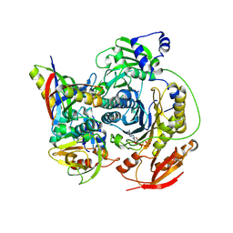
Crystal structure of the dATP inhibited E. coli class Ia ribonucleotide reductase complex
Descriptor: 2'-DEOXYADENOSINE 5'-TRIPHOSPHATE, FE (III) ION, Ribonucleoside-diphosphate reductase 1 subunit alpha, ...
Authors:Zimanyi, C.M, Drennan, C.L.
Deposit date:2011-11-28
Release date:2011-12-21
Last modified:2024-11-20
Method:X-RAY DIFFRACTION (5.65 Å)
Cite:Structural interconversions modulate activity of Escherichia coli ribonucleotide reductase.
Proc.Natl.Acad.Sci.USA, 108, 2011
4ERM
DownloadVisualize
BU of 4erm by Molmil
Crystal structure of the dATP inhibited E. coli class Ia ribonucleotide reductase complex at 4 Angstroms resolution
Descriptor: 2'-DEOXYADENOSINE 5'-TRIPHOSPHATE, 2'-DEOXYADENOSINE-5'-DIPHOSPHATE, MAGNESIUM ION, ...
Authors:Zimanyi, C.M, Drennan, C.L.
Deposit date:2012-04-20
Release date:2012-07-04
Last modified:2023-09-13
Method:X-RAY DIFFRACTION (3.95 Å)
Cite:Tangled up in knots: structures of inactivated forms of E. coli class Ia ribonucleotide reductase.
Structure, 20, 2012
6CGM
DownloadVisualize
BU of 6cgm by Molmil
X-ray crystal structure of Bacillus subtilis ribonucleotide reductase NrdE alpha subunit (nucleotide free)
Descriptor: 1,2-ETHANEDIOL, Ribonucleoside-diphosphate reductase
Authors:Maggiolo, A.O, Boal, A.K.
Deposit date:2018-02-20
Release date:2018-05-09
Last modified:2024-03-13
Method:X-RAY DIFFRACTION (2 Å)
Cite:An endogenous dAMP ligand inBacillus subtilisclass Ib RNR promotes assembly of a noncanonical dimer for regulation by dATP.
Proc. Natl. Acad. Sci. U.S.A., 115, 2018
6CGN
DownloadVisualize
BU of 6cgn by Molmil
X-ray crystal structure of Bacillus subtilis ribonucleotide reductase NrdE alpha subunit dAMP-bound (pH 7)
Descriptor: 1,2-ETHANEDIOL, 2'-DEOXYADENOSINE-5'-MONOPHOSPHATE, PHOSPHATE ION, ...
Authors:Maggiolo, A.O, Boal, A.K.
Deposit date:2018-02-20
Release date:2018-05-09
Last modified:2024-05-29
Method:X-RAY DIFFRACTION (2.26 Å)
Cite:An endogenous dAMP ligand inBacillus subtilisclass Ib RNR promotes assembly of a noncanonical dimer for regulation by dATP.
Proc. Natl. Acad. Sci. U.S.A., 115, 2018

227561

PDB entries from 2024-11-20

PDB statisticsPDBj update infoContact PDBjnumon