Loading
PDBj
MenuPDBj@FacebookPDBj@TwitterPDBj@YouTubewwPDB FoundationwwPDB
RCSB PDBPDBeBMRBAdv. SearchSearch help
Search by PDB author
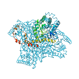
3KBM
DownloadVisualize
BU of 3kbm by Molmil
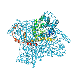
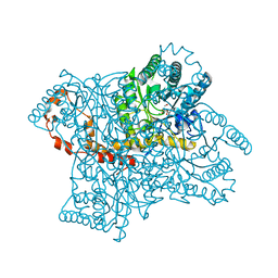
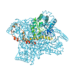
Room Temperature X-ray structure of D-Xylose Isomerase complexed with 2Cd(2+) co-factors and d12-D-alpha-glucose in the cyclic form
Descriptor: CADMIUM ION, Xylose isomerase, alpha-D-glucopyranose
Authors:Kovalevsky, A.Y, Hanson, L, Langan, P.
Deposit date:2009-10-20
Release date:2010-06-16
Last modified:2023-09-06
Method:X-RAY DIFFRACTION (2 Å)
Cite:Metal ion roles and the movement of hydrogen during reaction catalyzed by D-xylose isomerase: a joint x-ray and neutron diffraction study.
Structure, 18, 2010
3KCJ
DownloadVisualize
BU of 3kcj by Molmil
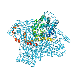
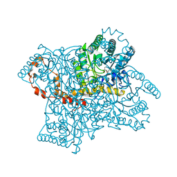
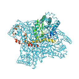
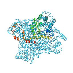
Room temperature neutron structure of apo-D-Xylose Isomerase (refined jointly with X-ray structure 3KBJ)
Descriptor: Xylose isomerase
Authors:Kovalevsky, A.Y, Langan, P.
Deposit date:2009-10-21
Release date:2010-09-29
Last modified:2024-04-03
Method:NEUTRON DIFFRACTION (1.8 Å), X-RAY DIFFRACTION
Cite:Identification of the Elusive Hydronium Ion Exchanging Roles with a Proton in an Enzyme at Lower pH Values
Angew.Chem.Int.Ed.Engl., 50, 2011
3KCL
DownloadVisualize
BU of 3kcl by Molmil
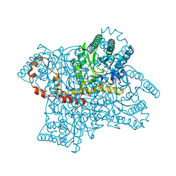
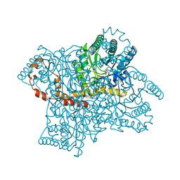
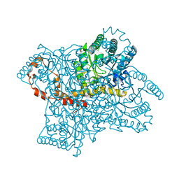
Room temperature neutron structure of D-Xylose Isomerase in complex with two Cd2+ cations and d12-D-alpha-glucose in the ring form (refined jointly with X-ray structure 3KBM)
Descriptor: CADMIUM ION, Xylose isomerase, alpha-D-glucopyranose
Authors:Kovalevsky, A.Y, Langan, P.
Deposit date:2009-10-21
Release date:2010-06-16
Last modified:2024-04-03
Method:NEUTRON DIFFRACTION (2 Å), X-RAY DIFFRACTION
Cite:Metal ion roles and the movement of hydrogen during reaction catalyzed by D-xylose isomerase: a joint x-ray and neutron diffraction study.
Structure, 18, 2010
3KBJ
DownloadVisualize
BU of 3kbj by Molmil
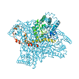
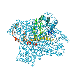
Room temperature X-ray structure of apo-D-Xylose Isomerase
Descriptor: Xylose isomerase
Authors:Kovalevsky, A.Y, Hanson, L, Langan, P.
Deposit date:2009-10-20
Release date:2010-09-29
Last modified:2024-02-21
Method:X-RAY DIFFRACTION (2 Å)
Cite:Identification of the Elusive Hydronium Ion Exchanging Roles with a Proton in an Enzyme at Lower pH Values.
Angew.Chem.Int.Ed.Engl., 50, 2011
3KCO
DownloadVisualize
BU of 3kco by Molmil
Room temperature neutron structure of D-Xylose Isomerase in complex with two Ni2+ cations and d12-D-glucose in the linear form (refined jointly with X-ray structure 3KBN)
Descriptor: D-glucose, NICKEL (II) ION, Xylose isomerase
Authors:Kovalevsky, A.Y, Langan, P.
Deposit date:2009-10-21
Release date:2010-06-16
Last modified:2024-04-03
Method:NEUTRON DIFFRACTION (1.8 Å), X-RAY DIFFRACTION
Cite:Metal ion roles and the movement of hydrogen during reaction catalyzed by D-xylose isomerase: a joint x-ray and neutron diffraction study.
Structure, 18, 2010
3KBN
DownloadVisualize
BU of 3kbn by Molmil
Room temperature structure of D-Xylose Isomerase in complex with 2Ni(2+) co-factors and d12-D-glucose in the linear form
Descriptor: D-glucose, NICKEL (II) ION, Xylose isomerase
Authors:Kovalevsky, A.Y, Hanson, L, Langan, P.
Deposit date:2009-10-20
Release date:2010-06-16
Last modified:2023-09-06
Method:X-RAY DIFFRACTION (1.53 Å)
Cite:Metal ion roles and the movement of hydrogen during reaction catalyzed by D-xylose isomerase: a joint x-ray and neutron diffraction study.
Structure, 18, 2010
4QE5
DownloadVisualize
BU of 4qe5 by Molmil
Room temperature X-ray structure of D-xylose isomerase in complex with two Mg2+ ions and L-ribulose
Descriptor: MAGNESIUM ION, Xylose isomerase, alpha-L-ribulofuranose
Authors:Kovalevsky, A.Y, Langan, P.
Deposit date:2014-05-15
Release date:2014-09-03
Last modified:2024-02-28
Method:X-RAY DIFFRACTION (1.56 Å)
Cite:L-Arabinose Binding, Isomerization, and Epimerization by D-Xylose Isomerase: X-Ray/Neutron Crystallographic and Molecular Simulation Study.
Structure, 22, 2014
4QE1
DownloadVisualize
BU of 4qe1 by Molmil
Room temperature X-ray structure of D-xylose isomerase in complex with two Cd2+ ions and L-ribulose
Descriptor: CADMIUM ION, Xylose isomerase, alpha-L-ribulofuranose
Authors:Kovalevsky, A.Y, Langan, P.
Deposit date:2014-05-15
Release date:2014-09-03
Last modified:2024-02-28
Method:X-RAY DIFFRACTION (1.55 Å)
Cite:L-Arabinose Binding, Isomerization, and Epimerization by D-Xylose Isomerase: X-Ray/Neutron Crystallographic and Molecular Simulation Study.
Structure, 22, 2014
4QDW
DownloadVisualize
BU of 4qdw by Molmil
Joint X-ray and neutron structure of Streptomyces rubiginosus D-xylose isomerase in complex with two Ni2+ ions and linear L-arabinose
Descriptor: L-arabinose, NICKEL (II) ION, Xylose isomerase
Authors:Kovalevsky, A.Y, Langan, P.
Deposit date:2014-05-14
Release date:2014-09-03
Last modified:2024-02-28
Method:NEUTRON DIFFRACTION (1.8 Å), X-RAY DIFFRACTION
Cite:L-Arabinose Binding, Isomerization, and Epimerization by D-Xylose Isomerase: X-Ray/Neutron Crystallographic and Molecular Simulation Study.
Structure, 22, 2014
4QEE
DownloadVisualize
BU of 4qee by Molmil
Room temperature X-ray structure of D-xylose isomerase in complex with two Ni2+ ions and L-ribose
Descriptor: NICKEL (II) ION, Xylose isomerase, alpha-L-ribofuranose
Authors:Kovalevsky, A.Y, Langan, P.
Deposit date:2014-05-15
Release date:2014-09-03
Last modified:2024-02-28
Method:X-RAY DIFFRACTION (1.6 Å)
Cite:L-Arabinose Binding, Isomerization, and Epimerization by D-Xylose Isomerase: X-Ray/Neutron Crystallographic and Molecular Simulation Study.
Structure, 22, 2014
4QE4
DownloadVisualize
BU of 4qe4 by Molmil
Room temperature X-ray structure of D-xylose isomerase in complex with two Ni2+ ions and L-ribulose
Descriptor: NICKEL (II) ION, Xylose isomerase, beta-L-ribulofuranose
Authors:Kovalevsky, A.Y, Langan, P.
Deposit date:2014-05-15
Release date:2014-09-03
Last modified:2024-02-28
Method:X-RAY DIFFRACTION (1.7 Å)
Cite:L-Arabinose Binding, Isomerization, and Epimerization by D-Xylose Isomerase: X-Ray/Neutron Crystallographic and Molecular Simulation Study.
Structure, 22, 2014
4QDP
DownloadVisualize
BU of 4qdp by Molmil
Joint X-ray and neutron structure of Streptomyces rubiginosus D-xylose isomerase in complex with two Cd2+ ions and cyclic beta-L-arabinose
Descriptor: CADMIUM ION, Xylose isomerase, beta-L-arabinopyranose
Authors:Kovalevsky, A.Y, Langan, P.
Deposit date:2014-05-14
Release date:2014-09-03
Last modified:2024-02-28
Method:NEUTRON DIFFRACTION (2 Å), X-RAY DIFFRACTION
Cite:L-Arabinose Binding, Isomerization, and Epimerization by D-Xylose Isomerase: X-Ray/Neutron Crystallographic and Molecular Simulation Study.
Structure, 22, 2014
4QEH
DownloadVisualize
BU of 4qeh by Molmil
Room temperature X-ray structure of D-xylose isomerase in complex with two Mg2+ ions and L-ribose
Descriptor: MAGNESIUM ION, Xylose isomerase, beta-L-ribofuranose
Authors:Kovalevsky, A.Y, Langan, P.
Deposit date:2014-05-16
Release date:2014-09-03
Last modified:2024-02-28
Method:X-RAY DIFFRACTION (1.55 Å)
Cite:L-Arabinose Binding, Isomerization, and Epimerization by D-Xylose Isomerase: X-Ray/Neutron Crystallographic and Molecular Simulation Study.
Structure, 22, 2014
5AI3
DownloadVisualize
BU of 5ai3 by Molmil
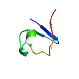
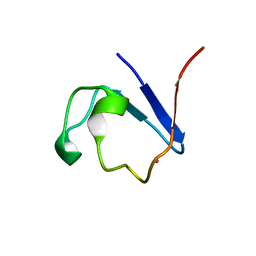
X-ray structure of 113Cd-substituted Perdeuterated Pyrococcus furiosus rubredoxin to 1.02A resolution at 295K in a quartz capillary
Descriptor: CADMIUM ION, RUBREDOXIN
Authors:Cuypers, M.G, Mitchell, E.P.
Deposit date:2015-02-11
Release date:2016-03-02
Last modified:2024-05-08
Method:X-RAY DIFFRACTION (1.02 Å)
Cite:Macromolecular Structure Phasing by Neutron Anomalous Diffraction.
Sci.Rep., 6, 2016
5AI2
DownloadVisualize
BU of 5ai2 by Molmil
Anomalous Neutron phased crystal structure of 113Cd-substituted Perdeuterated Pyrococcus furiosus rubredoxin to 1.75A resolution at 295K
Descriptor: CADMIUM ION, RUBREDOXIN, deuterium(1+)
Authors:Cuypers, M.G, Mossou, E, Mason, S.A.
Deposit date:2015-02-11
Release date:2016-03-02
Last modified:2024-05-08
Method:NEUTRON DIFFRACTION (1.75 Å)
Cite:Macromolecular Structure Phasing by Neutron Anomalous Diffraction.
Sci.Rep., 6, 2016
<123


222926

PDB entries from 2024-07-24

PDB statisticsPDBj update infoContact PDBjnumon