Loading
PDBj
MenuPDBj@FacebookPDBj@TwitterPDBj@YouTubewwPDB FoundationwwPDB
RCSB PDBPDBeBMRBAdv. SearchSearch help
Search by PDB author
5CXT
DownloadVisualize
BU of 5cxt by Molmil
Crystal structure of a RNA-binding protein 39 (RBM39) in complex with fragment of splicing factor (U2AF) from Unknown at 2.20 A resolution
Descriptor: RNA-binding protein 39, Splicing factor U2AF 65 kDa subunit
Authors:Joint Center for Structural Genomics (JCSG), Partnership for T-Cell Biology (TCELL)
Deposit date:2015-07-29
Release date:2015-10-14
Last modified:2023-09-27
Method:X-RAY DIFFRACTION (2.2 Å)
Cite:UHM-ULM interactions in the RBM39-U2AF65 splicing-factor complex.
Acta Crystallogr D Struct Biol, 72, 2016
3BY7
DownloadVisualize
BU of 3by7 by Molmil
CRYSTAL STRUCTURE OF A PROTEIN STRUCTURALLY SIMILAR TO SM/LSM-LIKE RNA-BINDING PROTEINS (JCVI_PEP_1096686650277) FROM UNCULTURED MARINE ORGANISM AT 2.60 A RESOLUTION
Descriptor: uncharacterized protein
Authors:Joint Center for Structural Genomics (JCSG)
Deposit date:2008-01-15
Release date:2008-01-29
Last modified:2023-02-01
Method:X-RAY DIFFRACTION (2.6 Å)
Cite:Crystal structure of a novel Sm-like protein of putative cyanophage origin at 2.60 A resolution.
Proteins, 75, 2009
6W61
DownloadVisualize
BU of 6w61 by Molmil
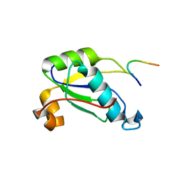
Crystal Structure of the methyltransferase-stimulatory factor complex of NSP16 and NSP10 from SARS CoV-2.
Descriptor: 1,2-ETHANEDIOL, 2'-O-methyltransferase, CHLORIDE ION, ...
Authors:Kim, Y, Jedrzejczak, R, Maltseva, N, Endres, M, Godzik, A, Joachimiak, A, Center for Structural Genomics of Infectious Diseases (CSGID)
Deposit date:2020-03-15
Release date:2020-03-25
Last modified:2023-11-15
Method:X-RAY DIFFRACTION (2 Å)
Cite:The crystal structure of nsp10-nsp16 heterodimer from SARS-CoV-2 in complex with S-adenosylmethionine
Biorxiv, 2020
3BYQ
DownloadVisualize
BU of 3byq by Molmil
Crystal structure of a duf1185 family protein (bb2672) from bordetella bronchiseptica rb50 at 1.70 A resolution
Descriptor: 1,2-ETHANEDIOL, CHLORIDE ION, TETRAETHYLENE GLYCOL, ...
Authors:Joint Center for Structural Genomics (JCSG)
Deposit date:2008-01-16
Release date:2008-01-29
Last modified:2023-02-01
Method:X-RAY DIFFRACTION (1.7 Å)
Cite:Structures of the first representatives of Pfam family PF06684 (DUF1185) reveal a novel variant of the Bacillus chorismate mutase fold and suggest a role in amino-acid metabolism.
Acta Crystallogr.,Sect.F, 66, 2010
3BOS
DownloadVisualize
BU of 3bos by Molmil
Crystal structure of a putative dna replication regulator HDA (SAMA_1916) from Shewanella amazonensis sb2b at 1.75 A resolution
Descriptor: 1,2-ETHANEDIOL, CHLORIDE ION, CYTIDINE-5'-DIPHOSPHATE, ...
Authors:Joint Center for Structural Genomics (JCSG)
Deposit date:2007-12-17
Release date:2008-01-15
Last modified:2023-01-25
Method:X-RAY DIFFRACTION (1.75 Å)
Cite:A structural basis for the regulatory inactivation of DnaA.
J.Mol.Biol., 385, 2009
3D00
DownloadVisualize
BU of 3d00 by Molmil
Crystal structure of a tungsten formylmethanofuran dehydrogenase subunit e (fmde)-like protein (syn_00638) from syntrophus aciditrophicus at 1.90 A resolution
Descriptor: CHLORIDE ION, Tungsten formylmethanofuran dehydrogenase subunit E, ZINC ION
Authors:Joint Center for Structural Genomics (JCSG)
Deposit date:2008-04-30
Release date:2008-05-20
Last modified:2023-02-01
Method:X-RAY DIFFRACTION (1.9 Å)
Cite:Structures of three members of Pfam PF02663 (FmdE) implicated in microbial methanogenesis reveal a conserved alpha+beta core domain and an auxiliary C-terminal treble-clef zinc finger.
Acta Crystallogr.,Sect.F, 66, 2010
3CGH
DownloadVisualize
BU of 3cgh by Molmil
Crystal structure of a susd homolog (bt_3984) from bacteroides thetaiotaomicron vpi-5482 at 1.70 A resolution
Descriptor: ACETATE ION, SusD homolog, ZINC ION
Authors:Joint Center for Structural Genomics (JCSG)
Deposit date:2008-03-05
Release date:2008-03-18
Last modified:2023-02-01
Method:X-RAY DIFFRACTION (1.7 Å)
Cite:Structure of BT_3984, a member of the SusD/RagB family of nutrient-binding molecules.
Acta Crystallogr.,Sect.F, 66, 2010
1ZKG
DownloadVisualize
BU of 1zkg by Molmil
Crystal structure of Transcriptional regulator, TETR family (tm1030) from Thermotoga maritima at 2.30 A resolution
Descriptor: UNKNOWN LIGAND, transcriptional regulator, TetR family
Authors:Joint Center for Structural Genomics (JCSG)
Deposit date:2005-05-02
Release date:2005-07-26
Last modified:2023-01-25
Method:X-RAY DIFFRACTION (2.3 Å)
Cite:Crystal structure of TM1030 from Thermotoga maritima at 2.3 A resolution reveals molecular details of its transcription repressor function.
Proteins, 68, 2007
2ETS
DownloadVisualize
BU of 2ets by Molmil
CRYSTAL STRUCTURE OF A BACTERIAL DOMAIN OF UNKNOWN FUNCTION FROM DUF1798 FAMILY (MW1337) FROM STAPHYLOCOCCUS AUREUS SUBSP. AUREUS AT 2.25 A RESOLUTION
Descriptor: CHLORIDE ION, PHOSPHATE ION, hypothetical protein
Authors:Joint Center for Structural Genomics (JCSG)
Deposit date:2005-10-27
Release date:2005-11-08
Last modified:2023-01-25
Method:X-RAY DIFFRACTION (2.25 Å)
Cite:Crystal structures of MW1337R and lin2004: representatives of a novel protein family that adopt a four-helical bundle fold.
Proteins, 71, 2008
2F46
DownloadVisualize
BU of 2f46 by Molmil
Crystal structure of a putative phosphatase (nma1982) from neisseria meningitidis z2491 at 1.41 A resolution
Descriptor: CHLORIDE ION, UNKNOWN LIGAND, hypothetical protein
Authors:Joint Center for Structural Genomics (JCSG)
Deposit date:2005-11-22
Release date:2006-02-07
Last modified:2023-01-25
Method:X-RAY DIFFRACTION (1.41 Å)
Cite:Crystal structure of NMA1982 from Neisseria meningitidis at 1.5 A resolution provides a structural scaffold for nonclassical, eukaryotic-like phosphatases.
Proteins, 69, 2007
2FEA
DownloadVisualize
BU of 2fea by Molmil
Crystal structure of MtnX phosphatase from Bacillus Subtilis at 2.00 A resolution
Descriptor: 1,2-ETHANEDIOL, 2-hydroxy-3-keto-5-methylthiopentenyl-1-phosphate phosphatase, MAGNESIUM ION, ...
Authors:Joint Center for Structural Genomics (JCSG)
Deposit date:2005-12-15
Release date:2005-12-27
Last modified:2023-01-25
Method:X-RAY DIFFRACTION (2 Å)
Cite:Crystal structure of MtnX phosphatase from Bacillus subtilis at 2.0 A resolution provides a structural basis for bipartite phosphomonoester hydrolysis of 2-hydroxy-3-keto-5-methylthiopentenyl-1-phosphate.
Proteins, 69, 2007
2FG0
DownloadVisualize
BU of 2fg0 by Molmil
Crystal structure of a putative gamma-d-glutamyl-l-diamino acid endopeptidase (npun_r0659) from nostoc punctiforme pcc 73102 at 1.79 A resolution
Descriptor: COG0791: Cell wall-associated hydrolases (invasion-associated proteins), GLYCEROL
Authors:Joint Center for Structural Genomics (JCSG)
Deposit date:2005-12-20
Release date:2006-01-10
Last modified:2023-01-25
Method:X-RAY DIFFRACTION (1.79 Å)
Cite:Structural Basis of Murein Peptide Specificity of a gamma-D-Glutamyl-L-Diamino Acid Endopeptidase.
Structure, 17, 2009
2EVR
DownloadVisualize
BU of 2evr by Molmil
CRYSTAL STRUCTURE OF A PUTATIVE GAMMA-D-GLUTAMYL-L-DIAMINO ACID ENDOPEPTIDASE (NPUN_R0659) FROM NOSTOC PUNCTIFORME PCC 73102 AT 1.60 A RESOLUTION
Descriptor: 1,2-ETHANEDIOL, CHLORIDE ION, COG0791: Cell wall-associated hydrolases (invasion-associated proteins), ...
Authors:Joint Center for Structural Genomics (JCSG)
Deposit date:2005-10-31
Release date:2005-11-22
Last modified:2023-01-25
Method:X-RAY DIFFRACTION (1.6 Å)
Cite:Structural Basis of Murein Peptide Specificity of a gamma-D-Glutamyl-L-Diamino Acid Endopeptidase.
Structure, 17, 2009
2FNA
DownloadVisualize
BU of 2fna by Molmil
Crystal structure of an archaeal aaa+ atpase (sso1545) from sulfolobus solfataricus p2 at 2.00 A resolution
Descriptor: 1,2-ETHANEDIOL, ADENOSINE-5'-DIPHOSPHATE, Conserved hypothetical protein, ...
Authors:Joint Center for Structural Genomics (JCSG)
Deposit date:2006-01-10
Release date:2006-02-07
Last modified:2023-01-25
Method:X-RAY DIFFRACTION (2 Å)
Cite:Crystal structure of a novel archaeal AAA+ ATPase SSO1545 from Sulfolobus solfataricus.
Proteins, 74, 2009
2G36
DownloadVisualize
BU of 2g36 by Molmil
Crystal structure of Tryptophanyl-tRNA synthetase (EC 6.1.1.2) (Tryptophan-tRNA ligase)(TrpRS) (tm0492) from THERMOTOGA MARITIMA at 2.50 A resolution
Descriptor: IRON/SULFUR CLUSTER, TRYPTOPHAN, Tryptophanyl-tRNA synthetase
Authors:Joint Center for Structural Genomics (JCSG)
Deposit date:2006-02-17
Release date:2006-03-28
Last modified:2024-04-03
Method:X-RAY DIFFRACTION (2.5 Å)
Cite:Structure of a tryptophanyl-tRNA synthetase containing an iron-sulfur cluster.
Acta Crystallogr.,Sect.F, 66, 2010
1ZCZ
DownloadVisualize
BU of 1zcz by Molmil
Crystal structure of Phosphoribosylaminoimidazolecarboxamide formyltransferase / IMP cyclohydrolase (TM1249) from THERMOTOGA MARITIMA at 1.88 A resolution
Descriptor: Bifunctional purine biosynthesis protein purH, POTASSIUM ION, TETRAETHYLENE GLYCOL
Authors:Joint Center for Structural Genomics (JCSG)
Deposit date:2005-04-13
Release date:2005-04-26
Last modified:2023-09-20
Method:X-RAY DIFFRACTION (1.88 Å)
Cite:Crystal structure of AICAR transformylase IMP cyclohydrolase (TM1249) from Thermotoga maritima at 1.88 A resolution.
Proteins, 71, 2008
2A6A
DownloadVisualize
BU of 2a6a by Molmil
Crystal structure of Glycoprotein endopeptidase (tm0874) from THERMOTOGA MARITIMA at 2.50 A resolution
Descriptor: UNKNOWN LIGAND, hypothetical protein TM0874
Authors:Joint Center for Structural Genomics (JCSG)
Deposit date:2005-07-02
Release date:2005-07-19
Last modified:2023-01-25
Method:X-RAY DIFFRACTION (2.5 Å)
Cite:Structure of an essential bacterial protein YeaZ (TM0874) from Thermotoga maritima at 2.5 A resolution.
Acta Crystallogr.,Sect.F, 66, 2010
3IRB
DownloadVisualize
BU of 3irb by Molmil
Crystal structure of protein with unknown function from DUF35 family (13815350) from SULFOLOBUS SOLFATARICUS at 1.80 A resolution
Descriptor: ACETIC ACID, SULFATE ION, ZINC ION, ...
Authors:Joint Center for Structural Genomics (JCSG)
Deposit date:2009-08-21
Release date:2009-09-01
Last modified:2023-02-01
Method:X-RAY DIFFRACTION (1.8 Å)
Cite:The structure of SSO2064, the first representative of Pfam family PF01796, reveals a novel two-domain zinc-ribbon OB-fold architecture with a potential acyl-CoA-binding role.
Acta Crystallogr.,Sect.F, 66, 2010
3L5O
DownloadVisualize
BU of 3l5o by Molmil
Crystal structure of protein with unknown function from DUF364 family (ZP_00559375.1) from Desulfitobacterium hafniense DCB-2 at 2.01 A resolution
Descriptor: 1,2-ETHANEDIOL, CHLORIDE ION, IMIDAZOLE, ...
Authors:Joint Center for Structural Genomics, Joint Center for Structural Genomics (JCSG)
Deposit date:2009-12-22
Release date:2010-02-02
Last modified:2023-02-01
Method:X-RAY DIFFRACTION (2.01 Å)
Cite:Structure of the first representative of Pfam family PF04016 (DUF364) reveals enolase and Rossmann-like folds that combine to form a unique active site with a possible role in heavy-metal chelation.
Acta Crystallogr.,Sect.F, 66, 2010
3K5J
DownloadVisualize
BU of 3k5j by Molmil
Crystal structure of Putative SUFU (suppressor of fused protein) homolog (YP_208451.1) from Neisseria gonorrhoeae FA 1090 at 1.40 A resolution
Descriptor: GLYCEROL, SULFATE ION, Suppressor of fused family protein
Authors:Joint Center for Structural Genomics (JCSG)
Deposit date:2009-10-07
Release date:2010-01-26
Last modified:2019-07-17
Method:X-RAY DIFFRACTION (1.4 Å)
Cite:The crystal structure of a bacterial Sufu-like protein defines a novel group of bacterial proteins that are similar to the N-terminal domain of human Sufu.
Protein Sci., 19, 2010
3HSA
DownloadVisualize
BU of 3hsa by Molmil
Crystal structure of pleckstrin homology domain (YP_926556.1) from SHEWANELLA AMAZONENSIS SB2B at 1.99 A resolution
Descriptor: DI(HYDROXYETHYL)ETHER, GLYCEROL, pleckstrin homology domain
Authors:Joint Center for Structural Genomics (JCSG)
Deposit date:2009-06-10
Release date:2009-06-23
Last modified:2023-02-01
Method:X-RAY DIFFRACTION (1.99 Å)
Cite:Bacterial pleckstrin homology domains: a prokaryotic origin for the PH domain.
J.Mol.Biol., 396, 2010
3KK7
DownloadVisualize
BU of 3kk7 by Molmil
Crystal structure of Putative cell invasion protein with MAC/Perforin domain (NP_812351.1) from BACTERIODES THETAIOTAOMICRON VPI-5482 at 2.46 A resolution
Descriptor: (4S)-2-METHYL-2,4-PENTANEDIOL, 1,2-ETHANEDIOL, CHLORIDE ION, ...
Authors:Joint Center for Structural Genomics (JCSG)
Deposit date:2009-11-04
Release date:2009-11-24
Last modified:2023-02-01
Method:X-RAY DIFFRACTION (2.46 Å)
Cite:Structure of a membrane-attack complex/perforin (MACPF) family protein from the human gut symbiont Bacteroides thetaiotaomicron.
Acta Crystallogr.,Sect.F, 66, 2010
4EPS
DownloadVisualize
BU of 4eps by Molmil
Crystal structure of a fimbrial protein (BACOVA_04982) from Bacteroides ovatus ATCC 8483 at 1.85 A resolution
Descriptor: CHLORIDE ION, GLYCEROL, SULFATE ION, ...
Authors:Joint Center for Structural Genomics (JCSG)
Deposit date:2012-04-17
Release date:2012-06-13
Last modified:2023-02-01
Method:X-RAY DIFFRACTION (1.85 Å)
Cite:A Distinct Type of Pilus from the Human Microbiome.
Cell, 165, 2016
2XMQ
DownloadVisualize
BU of 2xmq by Molmil
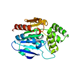
Crystal structure of human NDRG2 protein provides insight into its role as a tumor suppressor
Descriptor: ACETATE ION, PROTEIN NDRG2
Authors:Hwang, J, Kim, Y, Lee, H, Kim, M.H.
Deposit date:2010-07-29
Release date:2011-01-19
Last modified:2023-12-20
Method:X-RAY DIFFRACTION (2.81 Å)
Cite:Crystal Structure of Human Ndrg2 Protein Provides Insight Into its Role as a Tumor Suppressor.
J.Biol.Chem., 286, 2011
2V1V
DownloadVisualize
BU of 2v1v by Molmil
3D STRUCTURE OF THE M8L MUTANT OF SQUASH TRYPSIN INHIBITOR CMTI-I
Descriptor: TRYPSIN INHIBITOR 1
Authors:Sledz, P, Bolewska, K, Bierzynski, A, Zhukov, I.
Deposit date:2007-05-30
Release date:2007-06-19
Last modified:2019-10-09
Method:SOLUTION NMR
Cite:Conservative mutation Met8 --> Leu affects the folding process and structural stability of squash trypsin inhibitor CMTI-I.
Protein Sci., 9, 2000

222624

PDB entries from 2024-07-17

PDB statisticsPDBj update infoContact PDBjnumon