Loading
PDBj
MenuPDBj@FacebookPDBj@TwitterPDBj@YouTubewwPDB FoundationwwPDB
RCSB PDBPDBeBMRBAdv. SearchSearch help
Search by PDB author
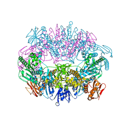
3N58
DownloadVisualize
BU of 3n58 by Molmil
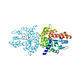
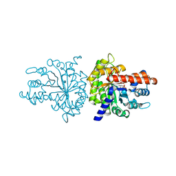
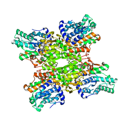
Crystal structure of S-ADENOSYL-L-HOMOCYSTEINE hydrolase from brucella melitensis in ternary complex with NAD and adenosine, orthorhombic form
Descriptor: ADENOSINE, Adenosylhomocysteinase, NICOTINAMIDE-ADENINE-DINUCLEOTIDE, ...
Authors:Seattle Structural Genomics Center for Infectious Disease (SSGCID)
Deposit date:2010-05-24
Release date:2010-06-02
Last modified:2023-09-06
Method:X-RAY DIFFRACTION (2.39 Å)
Cite:Crystal structure of S-adenosyl-L-homocysteine hydrolase from brucella melitensis in ternary complex with NAD and adenosine, orthorhombic form
To be Published
6O3V
DownloadVisualize
BU of 6o3v by Molmil
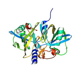
Crystal structure for RVA-VP3
Descriptor: 1,2-ETHANEDIOL, GUANOSINE-5'-MONOPHOSPHATE, Protein VP3, ...
Authors:Kumar, D, Yu, X, Wang, Z, Hu, L, Prasad, V.
Deposit date:2019-02-27
Release date:2020-03-04
Last modified:2024-03-13
Method:X-RAY DIFFRACTION (3.5 Å)
Cite:2.7 angstrom cryo-EM structure of rotavirus core protein VP3, a unique capping machine with a helicase activity.
Sci Adv, 6, 2020
6O01
DownloadVisualize
BU of 6o01 by Molmil
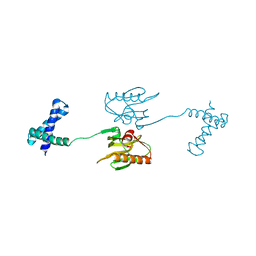
X-ray structure of H5N1-NS1 R38A K41A G71E mutant
Descriptor: Non-structural protein 1
Authors:Mitra, S, Kumar, D, Hu, L, Prasad, B.V.V.
Deposit date:2019-02-14
Release date:2019-07-31
Last modified:2023-10-11
Method:X-RAY DIFFRACTION (3 Å)
Cite:Influenza A Virus Protein NS1 Exhibits Strain-Independent Conformational Plasticity.
J.Virol., 93, 2019
6NQ4
DownloadVisualize
BU of 6nq4 by Molmil
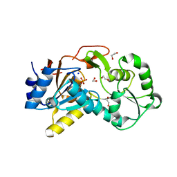
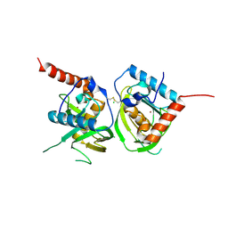
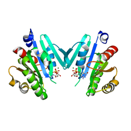
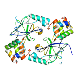
Crystal structure of a Hydrolase, haloacid dehalogenase-like family from Brucella suis 1330
Descriptor: 1,2-ETHANEDIOL, HAD superfamily hydrolase, PHOSPHATE ION, ...
Authors:Seattle Structural Genomics Center for Infectious Disease (SSGCID)
Deposit date:2019-01-19
Release date:2019-11-27
Last modified:2024-04-03
Method:X-RAY DIFFRACTION (1.45 Å)
Cite:Crystal structure of a Hydrolase, haloacid dehalogenase-like family from Brucella suis 1330
to be published
6NRL
DownloadVisualize
BU of 6nrl by Molmil
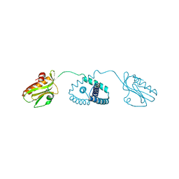
X-ray structure of H6N6-NS1 delta(80-84) R38A K41A E71G mutant
Descriptor: Non-structural protein 1
Authors:Mitra, S, Kumar, D, Hu, L, Prasad, B.V.V.
Deposit date:2019-01-23
Release date:2019-07-31
Last modified:2023-10-11
Method:X-RAY DIFFRACTION (3.2 Å)
Cite:Influenza A Virus Protein NS1 Exhibits Strain-Independent Conformational Plasticity.
J.Virol., 93, 2019
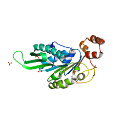
3MBD
DownloadVisualize
BU of 3mbd by Molmil
Crystal structure of fructose bisphosphate aldolase from Encephalitozoon cuniculi, bound to phosphate
Descriptor: CHLORIDE ION, Fructose-bisphosphate aldolase, PHOSPHATE ION
Authors:Seattle Structural Genomics Center for Infectious Disease (SSGCID)
Deposit date:2010-03-25
Release date:2010-04-14
Last modified:2023-09-06
Method:X-RAY DIFFRACTION (2 Å)
Cite:Structure of fructose bisphosphate aldolase from Encephalitozoon cuniculi.
Acta Crystallogr.,Sect.F, 67, 2011
3NFW
DownloadVisualize
BU of 3nfw by Molmil
Crystal structure of nitrilotriacetate monooxygenase component B (A0R521 homolog) from Mycobacterium thermoresistibile
Descriptor: Flavin reductase-like, FMN-binding protein, GLYCEROL
Authors:Seattle Structural Genomics Center for Infectious Disease (SSGCID)
Deposit date:2010-06-10
Release date:2010-06-23
Last modified:2023-09-06
Method:X-RAY DIFFRACTION (1.6 Å)
Cite:Structure of nitrilotriacetate monooxygenase component B from Mycobacterium thermoresistibile.
Acta Crystallogr.,Sect.F, 67, 2011
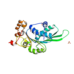
3MBF
DownloadVisualize
BU of 3mbf by Molmil
Crystal structure of fructose bisphosphate aldolase from Encephalitozoon cuniculi, bound to fructose 1,6-bisphosphate
Descriptor: 1,6-FRUCTOSE DIPHOSPHATE (LINEAR FORM), Fructose-bisphosphate aldolase
Authors:Seattle Structural Genomics Center for Infectious Disease (SSGCID)
Deposit date:2010-03-25
Release date:2010-04-07
Last modified:2023-09-06
Method:X-RAY DIFFRACTION (2.37 Å)
Cite:Structure of fructose bisphosphate aldolase from Encephalitozoon cuniculi.
Acta Crystallogr.,Sect.F, 67, 2011
3OCA
DownloadVisualize
BU of 3oca by Molmil
Crystal structure of peptide deformylase from Ehrlichia chaffeensis
Descriptor: CHLORIDE ION, Peptide deformylase, ZINC ION
Authors:Seattle Structural Genomics Center for Infectious Disease (SSGCID)
Deposit date:2010-08-09
Release date:2010-09-08
Last modified:2024-10-16
Method:X-RAY DIFFRACTION (2.4 Å)
Cite:Crystal structure of peptide deformylase from Ehrlichia chaffeensis
To be Published
4YG0
DownloadVisualize
BU of 4yg0 by Molmil
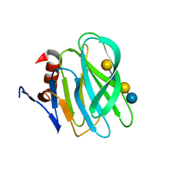
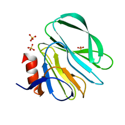
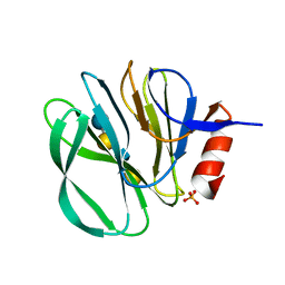
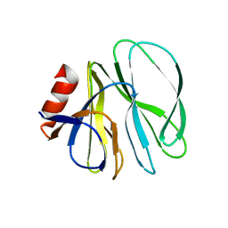
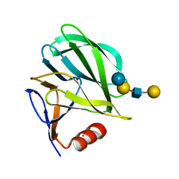
Structural basis of glycan recognition in neonate-specific rotaviruses
Descriptor: Outer capsid protein VP4, beta-D-galactopyranose-(1-4)-2-acetamido-2-deoxy-beta-D-glucopyranose-(1-3)-beta-D-galactopyranose-(1-4)-beta-D-glucopyranose
Authors:Hu, L, Prasad, B.V.V.
Deposit date:2015-02-25
Release date:2015-09-30
Last modified:2023-09-27
Method:X-RAY DIFFRACTION (1.285 Å)
Cite:Structural basis of glycan specificity in neonate-specific bovine-human reassortant rotavirus.
Nat Commun, 6, 2015
4YG3
DownloadVisualize
BU of 4yg3 by Molmil
Structural basis of glycan recognition in neonate-specific rotaviruses
Descriptor: Outer capsid protein VP4, SULFATE ION
Authors:Hu, L, Prasad, B.V.V.
Deposit date:2015-02-25
Release date:2015-09-30
Last modified:2023-09-27
Method:X-RAY DIFFRACTION (2.285 Å)
Cite:Structural basis of glycan specificity in neonate-specific bovine-human reassortant rotavirus.
Nat Commun, 6, 2015
4YG6
DownloadVisualize
BU of 4yg6 by Molmil
Structural basis of glycan recognition in neonate-specific rotaviruses
Descriptor: Outer capsid protein VP8*, PHOSPHATE ION, beta-D-galactopyranose-(1-4)-2-acetamido-2-deoxy-beta-D-glucopyranose, ...
Authors:Hu, L, Prasad, B.V.V.
Deposit date:2015-02-25
Release date:2015-09-30
Last modified:2023-09-27
Method:X-RAY DIFFRACTION (1.46 Å)
Cite:Structural basis of glycan specificity in neonate-specific bovine-human reassortant rotavirus.
Nat Commun, 6, 2015
4YFW
DownloadVisualize
BU of 4yfw by Molmil
Structural basis of glycan recognition in neonate-specific rotaviruses
Descriptor: Outer capsid protein VP4
Authors:Hu, L, Prasad, B.V.V.
Deposit date:2015-02-25
Release date:2015-09-30
Last modified:2023-09-27
Method:X-RAY DIFFRACTION (1.66 Å)
Cite:Structural basis of glycan specificity in neonate-specific bovine-human reassortant rotavirus.
Nat Commun, 6, 2015
4YLG
DownloadVisualize
BU of 4ylg by Molmil
Structure of an ADP ribosylation factor from Entamoeba histolytica HM-1:IMSS bound to Mg-GDP
Descriptor: ADP-ribosylation factor, GUANOSINE-5'-DIPHOSPHATE, MAGNESIUM ION
Authors:Seattle Structural Genomics Center for Infectious Disease, Seattle Structural Genomics Center for Infectious Disease (SSGCID)
Deposit date:2015-03-05
Release date:2015-05-20
Last modified:2023-09-27
Method:X-RAY DIFFRACTION (1.8 Å)
Cite:Structure of an ADP-ribosylation factor, ARF1, from Entamoeba histolytica bound to Mg(2+)-GDP.
Acta Crystallogr.,Sect.F, 71, 2015
6VFJ
DownloadVisualize
BU of 6vfj by Molmil
De novo designed icosahedral nanoparticle I53_dn5
Descriptor: I53_dn5A, I53_dn5B
Authors:Antanasijevic, A, Ward, A.B.
Deposit date:2020-01-05
Release date:2020-08-12
Last modified:2024-03-06
Method:ELECTRON MICROSCOPY (5.35 Å)
Cite:Tailored design of protein nanoparticle scaffolds for multivalent presentation of viral glycoprotein antigens.
Elife, 9, 2020
6VFH
DownloadVisualize
BU of 6vfh by Molmil
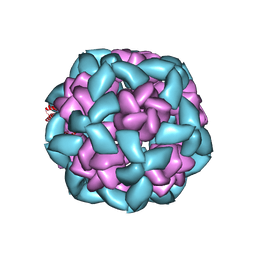
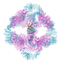
De novo designed tetrahedral nanoparticle T33_dn10
Descriptor: T33_dn10A, T33_dn10B
Authors:Antanasijevic, A, Ward, A.B.
Deposit date:2020-01-05
Release date:2020-08-12
Last modified:2024-03-06
Method:ELECTRON MICROSCOPY (3.86 Å)
Cite:Tailored design of protein nanoparticle scaffolds for multivalent presentation of viral glycoprotein antigens.
Elife, 9, 2020
6VFI
DownloadVisualize
BU of 6vfi by Molmil
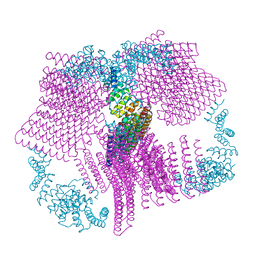
De novo designed octahedral nanoparticle O43_dn18
Descriptor: O43_dn18A, O43_dn18B
Authors:Antanasijevic, A, Ward, A.B.
Deposit date:2020-01-05
Release date:2020-08-12
Last modified:2024-10-23
Method:ELECTRON MICROSCOPY (4.54 Å)
Cite:Tailored design of protein nanoparticle scaffolds for multivalent presentation of viral glycoprotein antigens.
Elife, 9, 2020
6UVU
DownloadVisualize
BU of 6uvu by Molmil
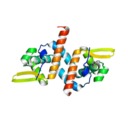
Crystal structure of the AntR antimony-specific transcriptional repressor
Descriptor: ArsR family transcriptional regulator
Authors:Thiruselvam, V, Banumathi, S, Palani, K, Manohar, R, Rosen, B.P.
Deposit date:2019-11-04
Release date:2020-11-04
Last modified:2023-10-11
Method:X-RAY DIFFRACTION (2.1 Å)
Cite:Functional and structural characterization of AntR, an Sb(III) responsive transcriptional repressor.
Mol.Microbiol., 116, 2021
4ZNJ
DownloadVisualize
BU of 4znj by Molmil
Thermus Phage P74-26 Large Terminase ATPase domain mutant R139A (I 2 3 space group)
Descriptor: Phage terminase large subunit, SULFATE ION
Authors:Hilbert, B.J, Hayes, J.A, Stone, N.P, Duffy, C.M, Kelch, B.A.
Deposit date:2015-05-04
Release date:2015-07-08
Last modified:2023-09-27
Method:X-RAY DIFFRACTION (2.532 Å)
Cite:Structure and mechanism of the ATPase that powers viral genome packaging.
Proc.Natl.Acad.Sci.USA, 112, 2015
4ZNK
DownloadVisualize
BU of 4znk by Molmil
Thermus Phage P74-26 Large Terminase ATPase domain from (P 32 2 1 space group)
Descriptor: Phage terminase large subunit, SULFATE ION
Authors:Hilbert, B.J, Hayes, J.A, Stone, N.P, Duffy, C.M, Kelch, B.A.
Deposit date:2015-05-04
Release date:2015-07-08
Last modified:2023-09-27
Method:X-RAY DIFFRACTION (1.931 Å)
Cite:Structure and mechanism of the ATPase that powers viral genome packaging.
Proc.Natl.Acad.Sci.USA, 112, 2015
6VH5
DownloadVisualize
BU of 6vh5 by Molmil
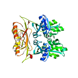
Crystal structure of prephenate dehydratase from brucella melitensis biovar abortus 2308 in complex with phenylalanine
Descriptor: 1,2-ETHANEDIOL, PHENYLALANINE, Prephenate dehydratase:Amino acid-binding ACT
Authors:Seattle Structural Genomics Center for Infectious Disease (SSGCID)
Deposit date:2020-01-09
Release date:2020-01-29
Last modified:2024-04-03
Method:X-RAY DIFFRACTION (2.4 Å)
Cite:Crystal structure of prephenate dehydratase from Brucella melitensis biovar abortus 2308 in complex with phenylalanine
to be published
6VEH
DownloadVisualize
BU of 6veh by Molmil
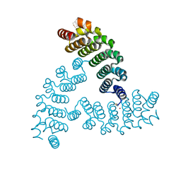
Computationally designed C3-symmetric homotrimer from HEAT repeat protein
Descriptor: HEAT repeat domain-containing protein
Authors:Bick, M.J, Ueda, G, Baker, D.
Deposit date:2020-01-02
Release date:2020-08-05
Last modified:2024-04-03
Method:X-RAY DIFFRACTION (2.303 Å)
Cite:Tailored design of protein nanoparticle scaffolds for multivalent presentation of viral glycoprotein antigens.
Elife, 9, 2020
4YFZ
DownloadVisualize
BU of 4yfz by Molmil
Structural basis of glycan recognition in neonate-specific rotaviruses
Descriptor: Outer capsid protein VP4, beta-D-galactopyranose-(1-3)-2-acetamido-2-deoxy-beta-D-glucopyranose-(1-3)-beta-D-galactopyranose-(1-4)-beta-D-glucopyranose
Authors:Hu, L, Prasad, B.V.V.
Deposit date:2015-02-25
Release date:2015-09-30
Last modified:2023-09-27
Method:X-RAY DIFFRACTION (1.5 Å)
Cite:Structural basis of glycan specificity in neonate-specific bovine-human reassortant rotavirus.
Nat Commun, 6, 2015
7JFL
DownloadVisualize
BU of 7jfl by Molmil
Crystal structure of human phosphorylated IRF-3 bound to CBP
Descriptor: CREB-binding protein, Interferon regulatory factor 3
Authors:Li, P, Jing, T, Zhao, B.
Deposit date:2020-07-17
Release date:2020-09-09
Last modified:2023-10-18
Method:X-RAY DIFFRACTION (1.68 Å)
Cite:The Structural Basis of IRF-3 Activation upon Phosphorylation.
J Immunol., 205, 2020
6VYJ
DownloadVisualize
BU of 6vyj by Molmil
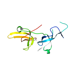
Human UHRF1 TTD domain in complex with a fragment
Descriptor: 2,4-dimethylpyridine, E3 ubiquitin-protein ligase UHRF1, beta-D-glucopyranose
Authors:Campbell, J.C, Chang, L, Young, D.W.
Deposit date:2020-02-26
Release date:2021-01-27
Last modified:2023-10-11
Method:X-RAY DIFFRACTION (1.39 Å)
Cite:Discovery of small molecules targeting the tandem tudor domain of the epigenetic factor UHRF1 using fragment-based ligand discovery.
Sci Rep, 11, 2021

226707

PDB entries from 2024-10-30

PDB statisticsPDBj update infoContact PDBjnumon