4HUM
 
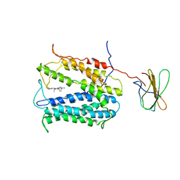 | | MATE transporter NorM-NG in complex with ethidium and monobody | | Descriptor: | ETHIDIUM, Multidrug efflux protein, Protein B | | Authors: | Lu, M. | | Deposit date: | 2012-11-02 | | Release date: | 2013-02-06 | | Last modified: | 2024-02-28 | | Method: | X-RAY DIFFRACTION (3.49 Å) | | Cite: | Structures of a Na+-coupled, substrate-bound MATE multidrug transporter.
Proc.Natl.Acad.Sci.USA, 110, 2013
|
|
3F7Q
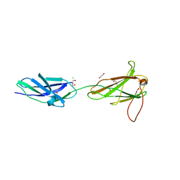 | |
5MYB
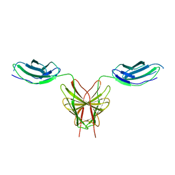 | | Homodimerization of Tie2 Fibronectin-like domains 2 and 3 in space group P21 | | Descriptor: | 2-acetamido-2-deoxy-beta-D-glucopyranose, 2-acetamido-2-deoxy-beta-D-glucopyranose-(1-4)-2-acetamido-2-deoxy-beta-D-glucopyranose, Angiopoietin-1 receptor, ... | | Authors: | Leppanen, V.-M, Saharinen, P, Alitalo, K. | | Deposit date: | 2017-01-26 | | Release date: | 2017-04-26 | | Last modified: | 2024-05-01 | | Method: | X-RAY DIFFRACTION (2.6 Å) | | Cite: | Structural basis of Tie2 activation and Tie2/Tie1 heterodimerization.
Proc. Natl. Acad. Sci. U.S.A., 114, 2017
|
|
5MYA
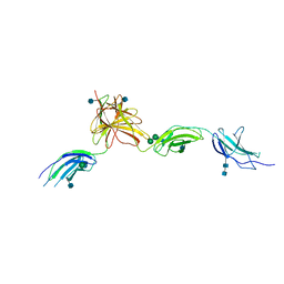 | | Homodimerization of Tie2 Fibronectin-like domains 1-3 in space group C2 | | Descriptor: | 2-acetamido-2-deoxy-beta-D-glucopyranose, 2-acetamido-2-deoxy-beta-D-glucopyranose-(1-4)-2-acetamido-2-deoxy-beta-D-glucopyranose, Angiopoietin-1 receptor, ... | | Authors: | Leppanen, V.-M, Saharinen, P, Alitalo, K. | | Deposit date: | 2017-01-26 | | Release date: | 2017-04-26 | | Last modified: | 2020-07-29 | | Method: | X-RAY DIFFRACTION (2.9 Å) | | Cite: | Structural basis of Tie2 activation and Tie2/Tie1 heterodimerization.
Proc. Natl. Acad. Sci. U.S.A., 114, 2017
|
|
5M0A
 | |
5N06
 | |
3F7R
 | |
6MFA
 | |
6MSV
 | |
5E4S
 | |
2VKX
 | | Human NCAM, FN3 domains 1 and 2, M610R mutant | | Descriptor: | NEURAL CELL ADHESION MOLECULE, SULFATE ION | | Authors: | Carafoli, F, Saffell, J.L, Hohenester, E. | | Deposit date: | 2008-01-04 | | Release date: | 2008-02-26 | | Last modified: | 2024-05-01 | | Method: | X-RAY DIFFRACTION (2.7 Å) | | Cite: | Structure of the Tandem Fibronectin Type 3 Domains of Neural Cell Adhesion Molecule
J.Mol.Biol., 377, 2008
|
|
1WK0
 | | Solution structure of Fibronectin type III domain derived from human KIAA0970 protein | | Descriptor: | KIAA0970 protein | | Authors: | Kobayashi, N, Koshiba, S, Inoue, M, Hayashi, F, Kigawa, T, Yokoyama, S, RIKEN Structural Genomics/Proteomics Initiative (RSGI) | | Deposit date: | 2004-05-29 | | Release date: | 2004-11-29 | | Last modified: | 2024-05-29 | | Method: | SOLUTION NMR | | Cite: | Solution structure of Fibronectin type III domain derived from human KIAA0970 protein
To be Published
|
|
1X5I
 | | The solution structure of the fourth fibronectin type III domain of human Neogenin | | Descriptor: | Neogenin | | Authors: | Tochio, N, Koshiba, S, Inoue, M, Kigawa, T, Yokoyama, S, RIKEN Structural Genomics/Proteomics Initiative (RSGI) | | Deposit date: | 2005-05-15 | | Release date: | 2005-11-15 | | Last modified: | 2024-05-29 | | Method: | SOLUTION NMR | | Cite: | The solution structure of the fourth fibronectin type III domain of human Neogenin
To be Published
|
|
1X5Y
 | | Solution structure of the fibronectin type-III domain of mouse myosin-binding protein C, Fast-type homolog | | Descriptor: | myosin binding protein C, fast-type | | Authors: | Yoneyama, M, Tochio, N, Koshiba, S, Inoue, M, Kigawa, T, Yokoyama, S, RIKEN Structural Genomics/Proteomics Initiative (RSGI) | | Deposit date: | 2005-05-17 | | Release date: | 2005-11-17 | | Last modified: | 2024-05-29 | | Method: | SOLUTION NMR | | Cite: | Solution structure of the fibronectin type-III domain of mouse myosin-binding protein C, Fast-type homolog
To be Published
|
|
5E52
 | | Crystal structure of human CNTN5 FN1-FN3 domains | | Descriptor: | Contactin-5, PHOSPHATE ION | | Authors: | Nikolaienko, R.M, Bouyain, S. | | Deposit date: | 2015-10-07 | | Release date: | 2016-08-31 | | Last modified: | 2023-09-27 | | Method: | X-RAY DIFFRACTION (2.685 Å) | | Cite: | Structural Basis for Interactions Between Contactin Family Members and Protein-tyrosine Phosphatase Receptor Type G in Neural Tissues.
J.Biol.Chem., 291, 2016
|
|
1X5A
 | | The solution structure of the second fibronectin type III domain of mouse Ephrin type-A receptor 1 | | Descriptor: | Ephrin type-A receptor 1 | | Authors: | Tochio, N, Sasagawa, A, Koshiba, S, Inoue, M, Kigawa, T, Yokoyama, S, RIKEN Structural Genomics/Proteomics Initiative (RSGI) | | Deposit date: | 2005-05-15 | | Release date: | 2005-11-15 | | Last modified: | 2024-05-29 | | Method: | SOLUTION NMR | | Cite: | The solution structure of the second fibronectin type III domain of mouse Ephrin type-A receptor 1
To be Published
|
|
1X5J
 | | The solution structure of the fifth fibronectin type III domain of human Neogenin | | Descriptor: | Neogenin | | Authors: | Tochio, N, Sasagawa, A, Koshiba, S, Inoue, M, Kigawa, T, Yokoyama, S, RIKEN Structural Genomics/Proteomics Initiative (RSGI) | | Deposit date: | 2005-05-15 | | Release date: | 2005-11-15 | | Last modified: | 2024-05-29 | | Method: | SOLUTION NMR | | Cite: | The solution structure of the fifth fibronectin type III domain of human Neogenin
To be Published
|
|
1X5Z
 | | Solution structure of the fibronectin type-III domain of human protein tyrosine phosphatase, receptor type, D isoform 4 variant | | Descriptor: | Receptor-type tyrosine-protein phosphatase delta | | Authors: | Yoneyama, M, Saito, K, Tochio, N, Koshiba, S, Inoue, M, Kigawa, T, Yokoyama, S, RIKEN Structural Genomics/Proteomics Initiative (RSGI) | | Deposit date: | 2005-05-17 | | Release date: | 2005-11-17 | | Last modified: | 2024-05-29 | | Method: | SOLUTION NMR | | Cite: | Solution structure of the fibronectin type-III domain of human protein tyrosine phosphatase, receptor type, D isoform 4 variant
To be Published
|
|
2VKW
 | | Human NCAM, FN3 domains 1 and 2 | | Descriptor: | NEURAL CELL ADHESION MOLECULE 1,140 KDA ISOFORM, SULFATE ION | | Authors: | Carafoli, F, Saffell, J.L, Hohenester, E. | | Deposit date: | 2008-01-04 | | Release date: | 2008-02-26 | | Last modified: | 2024-05-08 | | Method: | X-RAY DIFFRACTION (2.3 Å) | | Cite: | Structure of the Tandem Fibronectin Type 3 Domains of Neural Cell Adhesion Molecule
J.Mol.Biol., 377, 2008
|
|
5FM8
 | |
3WIH
 | | Crystal structure of the third fibronectin domain (Fn3) of human ROBO1 in complex with the Fab fragment of murine monoclonal antibody B2212A. | | Descriptor: | GLYCEROL, Roundabout homolog 1, anti-human ROBO1 antibody B2212A Fab heavy chain, ... | | Authors: | Nakayama, T, Mizohata, E, Yamashita, T, Nagatoishi, M, Iwanari, H, Mochizuki, Y, Kado, Y, Yokota, Y, Sato, R, Tsumoto, K, Fujitani, H, Kodama, T, Hamakubo, T, Inoue, T. | | Deposit date: | 2013-09-12 | | Release date: | 2015-01-21 | | Last modified: | 2023-11-08 | | Method: | X-RAY DIFFRACTION (1.701 Å) | | Cite: | Structural features of interfacial tyrosine residue in ROBO1 fibronectin domain-antibody complex: Crystallographic, thermodynamic, and molecular dynamic analyses
Protein Sci., 24, 2015
|
|
5FN8
 | | Crystal structure of rat CD45 extracellular region, domains d3-d4 | | Descriptor: | 2-acetamido-2-deoxy-beta-D-glucopyranose, CITRATE ANION, RECEPTOR-TYPE TYROSINE-PROTEIN PHOSPHATASE C | | Authors: | Chang, V.T, Fernandes, R.A, Ganzinger, K.A, Lee, S.F, Siebold, C, McColl, J, Jonsson, P, Palayret, M, Harlos, K, Coles, C.H, Jones, E.Y, Lui, Y, Huang, E, Gilbert, R.J.C, Klenerman, D, Aricescu, A.R, Davis, S.J. | | Deposit date: | 2015-11-11 | | Release date: | 2016-03-23 | | Last modified: | 2020-07-29 | | Method: | X-RAY DIFFRACTION (2.45 Å) | | Cite: | Initiation of T Cell Signaling by Cd45 Segregation at 'Close Contacts'.
Nat.Immunol., 17, 2016
|
|
5FM4
 | |
5DFT
 | |
5E7L
 | |


