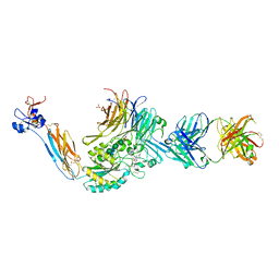
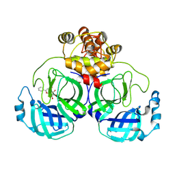
7U9V
 
 | | Integrin alpha IIB beta3 complex with BMS4-1 | | 分子名称: | (4-{[(5S)-3-(4-carbamimidoylphenyl)-4,5-dihydro-1,2-oxazol-5-yl]methyl}piperazin-1-yl)acetic acid, 10E5 Fab heavy chain, 10E5 light chain, ... | | 著者 | Zhu, J, Lin, F.-Y, Zhu, J, Springer, T.A. | | 登録日 | 2022-03-11 | | 公開日 | 2022-08-17 | | 最終更新日 | 2024-10-02 | | 実験手法 | X-RAY DIFFRACTION (2.25492157575 Å) | | 主引用文献 | A general chemical principle for creating closure-stabilizing integrin inhibitors.
Cell, 185, 2022
|
|
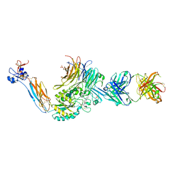
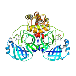
7UBR
 | | Integrin alpha IIB beta3 complex with GR144053 | | 分子名称: | 10E5 Fab heavy chain, 10E5 Fab light chain, 2-acetamido-2-deoxy-beta-D-glucopyranose, ... | | 著者 | Lin, F.-Y, Zhu, J, Zhu, J, Springer, T.A. | | 登録日 | 2022-03-15 | | 公開日 | 2022-08-17 | | 最終更新日 | 2024-10-02 | | 実験手法 | X-RAY DIFFRACTION (2.04992751372 Å) | | 主引用文献 | A general chemical principle for creating closure-stabilizing integrin inhibitors.
Cell, 185, 2022
|
|
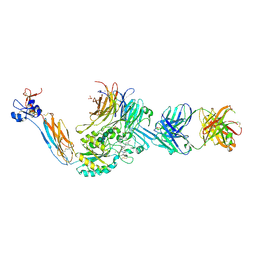
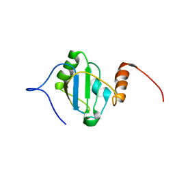
7UCY
 | | Integrin alpha IIB beta3 complex with gantofiban | | 分子名称: | (4-{[(5R)-3-(4-carbamimidoylphenyl)-2-oxo-1,3-oxazolidin-5-yl]methyl}piperazin-1-yl)acetic acid, 10E5 Fab heavy chain, 10E5 Fab light chain, ... | | 著者 | Lin, F.-Y, Zhu, J, Zhu, J, Springer, T.A. | | 登録日 | 2022-03-17 | | 公開日 | 2022-08-17 | | 最終更新日 | 2024-10-02 | | 実験手法 | X-RAY DIFFRACTION (2.34996314573 Å) | | 主引用文献 | A general chemical principle for creating closure-stabilizing integrin inhibitors.
Cell, 185, 2022
|
|
7UDG
 | | Integrin alpha IIB beta3 complex with lotrafiban | | 分子名称: | 10E5 Fab heavy chain, 10E5 Fab light chain, 2-acetamido-2-deoxy-beta-D-glucopyranose, ... | | 著者 | Lin, F.-Y, Zhu, J, Zhu, J, Springer, T.A. | | 登録日 | 2022-03-19 | | 公開日 | 2022-08-17 | | 最終更新日 | 2024-10-02 | | 実験手法 | X-RAY DIFFRACTION (2.80006901797 Å) | | 主引用文献 | A general chemical principle for creating closure-stabilizing integrin inhibitors.
Cell, 185, 2022
|
|
7UDH
 | | Integrin alpha IIB beta3 complex with BMS4-3 | | 分子名称: | (4-{[(5S)-3-(piperidin-4-yl)-4,5-dihydro-1,2-oxazol-5-yl]methyl}piperazin-1-yl)acetic acid, 10E5 Fab heavy chain, 10E5 Fab light chain, ... | | 著者 | Lin, F.-Y, Zhu, J, Zhu, J, Springer, T.A. | | 登録日 | 2022-03-19 | | 公開日 | 2022-08-17 | | 最終更新日 | 2024-10-02 | | 実験手法 | X-RAY DIFFRACTION (1.99998841007 Å) | | 主引用文献 | A general chemical principle for creating closure-stabilizing integrin inhibitors.
Cell, 185, 2022
|
|
7UE0
 | | Integrin alpha IIB beta3 complex with fradafiban | | 分子名称: | 10E5 Fab heavy chain, 10E5 Fab light chain, 2-acetamido-2-deoxy-beta-D-glucopyranose, ... | | 著者 | Lin, F.-Y, Zhu, J, Zhu, J, Springer, T.A. | | 登録日 | 2022-03-20 | | 公開日 | 2022-08-17 | | 最終更新日 | 2024-10-02 | | 実験手法 | X-RAY DIFFRACTION (2.74196193977 Å) | | 主引用文献 | A general chemical principle for creating closure-stabilizing integrin inhibitors.
Cell, 185, 2022
|
|
7UFH
 | | Integrin alpha IIB beta3 complex with fradafiban (Mn/Ca) | | 分子名称: | 10E5 Fab heavy chain, 10E5 Fab light chain, 2-acetamido-2-deoxy-beta-D-glucopyranose, ... | | 著者 | Lin, F.-Y, Zhu, J, Zhu, J, Springer, T.A. | | 登録日 | 2022-03-22 | | 公開日 | 2022-08-17 | | 最終更新日 | 2024-10-02 | | 実験手法 | X-RAY DIFFRACTION (2.99768170407 Å) | | 主引用文献 | A general chemical principle for creating closure-stabilizing integrin inhibitors.
Cell, 185, 2022
|
|
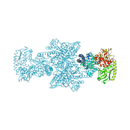
7UGW
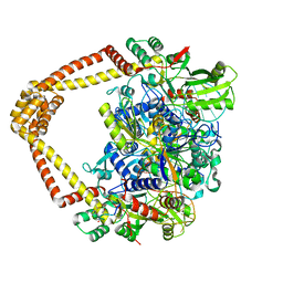 | | M. tuberculosis DNA gyrase cleavage core bound to DNA and evybactin | | 分子名称: | DNA (46-MER), DNA gyrase subunit A, DNA gyrase subunit B, ... | | 著者 | Hauk, G, Imai, Y, Lewis, K, Berger, J.M. | | 登録日 | 2022-03-25 | | 公開日 | 2022-08-17 | | 最終更新日 | 2024-10-02 | | 実験手法 | X-RAY DIFFRACTION (3 Å) | | 主引用文献 | Evybactin is a DNA gyrase inhibitor that selectively kills Mycobacterium tuberculosis.
Nat.Chem.Biol., 18, 2022
|
|
7UH8
 | | Integrin alpha IIB beta3 complex with roxifiban (Mn/Ca) | | 分子名称: | 10E5 Fab heavy chain, 10E5 Fab light chain, 2-acetamido-2-deoxy-beta-D-glucopyranose, ... | | 著者 | Lin, F.-Y, Zhu, J, Zhu, J, Springer, T.A. | | 登録日 | 2022-03-25 | | 公開日 | 2022-08-17 | | 最終更新日 | 2024-10-02 | | 実験手法 | X-RAY DIFFRACTION (2.75002413567 Å) | | 主引用文献 | A general chemical principle for creating closure-stabilizing integrin inhibitors.
Cell, 185, 2022
|
|
7UJE
 | | Integrin alpha IIB beta3 complex with UR2922 in Mn2+ | | 分子名称: | 2-acetamido-2-deoxy-beta-D-glucopyranose, 2-acetamido-2-deoxy-beta-D-glucopyranose-(1-4)-2-acetamido-2-deoxy-beta-D-glucopyranose, CALCIUM ION, ... | | 著者 | Lin, F.-Y, Zhu, J, Zhu, J, Springer, T.A. | | 登録日 | 2022-03-30 | | 公開日 | 2022-08-17 | | 最終更新日 | 2024-10-02 | | 実験手法 | X-RAY DIFFRACTION (2.4999681528 Å) | | 主引用文献 | A general chemical principle for creating closure-stabilizing integrin inhibitors.
Cell, 185, 2022
|
|
7UJK
 | | Integrin alpha IIB beta3 complex with lamifiban | | 分子名称: | 10E5 Fab heavy chain, 10E5 Fab light chain, 2-acetamido-2-deoxy-beta-D-glucopyranose, ... | | 著者 | Lin, F.-Y, Zhu, J, Zhu, J, Springer, T.A. | | 登録日 | 2022-03-30 | | 公開日 | 2022-08-17 | | 最終更新日 | 2024-10-02 | | 実験手法 | X-RAY DIFFRACTION (2.43265583434 Å) | | 主引用文献 | A general chemical principle for creating closure-stabilizing integrin inhibitors.
Cell, 185, 2022
|
|
7UK9
 | | Integrin alpha IIB beta3 complex with lamifiban (Mn) | | 分子名称: | 10E5 Fab heavy chain, 10E5 Fab light chain, 2-acetamido-2-deoxy-beta-D-glucopyranose, ... | | 著者 | Lin, F.-Y, Zhu, J, Zhu, J, Springer, T.A. | | 登録日 | 2022-03-31 | | 公開日 | 2022-08-17 | | 最終更新日 | 2024-10-02 | | 実験手法 | X-RAY DIFFRACTION (2.60001966299 Å) | | 主引用文献 | A general chemical principle for creating closure-stabilizing integrin inhibitors.
Cell, 185, 2022
|
|
7UKO
 | | Integrin alpha IIB beta3 complex with sibrafiban (Mn) | | 分子名称: | 10E5 Fab heavy chain, 10E5 Fab light chain, 2-acetamido-2-deoxy-beta-D-glucopyranose, ... | | 著者 | Lin, F.-Y, Zhu, J, Zhu, J, Springer, T.A. | | 登録日 | 2022-04-01 | | 公開日 | 2022-08-17 | | 最終更新日 | 2024-10-02 | | 実験手法 | X-RAY DIFFRACTION (2.604 Å) | | 主引用文献 | A general chemical principle for creating closure-stabilizing integrin inhibitors.
Cell, 185, 2022
|
|
7UKP
 | | Integrin alpha IIB beta3 complex with a gantofiban analog | | 分子名称: | 10E5 Fab heavy chain, 10E5 Fab light chain, 2-acetamido-2-deoxy-beta-D-glucopyranose, ... | | 著者 | Lin, F.-Y, Zhu, J, Zhu, J, Springer, T.A. | | 登録日 | 2022-04-01 | | 公開日 | 2022-08-17 | | 最終更新日 | 2024-10-02 | | 実験手法 | X-RAY DIFFRACTION (2.80112979054 Å) | | 主引用文献 | A general chemical principle for creating closure-stabilizing integrin inhibitors.
Cell, 185, 2022
|
|
7UKT
 | | Integrin alpha IIB beta3 complex with BMS4.2 | | 分子名称: | (1-{[(5S)-3-(4-carbamimidoylphenyl)-4,5-dihydro-1,2-oxazol-5-yl]methyl}piperidin-4-yl)acetic acid, 10E5 Fab heavy chain, 10E5 Fab light chain, ... | | 著者 | Lin, F.-Y, Zhu, J, Zhu, J, Springer, T.A. | | 登録日 | 2022-04-01 | | 公開日 | 2022-08-17 | | 最終更新日 | 2024-10-02 | | 実験手法 | X-RAY DIFFRACTION (2.36994437552 Å) | | 主引用文献 | A general chemical principle for creating closure-stabilizing integrin inhibitors.
Cell, 185, 2022
|
|
7XAX
 | | Crystal structure of SARS coronavirus main protease in complex with Baicalei | | 分子名称: | 3C-like proteinase nsp5, 5,6,7-trihydroxy-2-phenyl-4H-chromen-4-one | | 著者 | Zhou, X.L, Li, J, Zhang, J. | | 登録日 | 2022-03-19 | | 公開日 | 2023-03-22 | | 最終更新日 | 2024-10-02 | | 実験手法 | X-RAY DIFFRACTION (2.25 Å) | | 主引用文献 | Crystal structure of SARS-CoV 3C-like protease with baicalein.
Biochem.Biophys.Res.Commun., 611, 2022
|
|
7XB4
 | | Crystal structure of SARS-Cov-2 main protease D48N mutant in complex with PF07321332 | | 分子名称: | (1R,2S,5S)-N-{(1E,2S)-1-imino-3-[(3S)-2-oxopyrrolidin-3-yl]propan-2-yl}-6,6-dimethyl-3-[3-methyl-N-(trifluoroacetyl)-L-valyl]-3-azabicyclo[3.1.0]hexane-2-carboxamide, Replicase polyprotein 1a | | 著者 | Hu, X.H, Li, J, Zhang, J. | | 登録日 | 2022-03-20 | | 公開日 | 2023-03-22 | | 最終更新日 | 2024-10-02 | | 実験手法 | X-RAY DIFFRACTION (2.07 Å) | | 主引用文献 | Structural Basis for the Inhibition of SARS-CoV-2 M pro D48N Mutant by Shikonin and PF-07321332.
Viruses, 16, 2023
|
|
8BGF
 | |
8CMW
 | | A225L variant of the CODH/ACS complex of C. hydrogenoformans | | 分子名称: | (4S)-2-METHYL-2,4-PENTANEDIOL, ACETATE ION, CO-methylating acetyl-CoA synthase, ... | | 著者 | Ruickoldt, J, Jeoung, J, Lennartz, F, Dobbek, H. | | 登録日 | 2023-02-21 | | 公開日 | 2024-03-06 | | 最終更新日 | 2024-10-02 | | 実験手法 | X-RAY DIFFRACTION (2.6 Å) | | 主引用文献 | Coupling CO 2 Reduction and Acetyl-CoA Formation: The Role of a CO Capturing Tunnel in Enzymatic Catalysis.
Angew.Chem.Int.Ed.Engl., 63, 2024
|
|
8EB5
 | |
8G0I
 | |
8G2K
 | |
8GKJ
 | |
8GKK
 | |
8GKL
 | |


