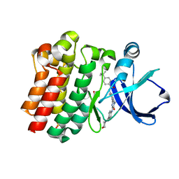
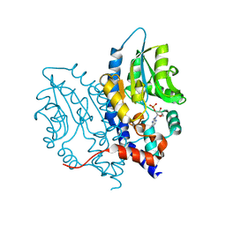
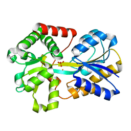
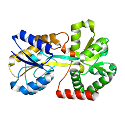
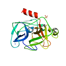
3OCR
 
 | | Crystal structure of aldolase II superfamily protein from Pseudomonas syringae | | 分子名称: | Class II aldolase/adducin domain protein, SULFATE ION | | 著者 | Chang, C, Kagan, O, Savchenko, A, Edwards, A, Joachimiak, A, Midwest Center for Structural Genomics (MCSG) | | 登録日 | 2010-08-10 | | 公開日 | 2010-08-25 | | 最終更新日 | 2024-10-09 | | 実験手法 | X-RAY DIFFRACTION (1.95 Å) | | 主引用文献 | Crystal structure of aldolase II superfamily protein from Pseudomonas syringae
To be Published
|
|
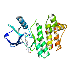
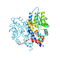
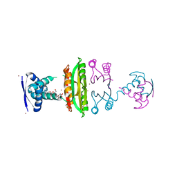
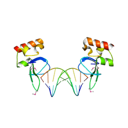
3OCS
 | | Crystal structure of bruton's tyrosine kinase in complex with inhibitor CGI1746 | | 分子名称: | 4-tert-butyl-N-[2-methyl-3-(4-methyl-6-{[4-(morpholin-4-ylcarbonyl)phenyl]amino}-5-oxo-4,5-dihydropyrazin-2-yl)phenyl]benzamide, BETA-MERCAPTOETHANOL, SULFATE ION, ... | | 著者 | Davies, D.R, Gallion, S.L, Staker, B.L. | | 登録日 | 2010-08-10 | | 公開日 | 2010-11-03 | | 最終更新日 | 2023-09-06 | | 実験手法 | X-RAY DIFFRACTION (1.8 Å) | | 主引用文献 | A novel, specific Btk inhibitor antagonizes BCR and Fc[gamma]R signaling and suppresses inflammatory arthritis
To be Published
|
|
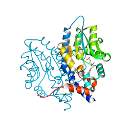
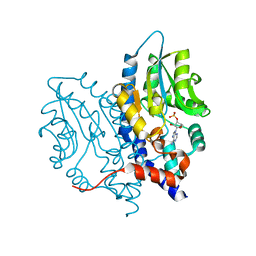
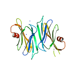
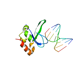
3OCT
 | |
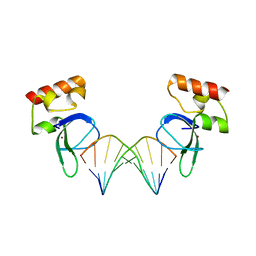
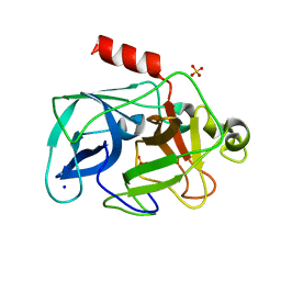
3OCU
 | | Structure of Recombinant Haemophilus Influenzae e(P4) Acid Phosphatase mutant D66N complexed with NMN | | 分子名称: | BETA-NICOTINAMIDE RIBOSE MONOPHOSPHATE, Lipoprotein E, MAGNESIUM ION | | 著者 | Singh, H, Schuermann, J, Reilly, T, Calcutt, M, Tanner, J. | | 登録日 | 2010-08-10 | | 公開日 | 2010-10-20 | | 最終更新日 | 2023-09-06 | | 実験手法 | X-RAY DIFFRACTION (1.35 Å) | | 主引用文献 | Recognition of nucleoside monophosphate substrates by Haemophilus influenzae class C acid phosphatase.
J.Mol.Biol., 404, 2010
|
|
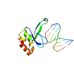
3OCV
 | | Structure of Recombinant Haemophilus Influenzae e(P4) Acid Phosphatase mutant D66N complexed with 5'-AMP | | 分子名称: | ADENOSINE MONOPHOSPHATE, Lipoprotein E, MAGNESIUM ION | | 著者 | Singh, H, Schuermann, J, Reilly, T, Calcutt, M, Tanner, J. | | 登録日 | 2010-08-10 | | 公開日 | 2010-10-20 | | 最終更新日 | 2023-09-06 | | 実験手法 | X-RAY DIFFRACTION (1.551 Å) | | 主引用文献 | Recognition of nucleoside monophosphate substrates by Haemophilus influenzae class C acid phosphatase.
J.Mol.Biol., 404, 2010
|
|
3OCW
 | | Structure of Recombinant Haemophilus influenzae e(P4) Acid Phosphatase mutant D66N complexed with 3'-AMP | | 分子名称: | Lipoprotein E, MAGNESIUM ION, [(2R,3S,4R,5R)-5-(6-aminopurin-9-yl)-4-hydroxy-2-(hydroxymethyl)oxolan-3-yl] dihydrogen phosphate | | 著者 | Singh, H, Schuermann, J, Reilly, T, Calcutt, M, Tanner, J. | | 登録日 | 2010-08-10 | | 公開日 | 2010-10-20 | | 最終更新日 | 2023-09-06 | | 実験手法 | X-RAY DIFFRACTION (1.85 Å) | | 主引用文献 | Recognition of nucleoside monophosphate substrates by Haemophilus influenzae class C acid phosphatase.
J.Mol.Biol., 404, 2010
|
|
3OCX
 | | Structure of Recombinant Haemophilus influenzae e(P4) Acid Phosphatase mutant D66N complexed with 2'-AMP | | 分子名称: | ADENOSINE-2'-MONOPHOSPHATE, Lipoprotein E, MAGNESIUM ION | | 著者 | Singh, H, Schuermann, J, Reilly, T, Calcutt, M, Tanner, J. | | 登録日 | 2010-08-10 | | 公開日 | 2010-10-20 | | 最終更新日 | 2023-09-06 | | 実験手法 | X-RAY DIFFRACTION (1.901 Å) | | 主引用文献 | Recognition of nucleoside monophosphate substrates by Haemophilus influenzae class C acid phosphatase.
J.Mol.Biol., 404, 2010
|
|
3OCY
 | | Structure of Recombinant Haemophilus Influenzae e(P4) Acid Phosphatase Complexed with inorganic phosphate | | 分子名称: | Lipoprotein E, MAGNESIUM ION, PHOSPHATE ION | | 著者 | Singh, H, Schuermann, J, Reilly, T, Calcutt, M, Tanner, J. | | 登録日 | 2010-08-10 | | 公開日 | 2010-10-20 | | 最終更新日 | 2023-09-06 | | 実験手法 | X-RAY DIFFRACTION (1.4 Å) | | 主引用文献 | Recognition of nucleoside monophosphate substrates by Haemophilus influenzae class C acid phosphatase.
J.Mol.Biol., 404, 2010
|
|
3OCZ
 | | Structure of Recombinant Haemophilus influenzae e(P4) Acid Phosphatase Complexed with the inhibitor adenosine 5-O-thiomonophosphate | | 分子名称: | ADENOSINE -5'-THIO-MONOPHOSPHATE, Lipoprotein E, MAGNESIUM ION | | 著者 | Singh, H, Schuermann, J, Reilly, T, Calcutt, M, Tanner, J. | | 登録日 | 2010-08-10 | | 公開日 | 2011-07-20 | | 最終更新日 | 2023-09-06 | | 実験手法 | X-RAY DIFFRACTION (1.35 Å) | | 主引用文献 | Structural basis of the inhibition of class C acid phosphatases by adenosine 5'-phosphorothioate.
Febs J., 278, 2011
|
|
3OD0
 | | Crystal structure of cGMP bound cGMP-dependent protein kinase(92-227) | | 分子名称: | CYCLIC GUANOSINE MONOPHOSPHATE, PRKG1 protein | | 著者 | Kim, J.J, Huang, G, Kwon, T.K, Zwart, P, Headd, J, Kim, C. | | 登録日 | 2010-08-10 | | 公開日 | 2011-05-11 | | 最終更新日 | 2024-02-21 | | 実験手法 | X-RAY DIFFRACTION (2.9 Å) | | 主引用文献 | Co-Crystal Structures of PKG Ibeta (92-227) with cGMP and cAMP Reveal the Molecular Details of Cyclic-Nucleotide Binding
Plos One, 6, 2011
|
|
3OD1
 | | The crystal structure of an ATP phosphoribosyltransferase regulatory subunit/histidyl-tRNA synthetase from Bacillus halodurans C | | 分子名称: | ATP phosphoribosyltransferase regulatory subunit, BETA-MERCAPTOETHANOL, DI(HYDROXYETHYL)ETHER | | 著者 | Tan, K, Bigelow, L, Hamilton, J, Bearden, J, Joachimiak, A, Midwest Center for Structural Genomics (MCSG) | | 登録日 | 2010-08-10 | | 公開日 | 2010-08-25 | | 最終更新日 | 2025-03-26 | | 実験手法 | X-RAY DIFFRACTION (1.97 Å) | | 主引用文献 | The crystal structure of aATP phosphoribosyltransferase regulatory subunit/histidyl-tRNA synthetase from Bacillus halodurans C
To be Published
|
|
3OD2
 | | E. coli NikR soaked with excess nickel ions | | 分子名称: | 3-CYCLOHEXYLPROPYL 4-O-ALPHA-D-GLUCOPYRANOSYL-BETA-D-GLUCOPYRANOSIDE, NICKEL (II) ION, Nickel-responsive regulatory protein | | 著者 | Phillips, C.M, Schreiter, E.R, Drennan, C.L. | | 登録日 | 2010-08-10 | | 公開日 | 2010-09-08 | | 最終更新日 | 2023-09-06 | | 実験手法 | X-RAY DIFFRACTION (2.6 Å) | | 主引用文献 | Structural basis of low-affinity nickel binding to the nickel-responsive transcription factor NikR from Escherichia coli.
Biochemistry, 49, 2010
|
|
3OD3
 | | CueO at 1.1 A resolution including residues in previously disordered region | | 分子名称: | 1,2-ETHANEDIOL, Blue copper oxidase cueO, COPPER (II) ION, ... | | 著者 | Montfort, W.R, Roberts, S.A, Singh, S.K. | | 登録日 | 2010-08-10 | | 公開日 | 2011-09-07 | | 最終更新日 | 2023-09-06 | | 実験手法 | X-RAY DIFFRACTION (1.1 Å) | | 主引用文献 | Crystal structures of multicopper oxidase CueO bound to copper(I) and silver(I): functional role of a methionine-rich sequence.
J. Biol. Chem., 286, 2011
|
|
3OD4
 | | Crystal Structure of Factor Inhibiting HIF-1 Alpha Complexed with Inhibitor | | 分子名称: | 8-hydroxyquinoline-5-carboxylic acid, GLYCEROL, Hypoxia-inducible factor 1-alpha inhibitor, ... | | 著者 | King, O.N.F, Bashford-Rogers, R, Maloney, D.J, Jadhav, A, Heightman, T.D, Simeonov, A, Clifton, I.J, McDonough, M.A, Schofield, C.J. | | 登録日 | 2010-08-10 | | 公開日 | 2011-07-06 | | 最終更新日 | 2024-03-20 | | 実験手法 | X-RAY DIFFRACTION (2.2 Å) | | 主引用文献 | Crystal Structure of Factor Inhibiting HIF-1 Alpha Complexed with Inhibitor
To be Published
|
|
3OD5
 | | Crystal structure of active caspase-6 bound with Ac-VEID-CHO | | 分子名称: | CACODYLATE ION, Caspase-6, peptide aldehyde inhibitor AC-VEID-CHO | | 著者 | Wang, X.-J, Liu, X, Wang, K.-T, Cao, Q, Su, X.-D. | | 登録日 | 2010-08-11 | | 公開日 | 2010-10-27 | | 最終更新日 | 2024-11-13 | | 実験手法 | X-RAY DIFFRACTION (1.6 Å) | | 主引用文献 | Crystal structures of human caspase 6 reveal a new mechanism for intramolecular cleavage self-activation
Embo Rep., 11, 2010
|
|
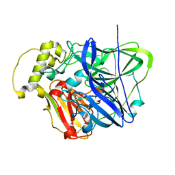
3OD6
 | | Crystal structure of p38alpha Y323T active mutant | | 分子名称: | Mitogen-activated protein kinase 14, octyl beta-D-glucopyranoside | | 著者 | Livnah, O, Tzarum, N. | | 登録日 | 2010-08-11 | | 公開日 | 2011-01-12 | | 最終更新日 | 2024-02-21 | | 実験手法 | X-RAY DIFFRACTION (2.682 Å) | | 主引用文献 | Active mutants of the TCR-mediated p38alpha alternative activation site show changes in the phosphorylation lip and DEF site formation.
J.Mol.Biol., 405, 2011
|
|
3OD7
 | | Haemophilus influenzae ferric binding protein A -Iron Loaded | | 分子名称: | FE (III) ION, Iron-utilization periplasmic protein, PHOSPHATE ION | | 著者 | Khambati, H.K, Moraes, T.F, Singh, J, Shouldice, S.R, Yu, R.H, Schryvers, A.B. | | 登録日 | 2010-08-11 | | 公開日 | 2010-09-15 | | 最終更新日 | 2023-11-01 | | 実験手法 | X-RAY DIFFRACTION (1.801 Å) | | 主引用文献 | The role of vicinal tyrosine residues in the function of Haemophilus influenzae ferric binding protein A.
Biochem.J., 2010
|
|
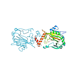
3OD8
 | | Human PARP-1 zinc finger 1 (Zn1) bound to DNA | | 分子名称: | 5'-D(*CP*CP*CP*AP*AP*GP*CP*GP*GP*C)-3', 5'-D(*GP*CP*CP*GP*CP*TP*TP*GP*GP*G)-3', Poly [ADP-ribose] polymerase 1, ... | | 著者 | Pascal, J.M, Langelier, M.-F. | | 登録日 | 2010-08-11 | | 公開日 | 2011-01-12 | | 最終更新日 | 2024-10-30 | | 実験手法 | X-RAY DIFFRACTION (2.4 Å) | | 主引用文献 | Crystal Structures of Poly(ADP-ribose) Polymerase-1 (PARP-1) Zinc Fingers Bound to DNA: STRUCTURAL AND FUNCTIONAL INSIGHTS INTO DNA-DEPENDENT PARP-1 ACTIVITY.
J.Biol.Chem., 286, 2011
|
|
3OD9
 | | Crystal structure of PliI-Ah, periplasmic lysozyme inhibitor of I-type lysozyme from Aeromonas hydrophyla | | 分子名称: | POTASSIUM ION, Putative exported protein, SODIUM ION | | 著者 | Leysen, S, Van Herreweghe, J.M, Callewaert, L, Heirbaut, M, Buntinx, P, Michiels, C.W, Strelkov, S.V. | | 登録日 | 2010-08-11 | | 公開日 | 2010-12-22 | | 最終更新日 | 2024-02-21 | | 実験手法 | X-RAY DIFFRACTION (1.411 Å) | | 主引用文献 | Molecular Basis of Bacterial Defense against Host Lysozymes: X-ray Structures of Periplasmic Lysozyme Inhibitors PliI and PliC.
J.Mol.Biol., 405, 2011
|
|
3ODA
 | | Human PARP-1 zinc finger 1 (Zn1) bound to DNA | | 分子名称: | 5'-D(*GP*CP*CP*TP*GP*CP*AP*GP*GP*C)-3', Poly [ADP-ribose] polymerase 1, ZINC ION | | 著者 | Pascal, J.M, Langelier, M.-F. | | 登録日 | 2010-08-11 | | 公開日 | 2011-01-12 | | 最終更新日 | 2024-02-21 | | 実験手法 | X-RAY DIFFRACTION (2.64 Å) | | 主引用文献 | Crystal Structures of Poly(ADP-ribose) Polymerase-1 (PARP-1) Zinc Fingers Bound to DNA: STRUCTURAL AND FUNCTIONAL INSIGHTS INTO DNA-DEPENDENT PARP-1 ACTIVITY.
J.Biol.Chem., 286, 2011
|
|
3ODB
 | | Haemophilus influenzae ferric binding protein A -Iron Loaded -open Conformation | | 分子名称: | FE (III) ION, Iron-utilization periplasmic protein | | 著者 | Khambati, H.K, Moraes, T.F, Singh, J, Shouldice, S.R, Yu, R.H, Schryvers, A.B. | | 登録日 | 2010-08-11 | | 公開日 | 2010-09-15 | | 最終更新日 | 2023-11-01 | | 実験手法 | X-RAY DIFFRACTION (1.62 Å) | | 主引用文献 | The role of vicinal tyrosine residues in the function of Haemophilus influenzae ferric binding protein A.
Biochem.J., 2010
|
|
3ODC
 | | Human PARP-1 zinc finger 2 (Zn2) bound to DNA | | 分子名称: | 5'-D(*CP*CP*CP*AP*GP*AP*CP*G)-3', 5'-D(*CP*GP*TP*CP*TP*GP*GP*G)-3', Poly [ADP-ribose] polymerase 1, ... | | 著者 | Pascal, J.M, Langelier, M.-F. | | 登録日 | 2010-08-11 | | 公開日 | 2011-01-12 | | 最終更新日 | 2024-02-21 | | 実験手法 | X-RAY DIFFRACTION (2.8 Å) | | 主引用文献 | Crystal Structures of Poly(ADP-ribose) Polymerase-1 (PARP-1) Zinc Fingers Bound to DNA: STRUCTURAL AND FUNCTIONAL INSIGHTS INTO DNA-DEPENDENT PARP-1 ACTIVITY.
J.Biol.Chem., 286, 2011
|
|
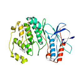
3ODD
 | | Comparison of the character and the speed of X-ray-induced structural changes of porcine pancreatic elastase at two temperatures, 100 and 15K. The data set was collected from region B of the crystal. Second step of radiation damage | | 分子名称: | Chymotrypsin-like elastase family member 1, SODIUM ION, SULFATE ION | | 著者 | Petrova, T, Ginell, S, Mitschler, A, Cousido-Siah, A, Hazemann, I, Podjarny, A, Joachimiak, A. | | 登録日 | 2010-08-11 | | 公開日 | 2010-08-25 | | 最終更新日 | 2023-09-06 | | 実験手法 | X-RAY DIFFRACTION (1.1 Å) | | 主引用文献 | X-ray-induced deterioration of disulfide bridges at atomic resolution.
Acta Crystallogr.,Sect.D, 66, 2010
|
|
3ODE
 | | Human PARP-1 zinc finger 2 (Zn2) bound to DNA | | 分子名称: | 5'-D(*CP*CP*CP*AP*AP*GP*CP*G)-3', 5'-D(*CP*GP*CP*TP*TP*GP*GP*G)-3', Poly [ADP-ribose] polymerase 1, ... | | 著者 | Pascal, J.M, Langelier, M.-F. | | 登録日 | 2010-08-11 | | 公開日 | 2011-01-12 | | 最終更新日 | 2024-02-21 | | 実験手法 | X-RAY DIFFRACTION (2.95 Å) | | 主引用文献 | Crystal Structures of Poly(ADP-ribose) Polymerase-1 (PARP-1) Zinc Fingers Bound to DNA: STRUCTURAL AND FUNCTIONAL INSIGHTS INTO DNA-DEPENDENT PARP-1 ACTIVITY.
J.Biol.Chem., 286, 2011
|
|
3ODF
 | | Comparison of the character and the speed of X-ray-induced structural changes of porcine pancreatic elastase at two temperatures, 100 and 15K. The data set was collected from region A of the crystal. Second step of radiation damage | | 分子名称: | Chymotrypsin-like elastase family member 1, SODIUM ION, SULFATE ION | | 著者 | Petrova, T, Ginell, S, Mitschler, A, Cousido-Siah, A, Hazemann, I, Podjarny, A, Joachimiak, A. | | 登録日 | 2010-08-11 | | 公開日 | 2010-08-25 | | 最終更新日 | 2023-09-06 | | 実験手法 | X-RAY DIFFRACTION (1.1 Å) | | 主引用文献 | X-ray-induced deterioration of disulfide bridges at atomic resolution.
Acta Crystallogr.,Sect.D, 66, 2010
|
|


