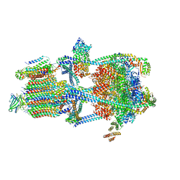
7UVY
 
 | |
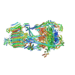
7UVZ
 | |
7UW1
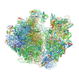
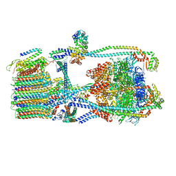
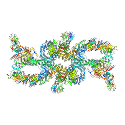
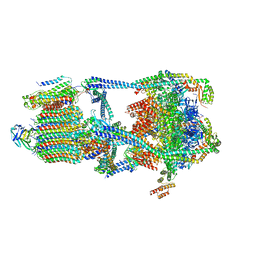
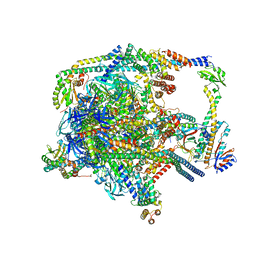
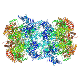
 | | A. baumannii 70S ribosome-Streptothricin-D complex | | 分子名称: | 16s Ribosomal RNA, 23s ribosomal RNA, 30S ribosomal protein S10, ... | | 著者 | Morgan, C.E, Yu, E.W. | | 登録日 | 2022-05-02 | | 公開日 | 2023-04-19 | | 最終更新日 | 2024-06-12 | | 実験手法 | ELECTRON MICROSCOPY (2.21 Å) | | 主引用文献 | Streptothricin F is a bactericidal antibiotic effective against highly drug-resistant gram-negative bacteria that interacts with the 30S subunit of the 70S ribosome.
Plos Biol., 21, 2023
|
|
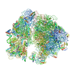
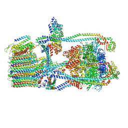
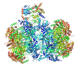
7UW9
 | | Citrus V-ATPase State 1, H in contact with subunit a | | 分子名称: | V-type proton ATPase catalytic subunit A, V-type proton ATPase subunit AP1 fragment, V-type proton ATPase subunit AP2 fragment, ... | | 著者 | Keon, K.A, Abdelaziz, R.A, Schulze, W.X, Schumacher, K, Rubinstein, J.L. | | 登録日 | 2022-05-03 | | 公開日 | 2022-07-06 | | 最終更新日 | 2024-06-12 | | 実験手法 | ELECTRON MICROSCOPY (4.2 Å) | | 主引用文献 | Structure of V-ATPase from citrus fruit.
Structure, 30, 2022
|
|
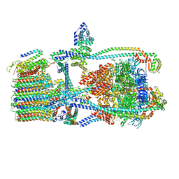
7UWA
 | | Citrus V-ATPase State 1, H in contact with subunits AB | | 分子名称: | V-type proton ATPase catalytic subunit A, V-type proton ATPase subunit AP1 fragment, V-type proton ATPase subunit AP2 fragment, ... | | 著者 | Abdelaziz, R.A, Keon, K.A, Schulze, W.X, Schumacher, K, Rubinstein, J.L. | | 登録日 | 2022-05-03 | | 公開日 | 2022-07-06 | | 最終更新日 | 2024-06-12 | | 実験手法 | ELECTRON MICROSCOPY (4.3 Å) | | 主引用文献 | Structure of V-ATPase from citrus fruit.
Structure, 30, 2022
|
|
7UWB
 | | Citrus V-ATPase State 2, Highest-Resolution Class | | 分子名称: | V-type proton ATPase catalytic subunit A, V-type proton ATPase subunit AP1 fragment, V-type proton ATPase subunit AP2 fragment, ... | | 著者 | Keon, K.A, Abdelaziz, R.A, Schulze, W.X, Schumacher, K, Rubinstein, J.L. | | 登録日 | 2022-05-03 | | 公開日 | 2022-07-06 | | 最終更新日 | 2024-01-17 | | 実験手法 | ELECTRON MICROSCOPY (3.9 Å) | | 主引用文献 | Structure of V-ATPase from citrus fruit.
Structure, 30, 2022
|
|
7UWC
 | | Citrus V-ATPase State 2, H in contact with subunit a | | 分子名称: | V-type proton ATPase catalytic subunit A, V-type proton ATPase subunit AP1 fragment, V-type proton ATPase subunit AP2 fragment, ... | | 著者 | Keon, K.A, Abdelaziz, R.A, Schulze, W.X, Schumacher, K, Rubinstein, J.L. | | 登録日 | 2022-05-03 | | 公開日 | 2022-07-06 | | 最終更新日 | 2024-01-17 | | 実験手法 | ELECTRON MICROSCOPY (4 Å) | | 主引用文献 | Structure of V-ATPase from citrus fruit.
Structure, 30, 2022
|
|
7UWD
 | | Citrus V-ATPase State 2, H in contact with subunits AB | | 分子名称: | V-type proton ATPase catalytic subunit A, V-type proton ATPase subunit, V-type proton ATPase subunit AP1 fragment, ... | | 著者 | Keon, K.A, Abdelaziz, R.A, Schulze, W.X, Schumacher, K, Rubinstein, J.L. | | 登録日 | 2022-05-03 | | 公開日 | 2022-07-06 | | 最終更新日 | 2024-01-17 | | 実験手法 | ELECTRON MICROSCOPY (4.1 Å) | | 主引用文献 | Structure of V-ATPase from citrus fruit.
Structure, 30, 2022
|
|
7UX3
 | | Asymmetric unit of AP-1, Arf1, Nef lattice on MHC-I lipopeptide incorporated narrow membrane tubes | | 分子名称: | ADP-ribosylation factor 1, AP-1 complex subunit beta-1, AP-1 complex subunit gamma-1, ... | | 著者 | Hooy, R.M, Hurley, J.H. | | 登録日 | 2022-05-04 | | 公開日 | 2023-05-10 | | 最終更新日 | 2024-06-12 | | 実験手法 | ELECTRON MICROSCOPY (9.6 Å) | | 主引用文献 | Self-assembly and structure of a clathrin-independent AP-1:Arf1 tubular membrane coat.
Sci Adv, 8, 2022
|
|
7UXH
 | |
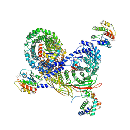
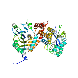
7UXX
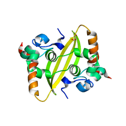 | | Crystal structure of SARS-CoV-2 nucleocapsid protein C-terminal domain | | 分子名称: | ACETATE ION, GLYCEROL, Nucleoprotein | | 著者 | Bezerra, E.H.S, Tonoli, C.C.C, Soprano, A.S, Franchini, K.G, Trivella, D.B.B, Benedetti, C.E. | | 登録日 | 2022-05-06 | | 公開日 | 2022-06-22 | | 最終更新日 | 2023-10-18 | | 実験手法 | X-RAY DIFFRACTION (1.85 Å) | | 主引用文献 | Discovery and structural characterization of chicoric acid as a SARS-CoV-2 nucleocapsid protein ligand and RNA binding disruptor.
Sci Rep, 12, 2022
|
|
7UXZ
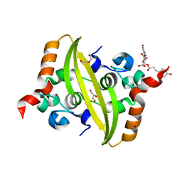 | | Crystal structure of SARS-CoV-2 nucleocapsid protein C-terminal domain complexed with Chicoric acid | | 分子名称: | (2R,3R)-2,3-bis{[(2E)-3-(3,4-dihydroxyphenyl)prop-2-enoyl]oxy}butanedioic acid, CHLORIDE ION, DI(HYDROXYETHYL)ETHER, ... | | 著者 | Bezerra, E.H.S, Tonoli, C.C.C, Soprano, A.S, Franchini, K.G, Trivella, D.B.B, Benedetti, C.E. | | 登録日 | 2022-05-06 | | 公開日 | 2022-06-22 | | 最終更新日 | 2023-10-18 | | 実験手法 | X-RAY DIFFRACTION (1.733 Å) | | 主引用文献 | Discovery and structural characterization of chicoric acid as a SARS-CoV-2 nucleocapsid protein ligand and RNA binding disruptor.
Sci Rep, 12, 2022
|
|
7UZF
 | |
7UZG
 | |
7UZH
 | |
7UZI
 | |
7UZJ
 | |
7UZK
 | |
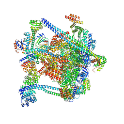
7V08
 | | Nucleoplasmic pre-60S intermediate of the Nog2 containing pre-rotation state from a Spb1 D52A suppressor 3 strain | | 分子名称: | 2-[3-(2-HYDROXY-1,1-DIHYDROXYMETHYL-ETHYLAMINO)-PROPYLAMINO]-2-HYDROXYMETHYL-PROPANE-1,3-DIOL, 25S rRNA, 5.8S rRNA, ... | | 著者 | Sekulski, K, Cruz, V.E, Weirich, C.S, Erzberger, J.P. | | 登録日 | 2022-05-10 | | 公開日 | 2023-03-15 | | 最終更新日 | 2024-06-12 | | 実験手法 | ELECTRON MICROSCOPY (2.36 Å) | | 主引用文献 | rRNA methylation by Spb1 regulates the GTPase activity of Nog2 during 60S ribosomal subunit assembly.
Nat Commun, 14, 2023
|
|
7V0K
 | | Consensus refinement of human erythrocyte ankyrin-1 complex (Composite map) | | 分子名称: | 2-acetamido-2-deoxy-beta-D-glucopyranose, Ammonium transporter Rh type A, Ankyrin-1, ... | | 著者 | Vallese, F, Kim, K, Yen, L.Y, Johnston, J.D, Noble, A.J, Cali, T, Clarke, O.B. | | 登録日 | 2022-05-10 | | 公開日 | 2022-07-20 | | 最終更新日 | 2022-07-27 | | 実験手法 | ELECTRON MICROSCOPY (2.4 Å) | | 主引用文献 | Architecture of the human erythrocyte ankyrin-1 complex.
Nat.Struct.Mol.Biol., 29, 2022
|
|
7V0T
 | | Local refinement of Band 3-I cytoplasmic domains, class 1 of erythrocyte ankyrin-1 complex | | 分子名称: | Band 3 anion transport protein | | 著者 | Vallese, F, Kim, K, Yen, L.Y, Johnston, J.D, Noble, A.J, Cali, T, Clarke, O.B. | | 登録日 | 2022-05-11 | | 公開日 | 2022-07-20 | | 最終更新日 | 2024-02-14 | | 実験手法 | ELECTRON MICROSCOPY (2.7 Å) | | 主引用文献 | Architecture of the human erythrocyte ankyrin-1 complex.
Nat.Struct.Mol.Biol., 29, 2022
|
|
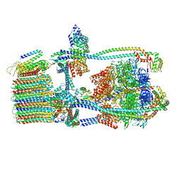
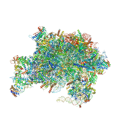
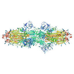
7V3U
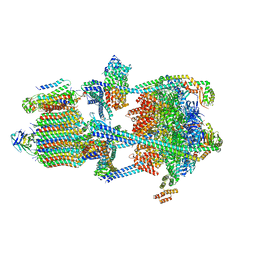
 | | Cryo-EM structure of MCM double hexamer with structured Mcm4-NSD | | 分子名称: | ADENOSINE-5'-DIPHOSPHATE, DNA replication licensing factor MCM2, DNA replication licensing factor MCM3, ... | | 著者 | Cheng, J, Li, N, Huo, Y, Dang, S, Tye, B, Gao, N, Zhai, Y. | | 登録日 | 2021-08-11 | | 公開日 | 2022-04-13 | | 実験手法 | ELECTRON MICROSCOPY (3.2 Å) | | 主引用文献 | Structural Insight into the MCM double hexamer activation by Dbf4-Cdc7 kinase.
Nat Commun, 13, 2022
|
|
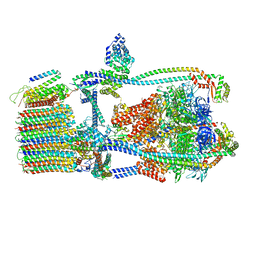
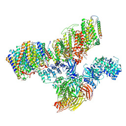
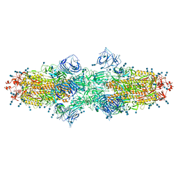
7V3V
 | | Cryo-EM structure of MCM double hexamer bound with DDK in State I | | 分子名称: | ADENOSINE-5'-DIPHOSPHATE, Cell division control protein 7, DDK kinase regulatory subunit DBF4, ... | | 著者 | Cheng, J, Li, N, Huo, Y, Dang, S, Tye, B, Gao, N, Zhai, Y. | | 登録日 | 2021-08-11 | | 公開日 | 2022-04-13 | | 実験手法 | ELECTRON MICROSCOPY (2.9 Å) | | 主引用文献 | Structural Insight into the MCM double hexamer activation by Dbf4-Cdc7 kinase.
Nat Commun, 13, 2022
|
|
7V7H
 | | Cryo-EM structure of SARS-CoV-2 S-Kappa variant (B.1.617.1), dimer of S trimer conformation 1 | | 分子名称: | 2-acetamido-2-deoxy-beta-D-glucopyranose, 2-acetamido-2-deoxy-beta-D-glucopyranose-(1-4)-2-acetamido-2-deoxy-beta-D-glucopyranose, Spike glycoprotein, ... | | 著者 | Yang, T.J, Yu, P.Y, Chang, Y.C, Hsu, S.T.D. | | 登録日 | 2021-08-21 | | 公開日 | 2021-10-06 | | 実験手法 | ELECTRON MICROSCOPY (3.2 Å) | | 主引用文献 | Cryo-EM structure of SARS-CoV-2 S-Kappa variant (B.1.617.1), dimer of S trimer conformation 1
To Be Published
|
|
7V7I
 | | Cryo-EM structure of SARS-CoV-2 S-Kappa variant (B.1.617.1), dimer of S trimer conformation 2 | | 分子名称: | 2-acetamido-2-deoxy-beta-D-glucopyranose, 2-acetamido-2-deoxy-beta-D-glucopyranose-(1-4)-2-acetamido-2-deoxy-beta-D-glucopyranose, Spike glycoprotein, ... | | 著者 | Yang, T.J, Yu, P.Y, Chang, Y.C, Hsu, S.T.D. | | 登録日 | 2021-08-21 | | 公開日 | 2021-10-06 | | 実験手法 | ELECTRON MICROSCOPY (3.3 Å) | | 主引用文献 | Cryo-EM structure of SARS-CoV-2 S-Kappa variant (B.1.617.1), dimer of S trimer conformation 2
To Be Published
|
|


