 Movie
Movie Controller

[English] 日本語
 Yorodumi
- PDB-6k3m: Application of anti-helix antibodies in protein structure determi... -
+ Open data

- Basic information
| Entry | Database: PDB / ID: 6k3m | |||||||||
|---|---|---|---|---|---|---|---|---|---|---|
| Title | Application of anti-helix antibodies in protein structure determination (8189-3LRH) | |||||||||
 Components Components |
| |||||||||
 Keywords Keywords |  STRUCTURAL PROTEIN / antibody / protein design STRUCTURAL PROTEIN / antibody / protein design | |||||||||
| Function / homology |  Function and homology information Function and homology information | |||||||||
| Biological species |   Staphylococcus aureus (bacteria) Staphylococcus aureus (bacteria) Homo sapiens (human) Homo sapiens (human) | |||||||||
| Method | X-RAY DIFFRACTION / SYNCHROTRON / MOLECULAR REPLACEMENT / Resolution: 1.8 Å | |||||||||
 Authors Authors | Lee, J.O. / Jin, M.S. / Kim, J.W. / Kim, S. / Lee, H. / Cho, G.Y. | |||||||||
| Funding support |  Korea, Republic Of, 2items Korea, Republic Of, 2items
| |||||||||
 Citation Citation | Journal: Proc.Natl.Acad.Sci.USA / Year: 2019 Title: Application of antihelix antibodies in protein structure determination. Authors: Kim, J.W. / Kim, S. / Lee, H. / Cho, G. / Kim, S.C. / Lee, H. / Jin, M.S. / Lee, J.O. | |||||||||
| History |
|
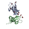
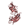
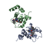
- Structure visualization
| Structure viewer | Molecule: MolmilJmol/JSmol |
|---|
- Downloads & links
-Download
| PDBx/mmCIF format | 6k3m.cif.gz | 51.7 KB | Display | PDBx/mmCIF format |
|---|---|---|---|---|
| PDB format | pdb6k3m.ent.gz | 34.3 KB | Display | PDB format |
| PDBx/mmJSON format | 6k3m.json.gz | Tree view | PDBx/mmJSON format | |
| Others |  Other downloads Other downloads |
-Validation report
| Arichive directory | https://data.pdbj.org/pub/pdb/validation_reports/k3/6k3mftp://data.pdbj.org/pub/pdb/validation_reports/k3/6k3m | HTTPS FTP |
|---|
-Related structure data
| Related structure data | 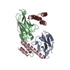 6k64C  6k65C  6k67C 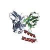 6k68C 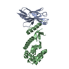 6k69C 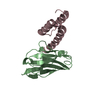 6k6aC 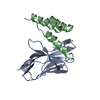 6k6bC  1deeS  3lrhS S: Starting model for refinement C: citing same article ( |
|---|---|
| Similar structure data |


-Links
 PDBj
PDBj


- Assembly
| Deposited unit | 
| ||||||||
|---|---|---|---|---|---|---|---|---|---|
| 1 |
| ||||||||
| Unit cell |
|
-Components
| #1: Protein | Mass: 9303.371 Da / Num. of mol.: 1 Source method: isolated from a genetically manipulated source Details: protein A(1DEE) and n-terminal of huntingtin peptide (3LRH) fusion,protein A(1DEE) and n-terminal of huntingtin peptide (3LRH) fusion Source: (gene. exp.) Staphylococcus aureus (bacteria) / Gene: spa / Production host: Escherichia coli (E. coli) / References: UniProt: D7RHB0, UniProt: P02976*PLUS |
|---|---|
| #2: Protein | Mass: 14203.453 Da / Num. of mol.: 1 Source method: isolated from a genetically manipulated source Source: (gene. exp.) Homo sapiens (human) / Production host: Escherichia coli (E. coli) |
| #3: Water | ChemComp-HOH / Water Mass: 18.015 Da / Num. of mol.: 149 / Source method: isolated from a natural source / Formula: H2O Mass: 18.015 Da / Num. of mol.: 149 / Source method: isolated from a natural source / Formula: H2O |
-Experimental details
-Experiment
| Experiment | Method: X-RAY DIFFRACTION / Number of used crystals: 1 |
|---|
- Sample preparation
| Crystal | Density Matthews: 1.57 Å3/Da / Density % sol: 21.55 % |
|---|---|
| Crystal grow | Temperature: 296 K / Method: vapor diffusion, sitting drop / Details: 30% PEG 4000, 0.2M MgCl2, 0.1M MOPS pH 6.5 |
-Data collection
| Diffraction | Mean temperature: 100 K / Serial crystal experiment: N |
|---|---|
| Diffraction source | Source: SYNCHROTRON / Site: PAL/PLS / Beamline: 7A (6B, 6C1) / Wavelength: 0.979 Å |
| Detector | Type: MAR CCD 130 mm / Detector: CCD / Date: May 12, 2016 |
| Radiation | Protocol: SINGLE WAVELENGTH / Monochromatic (M) / Laue (L): M / Scattering type: x-ray |
| Radiation wavelength | Wavelength: 0.979 Å / Relative weight: 1 |
| Reflection | Resolution: 1.8→50 Å / Num. obs: 14114 / % possible obs: 98.4 % / Redundancy: 6 % / Rpim(I) all: 0.022 / Net I/σ(I): 39.1 |
| Reflection shell | Resolution: 1.8→1.86 Å / Num. unique obs: 14114 / Rpim(I) all: 0.037 |
- Processing
| Software |
| ||||||||||||||||||||||||||||||||||||||||||||||||||||||||||||||||||||||||||||||||||||||||||||||||||||||||||||||||||||||||||||||||||||||||||||||||||||||||||||||||||||||||||||||||||||||
|---|---|---|---|---|---|---|---|---|---|---|---|---|---|---|---|---|---|---|---|---|---|---|---|---|---|---|---|---|---|---|---|---|---|---|---|---|---|---|---|---|---|---|---|---|---|---|---|---|---|---|---|---|---|---|---|---|---|---|---|---|---|---|---|---|---|---|---|---|---|---|---|---|---|---|---|---|---|---|---|---|---|---|---|---|---|---|---|---|---|---|---|---|---|---|---|---|---|---|---|---|---|---|---|---|---|---|---|---|---|---|---|---|---|---|---|---|---|---|---|---|---|---|---|---|---|---|---|---|---|---|---|---|---|---|---|---|---|---|---|---|---|---|---|---|---|---|---|---|---|---|---|---|---|---|---|---|---|---|---|---|---|---|---|---|---|---|---|---|---|---|---|---|---|---|---|---|---|---|---|---|---|---|---|
| Refinement | Method to determine structure: MOLECULAR REPLACEMENT Starting model: 3LRH, 1DEE Resolution: 1.8→28 Å / Cor.coef. Fo:Fc: 0.918 / Cor.coef. Fo:Fc free: 0.896 / SU B: 2.45 / SU ML: 0.079 / Cross valid method: THROUGHOUT / ESU R: 0.154 / ESU R Free: 0.141 / Details: HYDROGENS HAVE BEEN ADDED IN THE RIDING POSITIONS
| ||||||||||||||||||||||||||||||||||||||||||||||||||||||||||||||||||||||||||||||||||||||||||||||||||||||||||||||||||||||||||||||||||||||||||||||||||||||||||||||||||||||||||||||||||||||
| Solvent computation | Ion probe radii: 0.8 Å / Shrinkage radii: 0.8 Å / VDW probe radii: 1.2 Å | ||||||||||||||||||||||||||||||||||||||||||||||||||||||||||||||||||||||||||||||||||||||||||||||||||||||||||||||||||||||||||||||||||||||||||||||||||||||||||||||||||||||||||||||||||||||
| Displacement parameters | Biso mean: 10.881 Å2
| ||||||||||||||||||||||||||||||||||||||||||||||||||||||||||||||||||||||||||||||||||||||||||||||||||||||||||||||||||||||||||||||||||||||||||||||||||||||||||||||||||||||||||||||||||||||
| Refinement step | Cycle: 1 / Resolution: 1.8→28 Å
| ||||||||||||||||||||||||||||||||||||||||||||||||||||||||||||||||||||||||||||||||||||||||||||||||||||||||||||||||||||||||||||||||||||||||||||||||||||||||||||||||||||||||||||||||||||||
| Refine LS restraints |
|