 Movie
Movie Controller

[English] 日本語
 Yorodumi
- PDB-6k68: Application of anti-helix antibodies in protein structure determi... -
+ Open data

- Basic information
| Entry | Database: PDB / ID: 6k68 | |||||||||
|---|---|---|---|---|---|---|---|---|---|---|
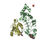
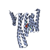
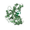
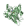
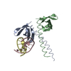
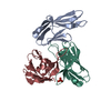
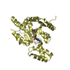
| Title | Application of anti-helix antibodies in protein structure determination (8420-3MNZ) | |||||||||
 Components Components |
| |||||||||
 Keywords Keywords | STRUCTURAL PROTEIN / antibody / protein design | |||||||||
| Function / homology |  Function and homology information Function and homology informationsymbiont-mediated suppression of host signal transduction pathway via antagonism of host cell surface receptor / IgG binding / extracellular region Similarity search - Function | |||||||||
| Biological species |    Staphylococcus aureus (bacteria) Staphylococcus aureus (bacteria) | |||||||||
| Method |  X-RAY DIFFRACTION / SYNCHROTRON / MOLECULAR REPLACEMENT / Resolution: 3.2 Å X-RAY DIFFRACTION / SYNCHROTRON / MOLECULAR REPLACEMENT / Resolution: 3.2 Å | |||||||||
 Authors Authors | Lee, J.O. / Jin, M.S. / Kim, J.W. / Kim, S. / Lee, H. / Cho, G.Y. | |||||||||
| Funding support |  Korea, Republic Of, 2items Korea, Republic Of, 2items
| |||||||||
 Citation Citation | Journal: Proc.Natl.Acad.Sci.USA / Year: 2019 Title: Application of antihelix antibodies in protein structure determination. Authors: Kim, J.W. / Kim, S. / Lee, H. / Cho, G. / Kim, S.C. / Lee, H. / Jin, M.S. / Lee, J.O. | |||||||||
| History |
|
- Structure visualization
| Structure viewer | Molecule: MolmilJmol/JSmol |
|---|
- Downloads & links
-Download
| PDBx/mmCIF format | 6k68.cif.gz | 252.1 KB | Display | PDBx/mmCIF format |
|---|---|---|---|---|
| PDB format | pdb6k68.ent.gz | 175.6 KB | Display | PDB format |
| PDBx/mmJSON format | 6k68.json.gz | Tree view | PDBx/mmJSON format | |
| Others |  Other downloads Other downloads |
-Validation report
| Arichive directory | https://data.pdbj.org/pub/pdb/validation_reports/k6/6k68ftp://data.pdbj.org/pub/pdb/validation_reports/k6/6k68 | HTTPS FTP |
|---|
-Related structure data
| Related structure data | 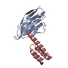 6k3mC 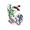 6k64C  6k65C  6k67C 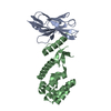 6k69C 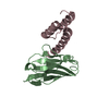 6k6aC 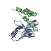 6k6bC  1deeS 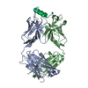 3mnzS S: Starting model for refinement C: citing same article ( |
|---|---|
| Similar structure data |


-Links
 PDBj
PDBj


- Assembly
| Deposited unit | 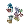
| ||||||||||
|---|---|---|---|---|---|---|---|---|---|---|---|
| 1 | 
| ||||||||||
| 2 | 
| ||||||||||
| 3 | 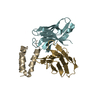
| ||||||||||
| 4 | 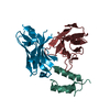
| ||||||||||
| Unit cell |
|
-Components
| #1: Antibody | Mass: 12752.225 Da / Num. of mol.: 4 Source method: isolated from a genetically manipulated source Source: (gene. exp.)  Trichoplusia ni (cabbage looper) Trichoplusia ni (cabbage looper)#2: Antibody | Mass: 12846.358 Da / Num. of mol.: 4 Source method: isolated from a genetically manipulated source Source: (gene. exp.) Trichoplusia ni (cabbage looper)#3: Protein | Mass: 8279.070 Da / Num. of mol.: 4 Source method: isolated from a genetically manipulated source Source: (gene. exp.) Staphylococcus aureus (bacteria) / Production host: Has protein modification | Y | |
|---|
-Experimental details
-Experiment
| Experiment | Method: X-RAY DIFFRACTION / Number of used crystals: 1 |
|---|
- Sample preparation
| Crystal | Density Matthews: 2.53 Å3/Da / Density % sol: 51.42 % |
|---|---|
| Crystal grow | Temperature: 296 K / Method: vapor diffusion, sitting drop / Details: 20% PEG MME 2000, 0.1M MOPS pH 6.5 |
-Data collection
| Diffraction | Mean temperature: 103 K / Serial crystal experiment: N |
|---|---|
| Diffraction source | Source: SYNCHROTRON / Site: PAL/PLS / Beamline: 7A (6B, 6C1) / Wavelength: 1 Å |
| Detector | Type: ADSC QUANTUM 315r / Detector: CCD / Date: Nov 18, 2015 |
| Radiation | Protocol: SINGLE WAVELENGTH / Monochromatic (M) / Laue (L): M / Scattering type: x-ray |
| Radiation wavelength | Wavelength: 1 Å / Relative weight: 1 |
| Reflection | Resolution: 3.2→50 Å / Num. obs: 22763 / % possible obs: 98.3 % / Redundancy: 3.2 % / Biso Wilson estimate: 52.84 Å2 / Rpim(I) all: 0.109 / Net I/σ(I): 7.4 |
| Reflection shell | Resolution: 3.2→3.3 Å / Num. unique obs: 22763 / Rpim(I) all: 0.346 |
- Processing
| Software |
| ||||||||||||||||||||||||||||||||||||||||||||||||||||||||||||||||||||||||||||||||||||||||||||||||||
|---|---|---|---|---|---|---|---|---|---|---|---|---|---|---|---|---|---|---|---|---|---|---|---|---|---|---|---|---|---|---|---|---|---|---|---|---|---|---|---|---|---|---|---|---|---|---|---|---|---|---|---|---|---|---|---|---|---|---|---|---|---|---|---|---|---|---|---|---|---|---|---|---|---|---|---|---|---|---|---|---|---|---|---|---|---|---|---|---|---|---|---|---|---|---|---|---|---|---|---|
| Refinement | Method to determine structure: MOLECULAR REPLACEMENT Starting model: 3MNZ, 1DEE Resolution: 3.2→33.42 Å / SU ML: 0.4453 / Cross valid method: THROUGHOUT / σ(F): 1.35 / Phase error: 31.3089
| ||||||||||||||||||||||||||||||||||||||||||||||||||||||||||||||||||||||||||||||||||||||||||||||||||
| Solvent computation | Shrinkage radii: 0.9 Å / VDW probe radii: 1.11 Å | ||||||||||||||||||||||||||||||||||||||||||||||||||||||||||||||||||||||||||||||||||||||||||||||||||
| Displacement parameters | Biso mean: 64.02 Å2 | ||||||||||||||||||||||||||||||||||||||||||||||||||||||||||||||||||||||||||||||||||||||||||||||||||
| Refinement step | Cycle: LAST / Resolution: 3.2→33.42 Å
| ||||||||||||||||||||||||||||||||||||||||||||||||||||||||||||||||||||||||||||||||||||||||||||||||||
| Refine LS restraints |
| ||||||||||||||||||||||||||||||||||||||||||||||||||||||||||||||||||||||||||||||||||||||||||||||||||
| LS refinement shell |
|
