 Movie
Movie Controller

[English] 日本語
 Yorodumi
- PDB-6pqb: Crystal structure of aminoglycoside-resistance methyltransferase ... -
+ Open data

- Basic information
| Entry | Database: PDB / ID: 6pqb | ||||||
|---|---|---|---|---|---|---|---|
| Title | Crystal structure of aminoglycoside-resistance methyltransferase RmtC bound to S-adenosylhomocysteine (SAH) | ||||||
 Components Components | 16S rRNA (guanine(1405)-N(7))-methyltransferase | ||||||
 Keywords Keywords | TRANSFERASE / methyltransferase / ribosome / aminoglycoside resistance / S-adenosylhomocysteine | ||||||
| Function / homology | 16S rRNA (guanine1405-N7)-methyltransferase / Ribosomal RNA aminoglycoside-resistance methyltransferase, Gram-negative bacteria / Ribosomal RNA aminoglycoside-resistance methyltransferase / Ribosomal RNA methyltransferase (FmrO) / rRNA methyltransferase activity / S-adenosyl-L-methionine-dependent methyltransferase superfamily / response to antibiotic / S-ADENOSYL-L-HOMOCYSTEINE / 16S rRNA (guanine(1405)-N(7))-methyltransferase Function and homology information Function and homology information | ||||||
| Biological species |  Proteus mirabilis (bacteria) Proteus mirabilis (bacteria) | ||||||
| Method |  X-RAY DIFFRACTION / SYNCHROTRON / MOLECULAR REPLACEMENT / Resolution: 3.14 Å X-RAY DIFFRACTION / SYNCHROTRON / MOLECULAR REPLACEMENT / Resolution: 3.14 Å | ||||||
 Authors Authors | Nosrati, M. / Hoffer, E.D. / Conn, G.L. | ||||||
| Funding support |  United States, 1items United States, 1items
| ||||||
 Citation Citation | Journal: J.Biol.Chem. / Year: 2019 Title: Functionally critical residues in the aminoglycoside resistance-associated methyltransferase RmtC play distinct roles in 30S substrate recognition. Authors: Nosrati, M. / Dey, D. / Mehrani, A. / Strassler, S.E. / Zelinskaya, N. / Hoffer, E.D. / Stagg, S.M. / Dunham, C.M. / Conn, G.L. | ||||||
| History |
|
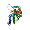
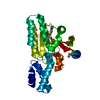
- Structure visualization
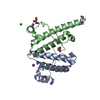
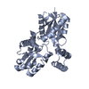
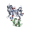
| Structure viewer | Molecule: MolmilJmol/JSmol |
|---|
- Downloads & links
-Download
| PDBx/mmCIF format | 6pqb.cif.gz | 446.6 KB | Display | PDBx/mmCIF format |
|---|---|---|---|---|
| PDB format | pdb6pqb.ent.gz | 371.4 KB | Display | PDB format |
| PDBx/mmJSON format | 6pqb.json.gz | Tree view | PDBx/mmJSON format | |
| Others |  Other downloads Other downloads |
-Validation report
| Arichive directory | https://data.pdbj.org/pub/pdb/validation_reports/pq/6pqbftp://data.pdbj.org/pub/pdb/validation_reports/pq/6pqb | HTTPS FTP |
|---|
-Related structure data
| Related structure data | 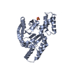 6cn0S S: Starting model for refinement |
|---|---|
| Similar structure data |


-Links
 PDBj
PDBj

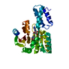
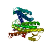
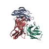
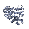
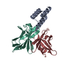
- Assembly
| Deposited unit | 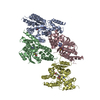
| ||||||||
|---|---|---|---|---|---|---|---|---|---|
| 1 | 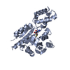
| ||||||||
| 2 | 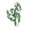
| ||||||||
| 3 | 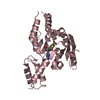
| ||||||||
| 4 | 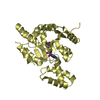
| ||||||||
| Unit cell |
|
-Components
| #1: Protein | Mass: 32290.963 Da / Num. of mol.: 4 Source method: isolated from a genetically manipulated source Source: (gene. exp.) Proteus mirabilis (bacteria) / Gene: rmtC / Plasmid: pET44 / Production host: References: UniProt: Q33DX5, 16S rRNA (guanine1405-N7)-methyltransferase #2: Chemical | ChemComp-SAH /   Type: L-peptide linking / Mass: 384.411 Da / Num. of mol.: 4 / Source method: obtained synthetically / Formula: C14H20N6O5S / Feature type: SUBJECT OF INVESTIGATION Type: L-peptide linking / Mass: 384.411 Da / Num. of mol.: 4 / Source method: obtained synthetically / Formula: C14H20N6O5S / Feature type: SUBJECT OF INVESTIGATION#3: Water | ChemComp-HOH / |  Mass: 18.015 Da / Num. of mol.: 9 / Source method: isolated from a natural source / Formula: H2O Mass: 18.015 Da / Num. of mol.: 9 / Source method: isolated from a natural source / Formula: H2OHas ligand of interest | Y | |
|---|
-Experimental details
-Experiment
| Experiment | Method: X-RAY DIFFRACTION / Number of used crystals: 1 |
|---|
- Sample preparation
| Crystal | Density Matthews: 3.84 Å3/Da / Density % sol: 67.97 % |
|---|---|
| Crystal grow | Temperature: 293 K / Method: vapor diffusion, hanging drop / pH: 7 Details: 2M ammonium sulfate, 0.1 M Na HEPES pH 7.0, 3 mM mellitic acid PH range: 6.8-7.8 |
-Data collection
| Diffraction | Mean temperature: 100 K / Serial crystal experiment: N |
|---|---|
| Diffraction source | Source: SYNCHROTRON / Site: APS / Beamline: 22-ID / Wavelength: 0.987 Å |
| Detector | Type: DECTRIS EIGER X 16M / Detector: PIXEL / Date: Jun 15, 2018 |
| Radiation | Protocol: SINGLE WAVELENGTH / Monochromatic (M) / Laue (L): M / Scattering type: x-ray |
| Radiation wavelength | Wavelength: 0.987 Å / Relative weight: 1 |
| Reflection | Resolution: 3.14→41 Å / Num. obs: 32552 / % possible obs: 100 % / Redundancy: 8.7 % / CC1/2: 0.99 / Rpim(I) all: 0.047 / Net I/σ(I): 10.82 |
| Reflection shell | Resolution: 3.14→3.25 Å / Redundancy: 8.2 % / Mean I/σ(I) obs: 1.15 / Num. unique obs: 3224 / CC1/2: 0.419 / Rpim(I) all: 0.87 / % possible all: 100 |
- Processing
| Software |
| ||||||||||||||||||||||||||||||||||||||||||||||||||||||||||||||||||||||||||||||
|---|---|---|---|---|---|---|---|---|---|---|---|---|---|---|---|---|---|---|---|---|---|---|---|---|---|---|---|---|---|---|---|---|---|---|---|---|---|---|---|---|---|---|---|---|---|---|---|---|---|---|---|---|---|---|---|---|---|---|---|---|---|---|---|---|---|---|---|---|---|---|---|---|---|---|---|---|---|---|---|
| Refinement | Method to determine structure: MOLECULAR REPLACEMENT Starting model: 6CN0 Resolution: 3.14→40.885 Å / SU ML: 0.48 / Cross valid method: THROUGHOUT / σ(F): 1.35 / Phase error: 28.25
| ||||||||||||||||||||||||||||||||||||||||||||||||||||||||||||||||||||||||||||||
| Solvent computation | Shrinkage radii: 0.9 Å / VDW probe radii: 1.11 Å | ||||||||||||||||||||||||||||||||||||||||||||||||||||||||||||||||||||||||||||||
| Displacement parameters | Biso max: 256.43 Å2 / Biso mean: 134.1971 Å2 / Biso min: 68.36 Å2 | ||||||||||||||||||||||||||||||||||||||||||||||||||||||||||||||||||||||||||||||
| Refinement step | Cycle: final / Resolution: 3.14→40.885 Å
| ||||||||||||||||||||||||||||||||||||||||||||||||||||||||||||||||||||||||||||||
| Refine LS restraints |
| ||||||||||||||||||||||||||||||||||||||||||||||||||||||||||||||||||||||||||||||
| LS refinement shell | Refine-ID: X-RAY DIFFRACTION / Rfactor Rfree error: 0
| ||||||||||||||||||||||||||||||||||||||||||||||||||||||||||||||||||||||||||||||
| Refinement TLS params. | Method: refined / Origin x: 136.1806 Å / Origin y: 124.3803 Å / Origin z: 23.0524 Å
| ||||||||||||||||||||||||||||||||||||||||||||||||||||||||||||||||||||||||||||||
| Refinement TLS group |
|
