- PDB-5ngv: CRYSTAL STRUCTURE OF THE Activin receptor type-2B LIGAND BINDING ... -
+
データを開く
IDまたはキーワード:
読み込み中...
-
基本情報
登録情報
データベース: PDB / ID: 5ngv
タイトル
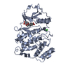
CRYSTAL STRUCTURE OF THE Activin receptor type-2B LIGAND BINDING DOMAIN IN COMPLEX WITH BIMAGRUMAB FV, ORTHORHOMBIC CRYSTAL FORM
要素
Activin receptor type-2B
anti-human ActRIIB mAb BYM338 heavy-chain
anti-human ActRIIB mAb BYM338 light-chain
キーワード
TRANSFERASE / three-finger toxin fold / antibody Fv fragment
機能・相同性
機能・相同性情報
Regulation of signaling by NODAL / activin receptor activity / activin receptor activity, type II / lymphatic endothelial cell differentiation / positive regulation of activin receptor signaling pathway / venous blood vessel development / lymphangiogenesis / trophoblast cell migration / retina vasculature development in camera-type eye / embryonic foregut morphogenesis ...Regulation of signaling by NODAL / activin receptor activity / activin receptor activity, type II / lymphatic endothelial cell differentiation / positive regulation of activin receptor signaling pathway / venous blood vessel development / lymphangiogenesis / trophoblast cell migration / retina vasculature development in camera-type eye / embryonic foregut morphogenesis / activin receptor complex / negative regulation of adipose tissue development / artery development / receptor protein serine/threonine kinase / activin binding / pattern specification process / Signaling by BMP / Signaling by Activin / activin receptor signaling pathway / Signaling by NODAL / pancreas development / gastrulation with mouth forming second / determination of left/right symmetry / negative regulation of ossification / anterior/posterior pattern specification / kinase activator activity / negative regulation of cold-induced thermogenesis / skeletal system morphogenesis / organ growth / cell surface receptor protein serine/threonine kinase signaling pathway / insulin secretion / growth factor binding / odontogenesis of dentin-containing tooth / mesoderm development / roof of mouth development / positive regulation of osteoblast differentiation / blood vessel remodeling / positive regulation of bone mineralization / BMP signaling pathway / response to glucose / protein serine/threonine/tyrosine kinase activity / lung development / post-embryonic development / kidney development / cellular response to growth factor stimulus / heart development / intracellular iron ion homeostasis / signaling receptor complex / protein serine/threonine kinase activity / regulation of DNA-templated transcription / negative regulation of transcription by RNA polymerase II / signal transduction / protein-containing complex / ATP binding / metal ion binding / plasma membrane / cytoplasm 類似検索 - 分子機能
Activin types I and II receptor domain / Activin types I and II receptor domain / CD59 / CD59 / Ser/Thr protein kinase, TGFB receptor / Snake toxin-like superfamily / Ribbon / Serine/threonine-protein kinase, active site / Serine/Threonine protein kinases active-site signature. / Protein kinase domain ...Activin types I and II receptor domain / Activin types I and II receptor domain / CD59 / CD59 / Ser/Thr protein kinase, TGFB receptor / Snake toxin-like superfamily / Ribbon / Serine/threonine-protein kinase, active site / Serine/Threonine protein kinases active-site signature. / Protein kinase domain / Immunoglobulins / Protein kinase domain profile. / Protein kinase domain / Protein kinase-like domain superfamily / Immunoglobulin-like / Sandwich / Mainly Beta 類似検索 - ドメイン・相同性
 ムービー
ムービー コントローラー


 データを開く
 基本情報
基本情報 要素
要素 キーワード
キーワード 機能・相同性情報
 Homo sapiens (ヒト)
 X線回折 /
  データ登録者
 引用
引用 構造の表示
構造の表示 ダウンロードとリンク
 その他のダウンロード
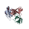


 PDBj
PDBj


 集合体
集合体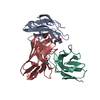


 分子量: 194.226 Da / 分子数: 1 / 由来タイプ: 合成 / 式: C8H18O5 / コメント: 沈殿剤*YM
 分子量: 18.015 Da / 分子数: 174 / 由来タイプ: 天然 / 式: H2O
 試料調製
試料調製 / ビームライン: X06DA / 波長: 1.00001 Å
 解析
解析