 Movie
Movie Controller

[English] 日本語
 Yorodumi
- PDB-5nhr: CRYSTAL STRUCTURE OF THE Activin receptor type-2B LIGAND BINDING ... -
+ Open data

- Basic information
| Entry | Database: PDB / ID: 5nhr | ||||||
|---|---|---|---|---|---|---|---|
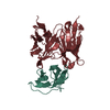
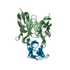
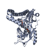
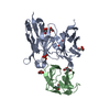
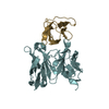
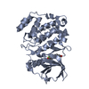
| Title | CRYSTAL STRUCTURE OF THE Activin receptor type-2B LIGAND BINDING DOMAIN IN COMPLEX WITH BIMAGRUMAB FV, CUBIC CRYSTAL FORM | ||||||
 Components Components |
| ||||||
 Keywords Keywords | IMMUNE SYSTEM / three-finger toxin fold / antibody Fv fragment | ||||||
| Function / homology |  Function and homology information Function and homology informationRegulation of signaling by NODAL / activin receptor activity / activin receptor activity, type II / lymphatic endothelial cell differentiation / positive regulation of activin receptor signaling pathway / venous blood vessel development / lymphangiogenesis / trophoblast cell migration / retina vasculature development in camera-type eye / embryonic foregut morphogenesis ...Regulation of signaling by NODAL / activin receptor activity / activin receptor activity, type II / lymphatic endothelial cell differentiation / positive regulation of activin receptor signaling pathway / venous blood vessel development / lymphangiogenesis / trophoblast cell migration / retina vasculature development in camera-type eye / embryonic foregut morphogenesis / activin receptor complex / negative regulation of adipose tissue development / artery development / receptor protein serine/threonine kinase / activin binding / pattern specification process / Signaling by BMP / Signaling by Activin / activin receptor signaling pathway / Signaling by NODAL / pancreas development / gastrulation with mouth forming second / determination of left/right symmetry / negative regulation of ossification / anterior/posterior pattern specification / kinase activator activity / negative regulation of cold-induced thermogenesis / skeletal system morphogenesis / organ growth / cell surface receptor protein serine/threonine kinase signaling pathway / insulin secretion / growth factor binding / odontogenesis of dentin-containing tooth / mesoderm development / roof of mouth development / positive regulation of osteoblast differentiation / blood vessel remodeling / positive regulation of bone mineralization / BMP signaling pathway / response to glucose / protein serine/threonine/tyrosine kinase activity / lung development / post-embryonic development / kidney development / cellular response to growth factor stimulus / heart development / intracellular iron ion homeostasis / signaling receptor complex / protein serine/threonine kinase activity / regulation of DNA-templated transcription / negative regulation of transcription by RNA polymerase II / signal transduction / protein-containing complex / ATP binding / metal ion binding / plasma membrane / cytoplasm Similarity search - Function | ||||||
| Biological species |  Homo sapiens (human) Homo sapiens (human) | ||||||
| Method |  X-RAY DIFFRACTION / SYNCHROTRON / MOLECULAR REPLACEMENT / molecular replacement / Resolution: 3.35 Å X-RAY DIFFRACTION / SYNCHROTRON / MOLECULAR REPLACEMENT / molecular replacement / Resolution: 3.35 Å | ||||||
| Model details | CUBIC CRYSTAL FORM | ||||||
 Authors Authors | Rondeau, J.-M. | ||||||
 Citation Citation | Journal: Proc. Natl. Acad. Sci. U.S.A. / Year: 2017 Title: Blockade of activin type II receptors with a dual anti-ActRIIA/IIB antibody is critical to promote maximal skeletal muscle hypertrophy. Authors: Morvan, F. / Rondeau, J.-M. / Zou, C. / Minetti, G. / Scheufler, C. / Scharenberg, M. / Jacobi, C. / Brebbia, P. / Ritter, V. / Toussaint, G. / Koelbing, C. / Leber, X. / Schilb, A. / Witte, ...Authors: Morvan, F. / Rondeau, J.-M. / Zou, C. / Minetti, G. / Scheufler, C. / Scharenberg, M. / Jacobi, C. / Brebbia, P. / Ritter, V. / Toussaint, G. / Koelbing, C. / Leber, X. / Schilb, A. / Witte, F. / Lehmann, S. / Koch, E. / Geisse, S. / Glass, D.J. / Lach-Trifilieff, E. | ||||||
| History |
|
- Structure visualization
| Structure viewer | Molecule: MolmilJmol/JSmol |
|---|
- Downloads & links
-Download
| PDBx/mmCIF format | 5nhr.cif.gz | 133.1 KB | Display | PDBx/mmCIF format |
|---|---|---|---|---|
| PDB format | pdb5nhr.ent.gz | 104.7 KB | Display | PDB format |
| PDBx/mmJSON format | 5nhr.json.gz | Tree view | PDBx/mmJSON format | |
| Others |  Other downloads Other downloads |
-Validation report
| Arichive directory | https://data.pdbj.org/pub/pdb/validation_reports/nh/5nhrftp://data.pdbj.org/pub/pdb/validation_reports/nh/5nhr | HTTPS FTP |
|---|
-Related structure data
| Related structure data |  5ngvC 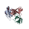 5nh3C  5nhwC  2h64S S: Starting model for refinement C: citing same article ( |
|---|---|
| Similar structure data |


-Links
 PDBj
PDBj


- Assembly
| Deposited unit | 
| ||||||||
|---|---|---|---|---|---|---|---|---|---|
| 1 | 
| ||||||||
| 2 | 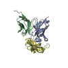
| ||||||||
| Unit cell |
|
-Components
| #1: Antibody | Mass: 12513.700 Da / Num. of mol.: 2 / Fragment: LIGAND BINDING domain Source method: isolated from a genetically manipulated source Details: VL domain / Source: (gene. exp.) Homo sapiens (human) / Details (production host): C-terminal Strep tagProduction host:  #2: Antibody | Mass: 13423.935 Da / Num. of mol.: 2 / Fragment: VH Source method: isolated from a genetically manipulated source Details: periplasmic expression / Source: (gene. exp.) Homo sapiens (human) / Details (production host): C-terminal His6 tagProduction host: #3: Protein | Mass: 11646.901 Da / Num. of mol.: 2 / Fragment: VL, UNP residues 24-117 Source method: isolated from a genetically manipulated source Source: (gene. exp.) Homo sapiens (human) / Gene: ACVR2B / Details (production host): Thioredoxin-fusion / Production host: References: UniProt: Q13705, receptor protein serine/threonine kinase Has protein modification | Y | |
|---|
-Experimental details
-Experiment
| Experiment | Method: X-RAY DIFFRACTION / Number of used crystals: 1 |
|---|
- Sample preparation
| Crystal | Density Matthews: 7.35 Å3/Da / Mosaicity: 0.707 ° / Mosaicity esd: 0.003 ° |
|---|---|
| Crystal grow | Temperature: 293 K / Method: vapor diffusion / pH: 4.6 / Details: 1.4M AMMONIUM SULFATE, 0.1M SODIUM CITRATE |
-Data collection
| Diffraction | Mean temperature: 100 K | ||||||||||||||||||||||||||||||||||||||||||||||||||||||||||||||||||
|---|---|---|---|---|---|---|---|---|---|---|---|---|---|---|---|---|---|---|---|---|---|---|---|---|---|---|---|---|---|---|---|---|---|---|---|---|---|---|---|---|---|---|---|---|---|---|---|---|---|---|---|---|---|---|---|---|---|---|---|---|---|---|---|---|---|---|---|
| Diffraction source | Source: SYNCHROTRON / Site: SLS  / Beamline: X10SA / Wavelength: 1 Å / Beamline: X10SA / Wavelength: 1 Å | ||||||||||||||||||||||||||||||||||||||||||||||||||||||||||||||||||
| Detector | Type: MARMOSAIC 225 mm CCD / Detector: CCD / Date: Sep 8, 2009 | ||||||||||||||||||||||||||||||||||||||||||||||||||||||||||||||||||
| Radiation | Protocol: SINGLE WAVELENGTH / Monochromatic (M) / Laue (L): M / Scattering type: x-ray | ||||||||||||||||||||||||||||||||||||||||||||||||||||||||||||||||||
| Radiation wavelength | Wavelength: 1 Å / Relative weight: 1 | ||||||||||||||||||||||||||||||||||||||||||||||||||||||||||||||||||
| Reflection | Resolution: 3.35→100 Å / Num. obs: 28995 / % possible obs: 90.6 % / Redundancy: 3.3 % / Rmerge(I) obs: 0.093 / Χ2: 1.066 / Net I/σ(I): 8.2 / Num. measured all: 96966 | ||||||||||||||||||||||||||||||||||||||||||||||||||||||||||||||||||
| Reflection shell |
|
-Phasing
| Phasing | Method: molecular replacement | |||||||||
|---|---|---|---|---|---|---|---|---|---|---|
| Phasing MR |
|
- Processing
| Software |
| ||||||||||||||||||||||||||||
|---|---|---|---|---|---|---|---|---|---|---|---|---|---|---|---|---|---|---|---|---|---|---|---|---|---|---|---|---|---|
| Refinement | Method to determine structure: MOLECULAR REPLACEMENT Starting model: 2H64 Resolution: 3.35→50 Å / Cross valid method: FREE R-VALUE / σ(F): 0
| ||||||||||||||||||||||||||||
| Displacement parameters | Biso max: 112.3 Å2 / Biso mean: 72.1553 Å2 / Biso min: 45.2 Å2
| ||||||||||||||||||||||||||||
| Refinement step | Cycle: final / Resolution: 3.35→50 Å
| ||||||||||||||||||||||||||||
| Refine LS restraints |
| ||||||||||||||||||||||||||||
| Xplor file |
|
