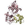
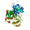
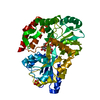
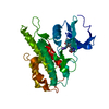
Entry Database : PDB / ID : 2wqbTitle Structure of the Tie2 kinase domain in complex with a thiazolopyrimidine inhibitor ANGIOPOIETIN-1 RECEPTOR Keywords / / / / / / / / / / / Function / homology Function Domain/homology Component
/ / / / / / / / / / / / / / / / / / / / / / / / / / / / / / / / / / / / / / / / / / / / / / / / / / / / / / / / / / / / / / / / / / / / / / / / / / / / / / / / / / / / / / / / / / / / / / / / / / / / / / / / / Biological species HOMO SAPIENS (human)Method / / / Resolution : 2.95 Å Authors Brassington, C. / Breed, J. / Buttar, D. / Fitzek, M. / Forder, C. / Hassall, L. / Hayter, B.R. / Jones, C.D. / Luke, R.W.A. / McCall, E. ...Brassington, C. / Breed, J. / Buttar, D. / Fitzek, M. / Forder, C. / Hassall, L. / Hayter, B.R. / Jones, C.D. / Luke, R.W.A. / McCall, E. / McCoull, W. / Norman, R. / Paterson, D. / McMiken, H. / Rowsell, S. / Tucker, J.A. Journal : Bioorg.Med.Chem.Lett. / Year : 2009Title : Novel Thienopyrimidine and Thiazolopyrimidine Kinase Inhibitors with Activity Against Tie-2 in Vitro and in Vivo.Authors : Luke, R.W. / Ballard, P. / Buttar, D. / Campbell, L. / Curwen, J. / Emery, S.C. / Griffen, A.M. / Hassall, L. / Hayter, B.R. / Jones, C.D. / Mccoull, W. / Mellor, M. / Swain, M.L. / Tucker, J.A. History Deposition Aug 18, 2009 Deposition site / Processing site Revision 1.0 Nov 3, 2009 Provider / Type Revision 1.1 Jul 13, 2011 Group / Refinement description / Version format complianceRevision 1.2 Apr 17, 2013 Group / Refinement descriptionRevision 1.3 Mar 6, 2019 Group / Experimental preparation / OtherCategory / pdbx_database_proc / pdbx_database_statusItem / _pdbx_database_status.recvd_author_approvalRevision 1.4 Dec 20, 2023 Group Data collection / Database references ... Data collection / Database references / Derived calculations / Other / Refinement description Category chem_comp_atom / chem_comp_bond ... chem_comp_atom / chem_comp_bond / database_2 / pdbx_database_status / pdbx_initial_refinement_model / struct_site Item _database_2.pdbx_DOI / _database_2.pdbx_database_accession ... _database_2.pdbx_DOI / _database_2.pdbx_database_accession / _pdbx_database_status.status_code_sf / _struct_site.pdbx_auth_asym_id / _struct_site.pdbx_auth_comp_id / _struct_site.pdbx_auth_seq_id Revision 1.5 Nov 13, 2024 Group / Category / pdbx_modification_feature / Item
Show all Show less
 Movie
Movie Controller

 Yorodumi
 Open data
 Basic information
 Components
 Keywords
 Function and homology information
 HOMO SAPIENS (human)
 X-RAY DIFFRACTION /
  Authors
 Citation
 Structure visualization
 Downloads & links
 Other downloads
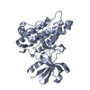


 PDBj
PDBj


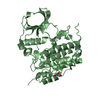
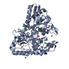
 Assembly
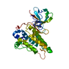

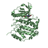
 SPODOPTERA FRUGIPERDA (fall armyworm) / Strain (production host): SF9
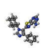
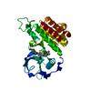
 Mass: 390.505 Da / Num. of mol.: 1 / Source method: obtained synthetically / Formula: C21H22N6S
 Mass: 18.015 Da / Num. of mol.: 19 / Source method: isolated from a natural source / Formula: H2O
 Sample preparation
 / Beamline: PX14.2 / Wavelength: 0.975
  Processing