 Movie
Movie Controller


+ Open data

- Basic information
| Entry | Database: PDB / ID: 1fvr | ||||||
|---|---|---|---|---|---|---|---|
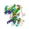
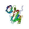
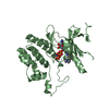
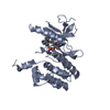
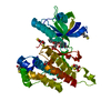
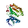
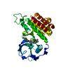
| Title | TIE2 KINASE DOMAIN | ||||||
 Components Components | TYROSINE-PROTEIN KINASE TIE-2 | ||||||
 Keywords Keywords | TRANSFERASE / tyrosine kinase | ||||||
| Function / homology |  Function and homology information Function and homology informationTie signaling pathway / glomerulus vasculature development / transmembrane receptor protein kinase activity / regulation of endothelial cell apoptotic process / regulation of establishment or maintenance of cell polarity / heart trabecula formation / definitive hemopoiesis / regulation of vascular permeability / sprouting angiogenesis / endothelial cell proliferation ...Tie signaling pathway / glomerulus vasculature development / transmembrane receptor protein kinase activity / regulation of endothelial cell apoptotic process / regulation of establishment or maintenance of cell polarity / heart trabecula formation / definitive hemopoiesis / regulation of vascular permeability / sprouting angiogenesis / endothelial cell proliferation / positive regulation of Rho protein signal transduction / microvillus / positive regulation of intracellular signal transduction / positive regulation of focal adhesion assembly / negative regulation of endothelial cell apoptotic process / positive regulation of Rac protein signal transduction / Tie2 Signaling / positive regulation of endothelial cell proliferation / transmembrane receptor protein tyrosine kinase activity / substrate adhesion-dependent cell spreading / positive regulation of endothelial cell migration / negative regulation of angiogenesis / cell surface receptor protein tyrosine kinase signaling pathway / basal plasma membrane / cellular response to mechanical stimulus / receptor protein-tyrosine kinase / negative regulation of inflammatory response / positive regulation of angiogenesis / cell-cell junction / cell-cell signaling / heart development / RAF/MAP kinase cascade / signaling receptor activity / angiogenesis / basolateral plasma membrane / protein kinase activity / positive regulation of MAPK cascade / positive regulation of ERK1 and ERK2 cascade / cell surface receptor signaling pathway / positive regulation of phosphatidylinositol 3-kinase/protein kinase B signal transduction / signaling receptor complex / apical plasma membrane / ciliary basal body / membrane raft / focal adhesion / centrosome / negative regulation of apoptotic process / cell surface / extracellular region / ATP binding / identical protein binding / plasma membrane Similarity search - Function | ||||||
| Biological species |  Homo sapiens (human) Homo sapiens (human) | ||||||
| Method |  X-RAY DIFFRACTION / SYNCHROTRON / Resolution: 2.2 Å X-RAY DIFFRACTION / SYNCHROTRON / Resolution: 2.2 Å | ||||||
 Authors Authors | Shewchuk, L.M. / Hassell, A.M. / Ellis, B. / Holmes, W.D. / Davis, R. / Horne, E.L. / Kadwell, S.H. / McKee, D.D. / Moore, J.T. | ||||||
 Citation Citation | Journal: Structure Fold.Des. / Year: 2000 Title: Structure of the Tie2 RTK domain: self-inhibition by the nucleotide binding loop, activation loop, and C-terminal tail. Authors: Shewchuk, L.M. / Hassell, A.M. / Ellis, B. / Holmes, W.D. / Davis, R. / Horne, E.L. / Kadwell, S.H. / McKee, D.D. / Moore, J.T. | ||||||
| History |
|
- Structure visualization
| Structure viewer | Molecule: MolmilJmol/JSmol |
|---|
- Downloads & links
-Download
| PDBx/mmCIF format | 1fvr.cif.gz | 138.2 KB | Display | PDBx/mmCIF format |
|---|---|---|---|---|
| PDB format | pdb1fvr.ent.gz | 107 KB | Display | PDB format |
| PDBx/mmJSON format | 1fvr.json.gz | Tree view | PDBx/mmJSON format | |
| Others |  Other downloads Other downloads |
-Validation report
| Arichive directory | https://data.pdbj.org/pub/pdb/validation_reports/fv/1fvrftp://data.pdbj.org/pub/pdb/validation_reports/fv/1fvr | HTTPS FTP |
|---|
-Related structure data
| Similar structure data |
|---|


-Links
 PDBj
PDBj


- Assembly
| Deposited unit | 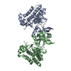
| ||||||||
|---|---|---|---|---|---|---|---|---|---|
| 1 | 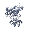
| ||||||||
| 2 | 
| ||||||||
| Unit cell |
|
-Components
| #1: Protein | Mass: 37528.812 Da / Num. of mol.: 2 / Fragment: KINASE DOMAIN Source method: isolated from a genetically manipulated source Source: (gene. exp.) Homo sapiens (human) / Production host:  unidentified baculovirus / References: UniProt: Q02763, EC: 2.7.1.112 unidentified baculovirus / References: UniProt: Q02763, EC: 2.7.1.112#2: Water | ChemComp-HOH / |  Mass: 18.015 Da / Num. of mol.: 441 / Source method: isolated from a natural source / Formula: H2O Mass: 18.015 Da / Num. of mol.: 441 / Source method: isolated from a natural source / Formula: H2O |
|---|
-Experimental details
-Experiment
| Experiment | Method: X-RAY DIFFRACTION / Number of used crystals: 1 |
|---|
- Sample preparation
| Crystal | Density Matthews: 2.75 Å3/Da / Density % sol: 55.35 % | ||||||||||||||||||||||||||||||||||||||||||||||||||||||
|---|---|---|---|---|---|---|---|---|---|---|---|---|---|---|---|---|---|---|---|---|---|---|---|---|---|---|---|---|---|---|---|---|---|---|---|---|---|---|---|---|---|---|---|---|---|---|---|---|---|---|---|---|---|---|---|
| Crystal grow | Temperature: 295 K / Method: vapor diffusion, hanging drop / pH: 7.5 Details: 2.5% PEG12000, 2.5% glycerol, 100mM Hepes, 10mM spermidine, pH 7.5, VAPOR DIFFUSION, HANGING DROP, temperature 22K | ||||||||||||||||||||||||||||||||||||||||||||||||||||||
| Crystal grow | *PLUS | ||||||||||||||||||||||||||||||||||||||||||||||||||||||
| Components of the solutions | *PLUS
|
-Data collection
| Diffraction | Mean temperature: 118 K |
|---|---|
| Diffraction source | Source: SYNCHROTRON / Site: APS  / Beamline: 17-ID / Wavelength: 1 / Beamline: 17-ID / Wavelength: 1 |
| Detector | Type: MARRESEARCH / Detector: CCD / Date: Jan 3, 2000 |
| Radiation | Protocol: SINGLE WAVELENGTH / Monochromatic (M) / Laue (L): M / Scattering type: x-ray |
| Radiation wavelength | Wavelength: 1 Å / Relative weight: 1 |
| Reflection | Resolution: 2.2→20 Å / Num. all: 39864 / Num. obs: 39864 / % possible obs: 96.4 % / Observed criterion σ(F): 0 / Observed criterion σ(I): 0 / Redundancy: 4 % / Biso Wilson estimate: 24.9 Å2 / Rmerge(I) obs: 0.075 / Net I/σ(I): 12 |
| Reflection shell | Resolution: 2.2→2.34 Å / Redundancy: 3 % / Rmerge(I) obs: 0.2 / Num. unique all: 5582 / % possible all: 91 |
| Reflection | *PLUS Lowest resolution: 20 Å |
| Reflection shell | *PLUS % possible obs: 95 % / Rmerge(I) obs: 0.2 |
- Processing
| Software |
| |||||||||||||||||||||||||
|---|---|---|---|---|---|---|---|---|---|---|---|---|---|---|---|---|---|---|---|---|---|---|---|---|---|---|
| Refinement | Resolution: 2.2→20 Å / σ(F): 0 / σ(I): 0 / Stereochemistry target values: Engh & Huber
| |||||||||||||||||||||||||
| Refinement step | Cycle: LAST / Resolution: 2.2→20 Å
| |||||||||||||||||||||||||
| Refine LS restraints |
| |||||||||||||||||||||||||
| Software | *PLUS Name: CNS / Classification: refinement | |||||||||||||||||||||||||
| Refinement | *PLUS Lowest resolution: 20 Å / σ(F): 0 | |||||||||||||||||||||||||
| Solvent computation | *PLUS | |||||||||||||||||||||||||
| Displacement parameters | *PLUS | |||||||||||||||||||||||||
| Refine LS restraints | *PLUS Type: c_bond_d / Dev ideal: 0.0058 |
