 Movie
Movie Controller


+ Open data

- Basic information
| Entry | Database: PDB / ID: 6d3l | ||||||
|---|---|---|---|---|---|---|---|
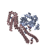
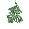
| Title | Crystal structure of unphosphorylated human PKR | ||||||
 Components Components | Interferon-induced, double-stranded RNA-activated protein kinase | ||||||
 Keywords Keywords | TRANSFERASE / unphosphorylated / kinase / activation loop swapping / apo | ||||||
| Function / homology |  Function and homology information Function and homology informationInhibition of PKR / regulation of NLRP3 inflammasome complex assembly / eukaryotic translation initiation factor 2alpha kinase activity / response to interferon-alpha / negative regulation of osteoblast proliferation / regulation of hematopoietic progenitor cell differentiation / positive regulation of stress-activated MAPK cascade / protein phosphatase regulator activity / SUMOylation of immune response proteins / regulation of hematopoietic stem cell proliferation ...Inhibition of PKR / regulation of NLRP3 inflammasome complex assembly / eukaryotic translation initiation factor 2alpha kinase activity / response to interferon-alpha / negative regulation of osteoblast proliferation / regulation of hematopoietic progenitor cell differentiation / positive regulation of stress-activated MAPK cascade / protein phosphatase regulator activity / SUMOylation of immune response proteins / regulation of hematopoietic stem cell proliferation / regulation of hematopoietic stem cell differentiation / regulation of translational initiation / negative regulation of viral genome replication / positive regulation of chemokine production / endoplasmic reticulum unfolded protein response / antiviral innate immune response / positive regulation of cytokine production / cellular response to amino acid starvation / : / non-specific protein-tyrosine kinase / non-membrane spanning protein tyrosine kinase activity / positive regulation of non-canonical NF-kappaB signal transduction / PKR-mediated signaling / Evasion by RSV of host interferon responses / ISG15 antiviral mechanism / response to virus / kinase activity / Interferon alpha/beta signaling / protein autophosphorylation / double-stranded RNA binding / defense response to virus / protein phosphorylation / protein kinase activity / positive regulation of MAPK cascade / non-specific serine/threonine protein kinase / negative regulation of translation / ribosome / translation / negative regulation of cell population proliferation / protein serine kinase activity / protein serine/threonine kinase activity / negative regulation of apoptotic process / perinuclear region of cytoplasm / RNA binding / nucleoplasm / ATP binding / membrane / identical protein binding / nucleus / cytoplasm / cytosol Similarity search - Function | ||||||
| Biological species |  Homo sapiens (human) Homo sapiens (human) | ||||||
| Method |  X-RAY DIFFRACTION / SYNCHROTRON / MOLECULAR REPLACEMENT / molecular replacement / Resolution: 3.1 Å X-RAY DIFFRACTION / SYNCHROTRON / MOLECULAR REPLACEMENT / molecular replacement / Resolution: 3.1 Å | ||||||
 Authors Authors | Erlandsen, H. / Mayo, C. / Robinson, V.L. / Cole, J.L. | ||||||
| Funding support |  United States, 1items United States, 1items
| ||||||
 Citation Citation | Journal: Biochemistry / Year: 2019 Title: Structural Basis of Protein Kinase R Autophosphorylation. Authors: Mayo, C.B. / Erlandsen, H. / Mouser, D.J. / Feinstein, A.G. / Robinson, V.L. / May, E.R. / Cole, J.L. | ||||||
| History |
|
- Structure visualization
| Structure viewer | Molecule: MolmilJmol/JSmol |
|---|
- Downloads & links
-Download
| PDBx/mmCIF format | 6d3l.cif.gz | 67.4 KB | Display | PDBx/mmCIF format |
|---|---|---|---|---|
| PDB format | pdb6d3l.ent.gz | 47.2 KB | Display | PDB format |
| PDBx/mmJSON format | 6d3l.json.gz | Tree view | PDBx/mmJSON format | |
| Others |  Other downloads Other downloads |
-Validation report
| Arichive directory | https://data.pdbj.org/pub/pdb/validation_reports/d3/6d3lftp://data.pdbj.org/pub/pdb/validation_reports/d3/6d3l | HTTPS FTP |
|---|
-Related structure data
| Related structure data |  6d3kC  2a19S S: Starting model for refinement C: citing same article ( |
|---|---|
| Similar structure data |


-Links
 PDBj
PDBj

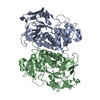
- Assembly
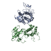
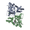
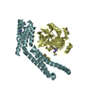
| Deposited unit | 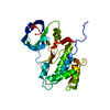
| ||||||||
|---|---|---|---|---|---|---|---|---|---|
| 1 | 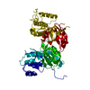
| ||||||||
| 2 | 
| ||||||||
| Unit cell |
|
-Components
| #1: Protein | Mass: 37445.773 Da / Num. of mol.: 1 / Fragment: kinase domain (229-551) Source method: isolated from a genetically manipulated source Source: (gene. exp.) Homo sapiens (human) / Gene: EIF2AK2, PKR, PRKR / Production host:  References: UniProt: P19525, non-specific serine/threonine protein kinase, non-specific protein-tyrosine kinase |
|---|
-Experimental details
-Experiment
| Experiment | Method: X-RAY DIFFRACTION / Number of used crystals: 1 |
|---|
- Sample preparation
| Crystal | Density Matthews: 2.04 Å3/Da / Density % sol: 39.76 % |
|---|---|
| Crystal grow | Temperature: 293 K / Method: vapor diffusion, sitting drop / pH: 5.5 Details: 0.1 M Bis-Tris, pH 5.5, 2.0 M ammonium sulfate, 1:3 molar ratio of dp8 (heparin) was added to protein prior to crystallization |
-Data collection
| Diffraction | Mean temperature: 100 K |
|---|---|
| Diffraction source | Source: SYNCHROTRON / Site: NSLS-II / Beamline: 17-ID-2 / Wavelength: 0.978971 Å |
| Detector | Type: DECTRIS EIGER X 16M / Detector: PIXEL / Date: Oct 22, 2016 |
| Radiation | Monochromator: double crystal Si(111) / Protocol: SINGLE WAVELENGTH / Monochromatic (M) / Laue (L): M / Scattering type: x-ray |
| Radiation wavelength | Wavelength: 0.978971 Å / Relative weight: 1 |
| Reflection | Resolution: 3.1→123.33 Å / Num. obs: 6130 / % possible obs: 100 % / Redundancy: 17.9 % / CC1/2: 0.997 / Rmerge(I) obs: 0.259 / Rpim(I) all: 0.063 / Rrim(I) all: 0.267 / Net I/σ(I): 9.1 |
| Reflection shell | Resolution: 3.1→3.31 Å / Redundancy: 19.3 % / Rmerge(I) obs: 1.133 / Num. unique obs: 1065 / CC1/2: 0.677 / Rpim(I) all: 0.263 / Rrim(I) all: 1.163 / % possible all: 100 |
-Phasing
| Phasing | Method: molecular replacement | |||||||||
|---|---|---|---|---|---|---|---|---|---|---|
| Phasing MR |
|
- Processing
| Software |
| ||||||||||||||||||||||||||||||||||||||||||||||||||||||||||||
|---|---|---|---|---|---|---|---|---|---|---|---|---|---|---|---|---|---|---|---|---|---|---|---|---|---|---|---|---|---|---|---|---|---|---|---|---|---|---|---|---|---|---|---|---|---|---|---|---|---|---|---|---|---|---|---|---|---|---|---|---|---|
| Refinement | Method to determine structure: MOLECULAR REPLACEMENT Starting model: PDB entry 2A19 Resolution: 3.1→123.33 Å / Cor.coef. Fo:Fc: 0.908 / Cor.coef. Fo:Fc free: 0.812 / SU B: 34.802 / SU ML: 0.573 / Cross valid method: THROUGHOUT / σ(F): 0 / ESU R Free: 0.65 / Details: HYDROGENS HAVE BEEN ADDED IN THE RIDING POSITIONS
| ||||||||||||||||||||||||||||||||||||||||||||||||||||||||||||
| Solvent computation | Ion probe radii: 0.8 Å / Shrinkage radii: 0.8 Å / VDW probe radii: 1.2 Å | ||||||||||||||||||||||||||||||||||||||||||||||||||||||||||||
| Displacement parameters | Biso max: 191.27 Å2 / Biso mean: 88.324 Å2 / Biso min: 41.4 Å2
| ||||||||||||||||||||||||||||||||||||||||||||||||||||||||||||
| Refinement step | Cycle: final / Resolution: 3.1→123.33 Å
| ||||||||||||||||||||||||||||||||||||||||||||||||||||||||||||
| Refine LS restraints |
| ||||||||||||||||||||||||||||||||||||||||||||||||||||||||||||
| LS refinement shell | Resolution: 3.1→3.181 Å / Rfactor Rfree error: 0 / Total num. of bins used: 20
|
