 Movie
Movie Controller


+ Open data

- Basic information
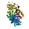
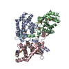
| Entry | Database: PDB / ID: 1ck7 | ||||||
|---|---|---|---|---|---|---|---|
| Title | GELATINASE A (FULL-LENGTH) | ||||||
 Components Components | PROTEIN (GELATINASE A) | ||||||
 Keywords Keywords | HYDROLASE / HYDROLASE (METALLOPROTEASE) / FULL-LENGTH / METALLOPROTEINASE / GELATINASE A | ||||||
| Function / homology |  Function and homology information Function and homology informationgelatinase A / intramembranous ossification / peripheral nervous system axon regeneration / luteinization / parturition / blood vessel maturation / bone trabecula formation / tissue remodeling / trophoblast cell migration / cellular response to UV-A ...gelatinase A / intramembranous ossification / peripheral nervous system axon regeneration / luteinization / parturition / blood vessel maturation / bone trabecula formation / tissue remodeling / trophoblast cell migration / cellular response to UV-A / ovulation from ovarian follicle / positive regulation of oxidative stress-induced neuron intrinsic apoptotic signaling pathway / prostate gland epithelium morphogenesis / negative regulation of cell adhesion / cellular response to fluid shear stress / face morphogenesis / negative regulation of vasoconstriction / Activation of Matrix Metalloproteinases / endodermal cell differentiation / response to amyloid-beta / Collagen degradation / collagen catabolic process / macrophage chemotaxis / fibronectin binding / extracellular matrix disassembly / EPH-ephrin mediated repulsion of cells / response to electrical stimulus / response to hyperoxia / ephrin receptor signaling pathway / cellular response to interleukin-1 / response to retinoic acid / response to mechanical stimulus / ovarian follicle development / positive regulation of vascular associated smooth muscle cell proliferation / Degradation of the extracellular matrix / extracellular matrix organization / response to activity / protein catabolic process / response to nicotine / sarcomere / cellular response to estradiol stimulus / response to hydrogen peroxide / cellular response to reactive oxygen species / cellular response to amino acid stimulus / metalloendopeptidase activity / response to estrogen / Regulation of Insulin-like Growth Factor (IGF) transport and uptake by Insulin-like Growth Factor Binding Proteins (IGFBPs) / metallopeptidase activity / cell migration / heart development / extracellular matrix / Interleukin-4 and Interleukin-13 signaling / angiogenesis / endopeptidase activity / response to hypoxia / Extra-nuclear estrogen signaling / positive regulation of cell migration / response to xenobiotic stimulus / serine-type endopeptidase activity / mitochondrion / proteolysis / : / extracellular region / zinc ion binding / nucleus / plasma membrane Similarity search - Function | ||||||
| Biological species |  Homo sapiens (human) Homo sapiens (human) | ||||||
| Method |  X-RAY DIFFRACTION / SYNCHROTRON / MOLECULAR REPLACEMENT / Resolution: 2.8 Å X-RAY DIFFRACTION / SYNCHROTRON / MOLECULAR REPLACEMENT / Resolution: 2.8 Å | ||||||
 Authors Authors | Morgunova, E. / Tuuttila, A. / Bergmann, U. / Isupov, M. / Lindqvist, Y. / Schneider, G. / Tryggvason, K. | ||||||
 Citation Citation | Journal: Science / Year: 1999 Title: Structure of human pro-matrix metalloproteinase-2: activation mechanism revealed. Authors: Morgunova, E. / Tuuttila, A. / Bergmann, U. / Isupov, M. / Lindqvist, Y. / Schneider, G. / Tryggvason, K. | ||||||
| History |
|
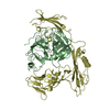
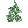
- Structure visualization
| Structure viewer | Molecule: MolmilJmol/JSmol |
|---|
- Downloads & links
-Download
| PDBx/mmCIF format | 1ck7.cif.gz | 142.3 KB | Display | PDBx/mmCIF format |
|---|---|---|---|---|
| PDB format | pdb1ck7.ent.gz | 109.8 KB | Display | PDB format |
| PDBx/mmJSON format | 1ck7.json.gz | Tree view | PDBx/mmJSON format | |
| Others |  Other downloads Other downloads |
-Validation report
| Arichive directory | https://data.pdbj.org/pub/pdb/validation_reports/ck/1ck7ftp://data.pdbj.org/pub/pdb/validation_reports/ck/1ck7 | HTTPS FTP |
|---|
-Related structure data


-Links
 PDBj
PDBj


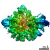
- Assembly
| Deposited unit | 
| ||||||||
|---|---|---|---|---|---|---|---|---|---|
| 1 |
| ||||||||
| Unit cell |
|
-Components
-Protein , 1 types, 1 molecules A
| #1: Protein | Mass: 70995.406 Da / Num. of mol.: 1 / Fragment: FULL-LENGTH / Mutation: E404A Source method: isolated from a genetically manipulated source Source: (gene. exp.) Homo sapiens (human) / Plasmid: PVL1393 / Cell line (production host): HIGH 5 / Gene (production host): CLG4 / Production host:  Trichoplusia ni (cabbage looper) / References: UniProt: P08253, gelatinase A Trichoplusia ni (cabbage looper) / References: UniProt: P08253, gelatinase A |
|---|
-Non-polymers , 6 types, 113 molecules 


| #2: Chemical |  Mass: 65.409 Da / Num. of mol.: 2 / Source method: obtained synthetically / Formula: Zn Mass: 65.409 Da / Num. of mol.: 2 / Source method: obtained synthetically / Formula: Zn#3: Chemical |  Mass: 40.078 Da / Num. of mol.: 3 / Source method: obtained synthetically / Formula: Ca Mass: 40.078 Da / Num. of mol.: 3 / Source method: obtained synthetically / Formula: Ca#4: Chemical | ChemComp-CL / |  Mass: 35.453 Da / Num. of mol.: 1 / Source method: obtained synthetically / Formula: Cl Mass: 35.453 Da / Num. of mol.: 1 / Source method: obtained synthetically / Formula: Cl#5: Chemical | ChemComp-NA / |  Mass: 22.990 Da / Num. of mol.: 1 / Source method: obtained synthetically / Formula: Na Mass: 22.990 Da / Num. of mol.: 1 / Source method: obtained synthetically / Formula: Na#6: Chemical |  Mass: 96.063 Da / Num. of mol.: 2 / Source method: obtained synthetically / Formula: SO4 Mass: 96.063 Da / Num. of mol.: 2 / Source method: obtained synthetically / Formula: SO4#7: Water | ChemComp-HOH / | Mass: 18.015 Da / Num. of mol.: 104 / Source method: isolated from a natural source / Formula: H2O |
|---|
-Details
| Has protein modification | Y |
|---|
-Experimental details
-Experiment
| Experiment | Method: X-RAY DIFFRACTION / Number of used crystals: 2 |
|---|
- Sample preparation
| Crystal | Density Matthews: 4.49 Å3/Da / Density % sol: 72 % | ||||||||||||||||||||||||||||||||||||
|---|---|---|---|---|---|---|---|---|---|---|---|---|---|---|---|---|---|---|---|---|---|---|---|---|---|---|---|---|---|---|---|---|---|---|---|---|---|
| Crystal grow | pH: 7.8 / Details: pH 7.8 | ||||||||||||||||||||||||||||||||||||
| Crystal | *PLUS | ||||||||||||||||||||||||||||||||||||
| Crystal grow | *PLUS Temperature: 4 ℃ / Method: vapor diffusion, hanging drop | ||||||||||||||||||||||||||||||||||||
| Components of the solutions | *PLUS
|
-Data collection
| Diffraction | Mean temperature: 100 K |
|---|---|
| Diffraction source | Source: SYNCHROTRON / Site: ESRF  / Beamline: BM02 / Wavelength: 0.97984 / Beamline: BM02 / Wavelength: 0.97984 |
| Detector | Detector: CCD / Date: Oct 1, 1997 |
| Radiation | Protocol: SINGLE WAVELENGTH / Monochromatic (M) / Laue (L): M / Scattering type: x-ray |
| Radiation wavelength | Wavelength: 0.97984 Å / Relative weight: 1 |
| Reflection | Resolution: 2.8→34 Å / Num. obs: 32057 / % possible obs: 99.7 % / Redundancy: 10.2 % / Biso Wilson estimate: 105 Å2 / Rsym value: 0.117 / Net I/σ(I): 20.9 |
| Reflection shell | Resolution: 2.8→2.98 Å / Redundancy: 10.5 % / Mean I/σ(I) obs: 2 / Rsym value: 0.403 / % possible all: 95.4 |
| Reflection | *PLUS Num. measured all: 333608 / Rmerge(I) obs: 0.117 |
| Reflection shell | *PLUS % possible obs: 99.7 % / Rmerge(I) obs: 0.402 |
- Processing
| Software |
| |||||||||||||||||||||||||||||||||||||||||||||||||||||||||||||||
|---|---|---|---|---|---|---|---|---|---|---|---|---|---|---|---|---|---|---|---|---|---|---|---|---|---|---|---|---|---|---|---|---|---|---|---|---|---|---|---|---|---|---|---|---|---|---|---|---|---|---|---|---|---|---|---|---|---|---|---|---|---|---|---|---|
| Refinement | Method to determine structure: MOLECULAR REPLACEMENT Starting model: PDB ENTRY 1GEN, PDB ENTRY 1FBL Resolution: 2.8→34 Å / σ(F): 0 Details: NO ELECTRON DENSITY WAS OBSERVED FOR THE N-TERMINAL RESIDUE ALA 30 AND FOR RESIDUES ASP 450 - THR 460. PRESUMABLY THEY ARE DISORDERED. THESE RESIDUES ARE PART OF A FLEXIBLE LINKAGE BETWEEN ...Details: NO ELECTRON DENSITY WAS OBSERVED FOR THE N-TERMINAL RESIDUE ALA 30 AND FOR RESIDUES ASP 450 - THR 460. PRESUMABLY THEY ARE DISORDERED. THESE RESIDUES ARE PART OF A FLEXIBLE LINKAGE BETWEEN CATALYTIC CORE AND C-TERMINAL HEMOPEXIN PARTS OF MMP-2. RESIDUES 108 - 116 WERE MODELED INTO POOR ELECTRON DENSITY. THE SIDE CHAIN ORIENTATIONS WERE TAKEN FROM THE ROTAMER LIBRARY. THESE RESIDUES ARE IN THE LOOP WHICH CONECTS PROPEPTIDE AND CATALYTIC DOMAIN. THE ELECTRON DENSITY FOR THE SURFACE LOOP 649 - 652 IS WEAK, AND THE INDIVIDUAL B-FACTORS ARE RATHER HIGH.
| |||||||||||||||||||||||||||||||||||||||||||||||||||||||||||||||
| Displacement parameters | Biso mean: 63.5 Å2 | |||||||||||||||||||||||||||||||||||||||||||||||||||||||||||||||
| Refinement step | Cycle: LAST / Resolution: 2.8→34 Å
| |||||||||||||||||||||||||||||||||||||||||||||||||||||||||||||||
| Refine LS restraints |
| |||||||||||||||||||||||||||||||||||||||||||||||||||||||||||||||
| Software | *PLUS Name: REFMAC / Classification: refinement | |||||||||||||||||||||||||||||||||||||||||||||||||||||||||||||||
| Refinement | *PLUS Highest resolution: 2.8 Å / Lowest resolution: 34 Å / % reflection Rfree: 5 % / Rfactor obs: 0.286 | |||||||||||||||||||||||||||||||||||||||||||||||||||||||||||||||
| Solvent computation | *PLUS | |||||||||||||||||||||||||||||||||||||||||||||||||||||||||||||||
| Displacement parameters | *PLUS Biso mean: 63.5 Å2 |
