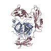
Mass: 27319.402 Da / Num. of mol.: 2 / Fragment: CATALYTIC DOMAIN / Mutation: S741A Source method: isolated from a genetically manipulated source Source: (gene. exp.) Homo sapiens (human) / Species (production host): Escherichia coli / Production host: Escherichia coli BL21 (bacteria) / Strain (production host): BL21 / References: UniProt: P00747, plasmin
#2: Protein
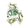
STREPTOKINASE
Mass: 41158.949 Da / Num. of mol.: 2 / Source method: isolated from a natural source Source: (natural) Streptococcus dysgalactiae subsp. equisimilis (bacteria) Species: Streptococcus dysgalactiae / Strain: subsp. equisimilis / References: UniProt: P00779
Has protein modification
Y
-
Experimental details
-
Experiment
Experiment
Method: X-RAY DIFFRACTION / Number of used crystals: 1
-
Sample preparation
Crystal
Density Matthews: 2.9 Å3/Da / Density % sol: 57 %
Crystal grow
pH: 8 / Details: pH 8.0
Crystal grow
*PLUS
Temperature: 20 ℃ / Method: vapor diffusion, sitting drop
Components of the solutions
*PLUS
ID
Conc.
Common name
Crystal-ID
Sol-ID
1
40mg/ml
protein
1
drop
2
1.0M
sodiumcitrate
1
reservoir
3
0.2M
HEPES
1
reservoir
4
1mM
magnesiumchloride
1
reservoir
-
Data collection
Diffraction
Mean temperature: 293 K
Diffraction source
Wavelength: 1.5418
Detector
Type: SIEMENS / Detector: AREA DETECTOR / Date: Aug 1, 1997
Radiation
Protocol: SINGLE WAVELENGTH / Monochromatic (M) / Laue (L): M / Scattering type: x-ray
Radiation wavelength
Wavelength: 1.5418 Å / Relative weight: 1
Reflection
Resolution: 2.9→45.4 Å / Num. obs: 33424 / % possible obs: 91.34 % / Redundancy: 1.9 % / Biso Wilson estimate: 47.7 Å2 / Rsym value: 0.053 / Net I/σ(I): 11.25
Reflection shell
Resolution: 2.9→3 Å / Redundancy: 1.43 % / Mean I/σ(I) obs: 2.17 / Rsym value: 0.252 / % possible all: 80.27
Reflection
*PLUS
% possible obs: 91.3 % / Rmerge(I) obs: 0.05
Reflection shell
*PLUS
% possible obs: 80.3 % / Rmerge(I) obs: 0.252
-
Processing
Software
Name
Version
Classification
SAINT
datascaling
SAINT
datareduction
MLPHARE
phasing
X-PLOR
3.8
refinement
Refinement
Method to determine structure: MIR / Resolution: 2.9→20 Å / Data cutoff high absF: 1000000 / Data cutoff low absF: 0.001 / Cross valid method: THROUGHOUT / σ(F): 0 / Details: A BULK SOLVENT CORRECTION WAS APPLIED.
Rfactor
Num. reflection
% reflection
Selection details
Rfree
0.291
3328
9.12 %
RANDOM
Rwork
0.201
-
-
-
obs
0.201
33304
91.34 %
-
Displacement parameters
Biso mean: 45.6 Å2
Refinement step
Cycle: LAST / Resolution: 2.9→20 Å
Protein
Nucleic acid
Ligand
Solvent
Total
Num. atoms
8940
0
0
0
8940
Refine LS restraints
Refine-ID
Type
Dev ideal
X-RAY DIFFRACTION
x_bond_d
0.009
X-RAY DIFFRACTION
x_bond_d_na
X-RAY DIFFRACTION
x_bond_d_prot
X-RAY DIFFRACTION
x_angle_d
X-RAY DIFFRACTION
x_angle_d_na
X-RAY DIFFRACTION
x_angle_d_prot
X-RAY DIFFRACTION
x_angle_deg
1.513
X-RAY DIFFRACTION
x_angle_deg_na
X-RAY DIFFRACTION
x_angle_deg_prot
X-RAY DIFFRACTION
x_dihedral_angle_d
28.77
X-RAY DIFFRACTION
x_dihedral_angle_d_na
X-RAY DIFFRACTION
x_dihedral_angle_d_prot
X-RAY DIFFRACTION
x_improper_angle_d
0.79
X-RAY DIFFRACTION
x_improper_angle_d_na
X-RAY DIFFRACTION
x_improper_angle_d_prot
X-RAY DIFFRACTION
x_mcbond_it
X-RAY DIFFRACTION
x_mcangle_it
X-RAY DIFFRACTION
x_scbond_it
X-RAY DIFFRACTION
x_scangle_it
Refine LS restraints NCS
NCS model details: RESTRAINTS
LS refinement shell
Resolution: 2.9→3.03 Å / Total num. of bins used: 8
In the structure databanks used in Yorodumi, some data are registered as the other names, "COVID-19 virus" and "2019-nCoV". Here are the details of the virus and the list of structure data.
Jan 31, 2019. EMDB accession codes are about to change! (news from PDBe EMDB page)
EMDB accession codes are about to change! (news from PDBe EMDB page)
The allocation of 4 digits for EMDB accession codes will soon come to an end. Whilst these codes will remain in use, new EMDB accession codes will include an additional digit and will expand incrementally as the available range of codes is exhausted. The current 4-digit format prefixed with “EMD-” (i.e. EMD-XXXX) will advance to a 5-digit format (i.e. EMD-XXXXX), and so on. It is currently estimated that the 4-digit codes will be depleted around Spring 2019, at which point the 5-digit format will come into force.
The EM Navigator/Yorodumi systems omit the EMD- prefix.
Related info.:Q: What is EMD? / ID/Accession-code notation in Yorodumi/EM Navigator
Yorodumi is a browser for structure data from EMDB, PDB, SASBDB, etc.
This page is also the successor to EM Navigator detail page, and also detail information page/front-end page for Omokage search.
The word "yorodu" (or yorozu) is an old Japanese word meaning "ten thousand". "mi" (miru) is to see.
Related info.:EMDB / PDB / SASBDB / Comparison of 3 databanks / Yorodumi Search / Aug 31, 2016. New EM Navigator & Yorodumi / Yorodumi Papers / Jmol/JSmol / Function and homology information / Changes in new EM Navigator and Yorodumi
 Movie
Movie Controller

 Yorodumi
 Open data
 Basic information
 Components
 Keywords
 Function and homology information
 Homo sapiens (human)
 Streptococcus dysgalactiae subsp. equisimilis (bacteria)
 X-RAY DIFFRACTION /
  Authors
 Citation
 Structure visualization
 Downloads & links
 Other downloads


 PDBj
PDBj


 Assembly
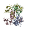
 Sample preparation
 Processing