 Movie
Movie Controller

[English] 日本語
 Yorodumi
- PDB-6ku9: Structure of the African swine fever virus major capsid protein p72 -
+ Open data

- Basic information
| Entry | Database: PDB / ID: 6ku9 | ||||||
|---|---|---|---|---|---|---|---|
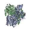
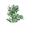
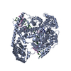
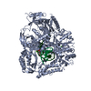
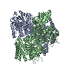
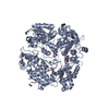
| Title | Structure of the African swine fever virus major capsid protein p72 | ||||||
 Components Components | B646L | ||||||
 Keywords Keywords | VIRAL PROTEIN / capsid protein | ||||||
| Function / homology | Major capsid protein, C-terminal / Major capsid protein, C-terminal domain superfamily / Large eukaryotic DNA virus major capsid protein / Group II dsDNA virus coat/capsid protein / viral capsid / structural molecule activity / B646L Function and homology information Function and homology information | ||||||
| Biological species |   African swine fever virus African swine fever virus | ||||||
| Method | ELECTRON MICROSCOPY / single particle reconstruction / cryo EM / Resolution: 2.67 Å | ||||||
 Authors Authors | Liu, Q. / Xiang, Y. | ||||||
| Funding support |  China, 1items China, 1items
| ||||||
 Citation Citation | Journal: Cell Res / Year: 2019 Title: Structure of the African swine fever virus major capsid protein p72. Authors: Qi Liu / Bingting Ma / Nianchao Qian / Fan Zhang / Xu Tan / Jianlin Lei / Ye Xiang / | ||||||
| History |
|
- Structure visualization
| Movie |
Movie viewer |
|---|---|
| Structure viewer | Molecule: MolmilJmol/JSmol |
- Downloads & links
-Download
| PDBx/mmCIF format | 6ku9.cif.gz | 270.4 KB | Display | PDBx/mmCIF format |
|---|---|---|---|---|
| PDB format | pdb6ku9.ent.gz | 215.8 KB | Display | PDB format |
| PDBx/mmJSON format | 6ku9.json.gz | Tree view | PDBx/mmJSON format | |
| Others |  Other downloads Other downloads |
-Validation report
| Arichive directory | https://data.pdbj.org/pub/pdb/validation_reports/ku/6ku9ftp://data.pdbj.org/pub/pdb/validation_reports/ku/6ku9 | HTTPS FTP |
|---|
-Related structure data
| Related structure data | 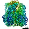 0776MC M: map data used to model this data C: citing same article ( |
|---|---|
| Similar structure data |


-Links
 PDBj
PDBj

- Assembly
| Deposited unit | 
|
|---|---|
| 1 |
|
-Components
| #1: Protein | Mass: 78930.531 Da / Num. of mol.: 3 Source method: isolated from a genetically manipulated source Source: (gene. exp.) African swine fever virus / Gene: p72, B646L, B646L CDS, ASFV-Georgia_4-106 / Cell line (production host): HEK293F / Production host:  Homo sapiens (human) / References: UniProt: Q5IZK2 Homo sapiens (human) / References: UniProt: Q5IZK2 |
|---|
-Experimental details
-Experiment
| Experiment | Method: ELECTRON MICROSCOPY |
|---|---|
| EM experiment | Aggregation state: PARTICLE / 3D reconstruction method: single particle reconstruction |
- Sample preparation
| Component | Name: p72 / Type: COMPLEX / Entity ID: all / Source: RECOMBINANT |
|---|---|
| Molecular weight | Value: 0.73 MDa / Experimental value: NO |
| Source (natural) | Organism: African swine fever virus |
| Source (recombinant) | Organism: Homo sapiens (human) / Cell: HEK293f |
| Details of virus | Isolate: STRAIN / Type: VIRION |
| Buffer solution | pH: 7.4 |
| Specimen | Conc.: 0.4 mg/ml / Embedding applied: NO / Shadowing applied: NO / Staining applied: NO / Vitrification applied: YES / Details: GraFix sample |
| Specimen support | Grid material: COPPER / Grid mesh size: 400 divisions/in. / Grid type: Quantifoil R1.2/1.3 |
| Vitrification | Instrument: FEI VITROBOT MARK IV / Cryogen name: ETHANE / Humidity: 100 % / Chamber temperature: 281.15 K / Details: blot 4.5s |
- Electron microscopy imaging
| Experimental equipment |  Model: Titan Krios / Image courtesy: FEI Company |
|---|---|
| Microscopy | Model: FEI TITAN KRIOS |
| Electron gun | Electron source:  FIELD EMISSION GUN / Accelerating voltage: 300 kV / Illumination mode: SPOT SCAN FIELD EMISSION GUN / Accelerating voltage: 300 kV / Illumination mode: SPOT SCAN |
| Electron lens | Mode: BRIGHT FIELD / Nominal defocus max: 3500 nm / Nominal defocus min: 1000 nm / Cs: 2.7 mm |
| Specimen holder | Specimen holder model: FEI TITAN KRIOS AUTOGRID HOLDER |
| Image recording | Electron dose: 50 e/Å2 / Detector mode: SUPER-RESOLUTION / Film or detector model: GATAN K2 SUMMIT (4k x 4k) |
- Processing
| CTF correction | Type: PHASE FLIPPING AND AMPLITUDE CORRECTION |
|---|---|
| Symmetry | Point symmetry: C3 (3 fold cyclic) |
| 3D reconstruction | Resolution: 2.67 Å / Resolution method: FSC 0.143 CUT-OFF / Num. of particles: 693388 / Symmetry type: POINT |
| Atomic model building | Protocol: AB INITIO MODEL |
