 Movie
Movie Controller

[English] 日本語
 Yorodumi
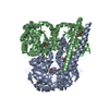
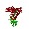
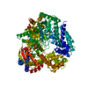
- EMDB-0776: Structure of the African swine fever virus major capsid protein p72 -
+ Open data

- Basic information
| Entry | Database: EMDB / ID: EMD-0776 | |||||||||
|---|---|---|---|---|---|---|---|---|---|---|
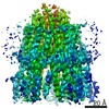
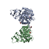
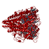
| Title | Structure of the African swine fever virus major capsid protein p72 | |||||||||
 Map data Map data | sharpened map | |||||||||
 Sample Sample |
| |||||||||
 Keywords Keywords | capsid protein / VIRAL PROTEIN | |||||||||
| Function / homology | Major capsid protein, C-terminal / Major capsid protein, C-terminal domain superfamily / Large eukaryotic DNA virus major capsid protein / Group II dsDNA virus coat/capsid protein / viral capsid / structural molecule activity / B646L Function and homology information Function and homology information | |||||||||
| Biological species |   African swine fever virus African swine fever virus | |||||||||
| Method | single particle reconstruction / cryo EM / Resolution: 2.67 Å | |||||||||
 Authors Authors | Liu Q / Xiang Y | |||||||||
| Funding support |  China, 1 items China, 1 items
| |||||||||
 Citation Citation | Journal: Cell Res / Year: 2019 Title: Structure of the African swine fever virus major capsid protein p72. Authors: Qi Liu / Bingting Ma / Nianchao Qian / Fan Zhang / Xu Tan / Jianlin Lei / Ye Xiang / | |||||||||
| History |
|
- Structure visualization
| Movie |
Movie viewer |
|---|---|
| Structure viewer | EM map: SurfViewMolmilJmol/JSmol |
| Supplemental images |

- Downloads & links
-EMDB archive
| Map data | emd_0776.map.gz | 4.9 MB | EMDB map data format | |
|---|---|---|---|---|
| Header (meta data) | emd-0776-v30.xmlemd-0776.xml | 17.1 KB 17.1 KB | Display Display | EMDB header |
| FSC (resolution estimation) | emd_0776_fsc.xml | 8.5 KB | Display | FSC data file |
| Images |  emd_0776.png emd_0776.png | 77.8 KB | ||
| Masks | emd_0776_msk_1.map | 52.7 MB | Mask map | |
| Filedesc metadata | emd-0776.cif.gz | 5.7 KB | ||
| Others | emd_0776_additional.map.gzemd_0776_additional_1.map.gzemd_0776_half_map_1.map.gzemd_0776_half_map_2.map.gz | 40.6 MB 40.6 MB 40.7 MB 40.7 MB | ||
| Archive directory |  http://ftp.pdbj.org/pub/emdb/structures/EMD-0776ftp://ftp.pdbj.org/pub/emdb/structures/EMD-0776 http://ftp.pdbj.org/pub/emdb/structures/EMD-0776ftp://ftp.pdbj.org/pub/emdb/structures/EMD-0776 | HTTPS FTP |
-Related structure data
| Related structure data | 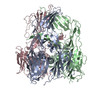 6ku9MC M: atomic model generated by this map C: citing same article ( |
|---|---|
| Similar structure data |


-Links
| EMDB pages | EMDB (EBI/PDBe) / EMDataResource |
|---|---|
| Related items in Molecule of the Month |


-Map
| File | Download / File: emd_0776.map.gz / Format: CCP4 / Size: 52.7 MB / Type: IMAGE STORED AS FLOATING POINT NUMBER (4 BYTES) | ||||||||||||||||||||||||||||||||||||||||||||||||||||||||||||||||||||
|---|---|---|---|---|---|---|---|---|---|---|---|---|---|---|---|---|---|---|---|---|---|---|---|---|---|---|---|---|---|---|---|---|---|---|---|---|---|---|---|---|---|---|---|---|---|---|---|---|---|---|---|---|---|---|---|---|---|---|---|---|---|---|---|---|---|---|---|---|---|
| Annotation | sharpened map | ||||||||||||||||||||||||||||||||||||||||||||||||||||||||||||||||||||
| Projections & slices | Image control
Images are generated by Spider. | ||||||||||||||||||||||||||||||||||||||||||||||||||||||||||||||||||||
| Voxel size | X=Y=Z: 1.091 Å | ||||||||||||||||||||||||||||||||||||||||||||||||||||||||||||||||||||
| Density |
| ||||||||||||||||||||||||||||||||||||||||||||||||||||||||||||||||||||
| Symmetry | Space group: 1 | ||||||||||||||||||||||||||||||||||||||||||||||||||||||||||||||||||||
| Details | EMDB XML:
CCP4 map header:
| ||||||||||||||||||||||||||||||||||||||||||||||||||||||||||||||||||||
 Z (Sec.)
 Y (Row.)
 X (Col.)


-Supplemental data
-Mask #1
| File | emd_0776_msk_1.map | ||||||||||||
|---|---|---|---|---|---|---|---|---|---|---|---|---|---|
| Projections & Slices |
| ||||||||||||
| Density Histograms |


-Additional map: unsharpened map
| File | emd_0776_additional.map | ||||||||||||
|---|---|---|---|---|---|---|---|---|---|---|---|---|---|
| Annotation | unsharpened map | ||||||||||||
| Projections & Slices |
| ||||||||||||
| Density Histograms |


-Additional map: unsharpened map
| File | emd_0776_additional_1.map | ||||||||||||
|---|---|---|---|---|---|---|---|---|---|---|---|---|---|
| Annotation | unsharpened map | ||||||||||||
| Projections & Slices |
| ||||||||||||
| Density Histograms |


-Half map: half map
| File | emd_0776_half_map_1.map | ||||||||||||
|---|---|---|---|---|---|---|---|---|---|---|---|---|---|
| Annotation | half map | ||||||||||||
| Projections & Slices |
| ||||||||||||
| Density Histograms |


-Half map: half map
| File | emd_0776_half_map_2.map | ||||||||||||
|---|---|---|---|---|---|---|---|---|---|---|---|---|---|
| Annotation | half map | ||||||||||||
| Projections & Slices |
| ||||||||||||
| Density Histograms |


- Sample components
-Entire : p72
| Entire | Name: p72 |
|---|---|
| Components |
|
-Supramolecule #1: p72
| Supramolecule | Name: p72 / type: complex / ID: 1 / Parent: 0 / Macromolecule list: all |
|---|---|
| Source (natural) | Organism: African swine fever virus |
| Molecular weight | Theoretical: 730 KDa |
-Macromolecule #1: B646L
| Macromolecule | Name: B646L / type: protein_or_peptide / ID: 1 / Number of copies: 3 / Enantiomer: LEVO |
|---|---|
| Source (natural) | Organism: African swine fever virus |
| Molecular weight | Theoretical: 78.930531 KDa |
| Recombinant expression | Organism:  Homo sapiens (human) Homo sapiens (human) |
| Sequence | String: MHHHHHHHHH HGSDYKDHDG DYKDHDIDYK DDDDKELENL YFQGAGSMAS GGAFCLIAND GKADKIILAQ DLLNSRISNI KNVNKSYGK PDPEPTLSQI EETHLVHFNA HFKPYVPVGF EYNKVRPHTG TPTLGNKLTF GIPQYGDFFH DMVGHHILGA C HSSWQDAP ...String: MHHHHHHHHH HGSDYKDHDG DYKDHDIDYK DDDDKELENL YFQGAGSMAS GGAFCLIAND GKADKIILAQ DLLNSRISNI KNVNKSYGK PDPEPTLSQI EETHLVHFNA HFKPYVPVGF EYNKVRPHTG TPTLGNKLTF GIPQYGDFFH DMVGHHILGA C HSSWQDAP IQGTSQMGAH GQLQTFPRNG YDWDNQTPLE GAVYTLVDPF GRPIVPGTKN AYRNLVYYCE YPGERLYENV RF DVNGNSL DEYSSDVTTL VRKFCIPGDK MTGYKHLVGQ EVSVEGTSGP LLCNIHDLHK PHQSKPILTD ENDTQRTCSH TNP KFLSQH FPENSHNIQT AGKQDITPIT DATYLDIRRN VHYSCNGPQT PKYYQPPLAL WIKLRFWFNE NVNLAIPSVS IPFG ERFIT IKLASQKDLV NEFPGLFVRQ SRFIAGRPSR RNIRFKPWFI PGVINEISLT NNELYINNLF VTPEIHNLFV KRVRF SLIR VHKTQVTHTN NNHHDEKLMS ALKWPIEYMF IGLKPTWNIS DQNPHQHRDW HKFGHVVNAI MQPTHHAEIS FQDRDT ALP DACSSISDIS PVTYPITLPI IKNISVTAHG INLIDKFPSK FCSSYIPFHY GGNAIKTPDD PGAMMITFAL KPREEYQ PS GHINVSRARE FYISWDTDYV GSITTADLVV SASAINFLLL QNGSAVLRYS T UniProtKB: B646L |
-Experimental details
-Structure determination
| Method | cryo EM |
|---|---|
 Processing Processing | single particle reconstruction |
| Aggregation state | particle |
-Sample preparation
| Concentration | 0.4 mg/mL |
|---|---|
| Buffer | pH: 7.4 |
| Grid | Model: Quantifoil R1.2/1.3 / Material: COPPER / Mesh: 400 / Support film - Material: CARBON / Support film - topology: HOLEY |
| Vitrification | Cryogen name: ETHANE / Chamber humidity: 100 % / Chamber temperature: 281.15 K / Instrument: FEI VITROBOT MARK IV / Details: blot 4.5s. |
| Details | GraFix sample |
- Electron microscopy
| Microscope | FEI TITAN KRIOS |
|---|---|
| Image recording | Film or detector model: GATAN K2 SUMMIT (4k x 4k) / Detector mode: SUPER-RESOLUTION / Average electron dose: 50.0 e/Å2 |
| Electron beam | Acceleration voltage: 300 kV / Electron source:  FIELD EMISSION GUN FIELD EMISSION GUN |
| Electron optics | Illumination mode: SPOT SCAN / Imaging mode: BRIGHT FIELD / Cs: 2.7 mm / Nominal defocus max: 3.5 µm / Nominal defocus min: 1.0 µm |
| Sample stage | Specimen holder model: FEI TITAN KRIOS AUTOGRID HOLDER |
| Experimental equipment |  Model: Titan Krios / Image courtesy: FEI Company |
+Image processing

-Atomic model buiding 1
| Refinement | Protocol: AB INITIO MODEL |
|---|---|
| Output model | PDB-6ku9: |
