 Movie
Movie Controller

[English] 日本語
 Yorodumi
- PDB-5rvz: PanDDA analysis group deposition -- Crystal Structure of DHTKD1 i... -
+ Open data

- Basic information
| Entry | Database: PDB / ID: 5rvz | ||||||
|---|---|---|---|---|---|---|---|
| Title | PanDDA analysis group deposition -- Crystal Structure of DHTKD1 in complex with Z1929757385 | ||||||
 Components Components | Probable 2-oxoglutarate dehydrogenase E1 component DHKTD1, mitochondrial | ||||||
 Keywords Keywords | OXIDOREDUCTASE / SGC - Diamond I04-1 fragment screening / PanDDA / XChemExplorer | ||||||
| Function / homology |  Function and homology information Function and homology informationOxidoreductases; Acting on the aldehyde or oxo group of donors; With a disulfide as acceptor / 2-oxoadipate dehydrogenase activity / OADH complex synthesizes glutaryl-CoA from 2-OA / oxoadipate dehydrogenase complex / thiamine pyrophosphate binding / hematopoietic progenitor cell differentiation / glycolytic process / generation of precursor metabolites and energy / mitochondrial matrix / mitochondrion Similarity search - Function | ||||||
| Biological species |  Homo sapiens (human) Homo sapiens (human) | ||||||
| Method |  X-RAY DIFFRACTION / SYNCHROTRON / FOURIER SYNTHESIS / molecular replacement / Resolution: 1.98 Å X-RAY DIFFRACTION / SYNCHROTRON / FOURIER SYNTHESIS / molecular replacement / Resolution: 1.98 Å | ||||||
 Authors Authors | Bezerra, G.A. / Foster, W.R. / Bailey, H.J. / Shrestha, L. / Krojer, T. / Brandao-Neto, J. / Douangamath, A. / Burgess-Brown, N. / von Delft, F. / Arrowsmith, C.H. ...Bezerra, G.A. / Foster, W.R. / Bailey, H.J. / Shrestha, L. / Krojer, T. / Brandao-Neto, J. / Douangamath, A. / Burgess-Brown, N. / von Delft, F. / Arrowsmith, C.H. / Edwards, A. / Bountra, C. / Yue, W.W. | ||||||
 Citation Citation | Journal: To Be Published Title: PanDDA analysis group deposition Authors: Bezerra, G.A. / Foster, W.R. / Bailey, H.J. / Shrestha, L. / Krojer, T. / Brandao-Neto, J. / Douangamath, A. / Burgess-Brown, N. / von Delft, F. / Arrowsmith, C.H. / Edwards, A. / Bountra, C. / Yue, W.W. | ||||||
| History |
|
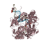
- Structure visualization
| Structure viewer | Molecule: MolmilJmol/JSmol |
|---|
- Downloads & links
-Download
| PDBx/mmCIF format | 5rvz.cif.gz | 401.6 KB | Display | PDBx/mmCIF format |
|---|---|---|---|---|
| PDB format | pdb5rvz.ent.gz | 310.8 KB | Display | PDB format |
| PDBx/mmJSON format | 5rvz.json.gz | Tree view | PDBx/mmJSON format | |
| Others |  Other downloads Other downloads |
-Validation report
| Arichive directory | https://data.pdbj.org/pub/pdb/validation_reports/rv/5rvzftp://data.pdbj.org/pub/pdb/validation_reports/rv/5rvz | HTTPS FTP |
|---|
-Group deposition
| ID | G_1002178 (5 entries) |
|---|---|
| Title | PanDDA analysis group deposition |
| Type | changed state |
| Description | human DHTKD1 screened against the DSI Fragment Library by X-ray Crystallography at the XChem facility of Diamond Light Source beamline I04-1 |


-Related structure data
| Related structure data |  6sy1S S: Starting model for refinement |
|---|---|
| Similar structure data |


-Links
 PDBj
PDBj


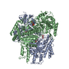
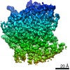
- Assembly
| Deposited unit | 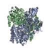
| ||||||||
|---|---|---|---|---|---|---|---|---|---|
| 1 |
| ||||||||
| Unit cell |
|
-Components
| #1: Protein | Mass: 100926.102 Da / Num. of mol.: 2 Source method: isolated from a genetically manipulated source Source: (gene. exp.) Homo sapiens (human) / Gene: DHTKD1, KIAA1630 / Production host:  References: UniProt: Q96HY7, oxoglutarate dehydrogenase (succinyl-transferring) #2: Chemical | ChemComp-WGA / ( | 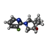  Mass: 210.248 Da / Num. of mol.: 1 / Source method: obtained synthetically / Formula: C11H15FN2O / Feature type: SUBJECT OF INVESTIGATION Mass: 210.248 Da / Num. of mol.: 1 / Source method: obtained synthetically / Formula: C11H15FN2O / Feature type: SUBJECT OF INVESTIGATION#3: Chemical |   Mass: 425.314 Da / Num. of mol.: 2 / Source method: obtained synthetically / Formula: C12H19N4O7P2S Mass: 425.314 Da / Num. of mol.: 2 / Source method: obtained synthetically / Formula: C12H19N4O7P2S#4: Chemical |   Mass: 24.305 Da / Num. of mol.: 3 / Source method: obtained synthetically / Formula: Mg Mass: 24.305 Da / Num. of mol.: 3 / Source method: obtained synthetically / Formula: Mg#5: Water | ChemComp-HOH / |  Mass: 18.015 Da / Num. of mol.: 1809 / Source method: isolated from a natural source / Formula: H2O Mass: 18.015 Da / Num. of mol.: 1809 / Source method: isolated from a natural source / Formula: H2OHas ligand of interest | Y | Has protein modification | N | |
|---|
-Experimental details
-Experiment
| Experiment | Method: X-RAY DIFFRACTION / Number of used crystals: 1 |
|---|
- Sample preparation
| Crystal | Density Matthews: 2.42 Å3/Da / Density % sol: 49.19 % / Mosaicity: 0.28 ° |
|---|---|
| Crystal grow | Temperature: 293 K / Method: vapor diffusion, sitting drop / pH: 7.1 Details: 0.1M Hepes, 0.1M Magnesium chloride, 20% PEG 6K, 10% ethylene glycol |
-Data collection
| Diffraction | Mean temperature: 100 K | ||||||||||||||||||||||||
|---|---|---|---|---|---|---|---|---|---|---|---|---|---|---|---|---|---|---|---|---|---|---|---|---|---|
| Diffraction source | Source: SYNCHROTRON / Site: Diamond  / Beamline: I04-1 / Wavelength: 0.9126 Å / Beamline: I04-1 / Wavelength: 0.9126 Å | ||||||||||||||||||||||||
| Detector | Type: DECTRIS PILATUS 6M / Detector: PIXEL / Date: Feb 21, 2020 | ||||||||||||||||||||||||
| Radiation | Protocol: SINGLE WAVELENGTH / Scattering type: x-ray | ||||||||||||||||||||||||
| Radiation wavelength | Wavelength: 0.9126 Å / Relative weight: 1 | ||||||||||||||||||||||||
| Reflection | Resolution: 1.989→85.122 Å / Num. obs: 78185 / % possible obs: 92.4 % / Redundancy: 3.7 % / CC1/2: 0.99 / Rpim(I) all: 0.097 / Rrim(I) all: 0.188 / Net I/σ(I): 6.9 / Num. measured all: 291693 / Scaling rejects: 0 | ||||||||||||||||||||||||
| Reflection shell | Diffraction-ID: 1 / Redundancy: 3.6 %
|
-Phasing
| Phasing | Method: molecular replacement |
|---|
- Processing
| Software |
| |||||||||||||||||||||||||||||||||||||||||||||||||||||||||||||||||||||||||||
|---|---|---|---|---|---|---|---|---|---|---|---|---|---|---|---|---|---|---|---|---|---|---|---|---|---|---|---|---|---|---|---|---|---|---|---|---|---|---|---|---|---|---|---|---|---|---|---|---|---|---|---|---|---|---|---|---|---|---|---|---|---|---|---|---|---|---|---|---|---|---|---|---|---|---|---|---|
| Refinement | Method to determine structure: FOURIER SYNTHESIS Starting model: 6SY1 Resolution: 1.98→85.12 Å / Cor.coef. Fo:Fc: 0.948 / Cor.coef. Fo:Fc free: 0.902 / SU B: 6.203 / SU ML: 0.162 / Cross valid method: THROUGHOUT / σ(F): 0 / ESU R: 0.439 / ESU R Free: 0.246 / Stereochemistry target values: MAXIMUM LIKELIHOOD Details: HYDROGENS HAVE BEEN ADDED IN THE RIDING POSITIONS U VALUES : REFINED INDIVIDUALLY
| |||||||||||||||||||||||||||||||||||||||||||||||||||||||||||||||||||||||||||
| Solvent computation | Ion probe radii: 0.8 Å / Shrinkage radii: 0.8 Å / VDW probe radii: 1.2 Å / Solvent model: MASK | |||||||||||||||||||||||||||||||||||||||||||||||||||||||||||||||||||||||||||
| Displacement parameters | Biso max: 157.05 Å2 / Biso mean: 24.78 Å2 / Biso min: 3.25 Å2
| |||||||||||||||||||||||||||||||||||||||||||||||||||||||||||||||||||||||||||
| Refinement step | Cycle: final / Resolution: 1.98→85.12 Å
| |||||||||||||||||||||||||||||||||||||||||||||||||||||||||||||||||||||||||||
| Refine LS restraints |
| |||||||||||||||||||||||||||||||||||||||||||||||||||||||||||||||||||||||||||
| LS refinement shell | Resolution: 1.976→2.027 Å / Total num. of bins used: 20
|
