 Movie
Movie Controller

[English] 日本語
 Yorodumi
- PDB-6vef: Cryo-EM Structure of Escherichia coli 2-oxoglutarate dehydrogenas... -
+ Open data

- Basic information
| Entry | Database: PDB / ID: 6vef | ||||||
|---|---|---|---|---|---|---|---|
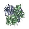
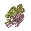
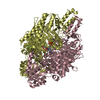
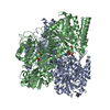
| Title | Cryo-EM Structure of Escherichia coli 2-oxoglutarate dehydrogenase E1 component sucA | ||||||
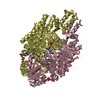
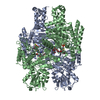
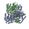
 Components Components | 2-oxoglutarate dehydrogenase E1 component | ||||||
 Keywords Keywords | OXIDOREDUCTASE / TCA cycle / 2-oxoglutarate dehydrogenase complex (OGDH) / E1 component / sucA / AMP / Oxaloacetate (OAA) / Dimer | ||||||
| Function / homology |  Function and homology information Function and homology informationoxoglutarate dehydrogenase (succinyl-transferring) / oxoglutarate dehydrogenase (succinyl-transferring) activity / oxoglutarate dehydrogenase complex / thiamine pyrophosphate binding / tricarboxylic acid cycle / nucleotide binding / magnesium ion binding / identical protein binding / cytosol / cytoplasm Similarity search - Function | ||||||
| Biological species |  | ||||||
| Method | ELECTRON MICROSCOPY / single particle reconstruction / cryo EM / Resolution: 4.08 Å | ||||||
 Authors Authors | Gao, H. | ||||||
 Citation Citation | Journal: To Be Published Title: Cryo-EM Structure of Escherichia coli 2-oxoglutarate dehydrogenase E1 component sucA Authors: Gao, H. | ||||||
| History |
|
- Structure visualization
| Movie |
Movie viewer |
|---|---|
| Structure viewer | Molecule: MolmilJmol/JSmol |
- Downloads & links
-Download
| PDBx/mmCIF format | 6vef.cif.gz | 308.9 KB | Display | PDBx/mmCIF format |
|---|---|---|---|---|
| PDB format | pdb6vef.ent.gz | 241.2 KB | Display | PDB format |
| PDBx/mmJSON format | 6vef.json.gz | Tree view | PDBx/mmJSON format | |
| Others |  Other downloads Other downloads |
-Validation report
| Arichive directory | https://data.pdbj.org/pub/pdb/validation_reports/ve/6vefftp://data.pdbj.org/pub/pdb/validation_reports/ve/6vef | HTTPS FTP |
|---|
-Related structure data
| Related structure data | 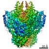 21156MC M: map data used to model this data C: citing same article ( |
|---|---|
| Similar structure data |


-Links
 PDBj
PDBj


- Assembly
| Deposited unit | 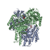
|
|---|---|
| 1 |
|
-Components
| #1: Protein | Mass: 94942.508 Da / Num. of mol.: 2 / Source method: isolated from a natural source / Source: (natural) References: UniProt: A0A403TZN2, UniProt: P0AFG3*PLUS, oxoglutarate dehydrogenase (succinyl-transferring) #2: Chemical |   Mass: 347.221 Da / Num. of mol.: 2 / Source method: obtained synthetically / Formula: C10H14N5O7P / Feature type: SUBJECT OF INVESTIGATION / Comment: AMP*YM Mass: 347.221 Da / Num. of mol.: 2 / Source method: obtained synthetically / Formula: C10H14N5O7P / Feature type: SUBJECT OF INVESTIGATION / Comment: AMP*YM#3: Chemical |   Mass: 131.064 Da / Num. of mol.: 2 / Source method: obtained synthetically / Formula: C4H3O5 / Feature type: SUBJECT OF INVESTIGATION Mass: 131.064 Da / Num. of mol.: 2 / Source method: obtained synthetically / Formula: C4H3O5 / Feature type: SUBJECT OF INVESTIGATIONHas ligand of interest | Y | |
|---|
-Experimental details
-Experiment
| Experiment | Method: ELECTRON MICROSCOPY |
|---|---|
| EM experiment | Aggregation state: PARTICLE / 3D reconstruction method: single particle reconstruction |
- Sample preparation
| Component | Name: Escherichia coli 2-oxoglutarate dehydrogenase E1 component sucA Type: COMPLEX / Entity ID: #1 / Source: NATURAL |
|---|---|
| Source (natural) | Organism: |
| Buffer solution | pH: 7.5 |
| Specimen | Embedding applied: NO / Shadowing applied: NO / Staining applied: NO / Vitrification applied: YES |
| Specimen support | Grid material: GOLD / Grid mesh size: 300 divisions/in. / Grid type: Quantifoil R1.2/1.3 |
| Vitrification | Instrument: FEI VITROBOT MARK IV / Cryogen name: ETHANE / Humidity: 100 % / Chamber temperature: 277 K |
- Electron microscopy imaging
| Experimental equipment |  Model: Talos Arctica / Image courtesy: FEI Company |
|---|---|
| Microscopy | Model: FEI TECNAI ARCTICA |
| Electron gun | Electron source:  FIELD EMISSION GUN / Accelerating voltage: 200 kV / Illumination mode: FLOOD BEAM FIELD EMISSION GUN / Accelerating voltage: 200 kV / Illumination mode: FLOOD BEAM |
| Electron lens | Mode: BRIGHT FIELD / Cs: 2.7 mm / C2 aperture diameter: 50 µm |
| Image recording | Average exposure time: 2 sec. / Electron dose: 80 e/Å2 / Film or detector model: GATAN K3 (6k x 4k) / Num. of grids imaged: 1 / Num. of real images: 800 |
- Processing
| EM software |
| ||||||||||||||||||||||||||||
|---|---|---|---|---|---|---|---|---|---|---|---|---|---|---|---|---|---|---|---|---|---|---|---|---|---|---|---|---|---|
| CTF correction | Type: PHASE FLIPPING AND AMPLITUDE CORRECTION | ||||||||||||||||||||||||||||
| Symmetry | Point symmetry: C2 (2 fold cyclic) | ||||||||||||||||||||||||||||
| 3D reconstruction | Resolution: 4.08 Å / Resolution method: FSC 0.143 CUT-OFF / Num. of particles: 86461 / Algorithm: SIMULTANEOUS ITERATIVE (SIRT) / Num. of class averages: 1 / Symmetry type: POINT | ||||||||||||||||||||||||||||
| Atomic model building | B value: 60 / Protocol: RIGID BODY FIT / Space: REAL | ||||||||||||||||||||||||||||
| Atomic model building | PDB-ID: 2JGD Accession code: 2JGD / Source name: PDB / Type: experimental model |

