 Movie
Movie Controller

[English] 日本語
 Yorodumi
- PDB-6ezr: Crystal structure of GH20 Exo beta-N-Acetylglucosaminidase from V... -
+ Open data

- Basic information
| Entry | Database: PDB / ID: 6ezr | ||||||||||||
|---|---|---|---|---|---|---|---|---|---|---|---|---|---|
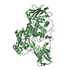
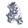
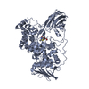
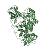
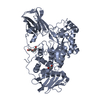
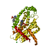
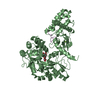
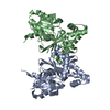
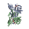
| Title | Crystal structure of GH20 Exo beta-N-Acetylglucosaminidase from Vibrio harveyi | ||||||||||||
 Components Components | Beta-N-acetylglucosaminidase Nag2 | ||||||||||||
 Keywords Keywords | HYDROLASE / N-acetylglucosamine | ||||||||||||
| Function / homology |  Function and homology information Function and homology informationglycosaminoglycan metabolic process / beta-N-acetylhexosaminidase / beta-N-acetylglucosaminidase activity / carbohydrate metabolic process / membrane Similarity search - Function | ||||||||||||
| Biological species |  Vibrio harveyi (bacteria) Vibrio harveyi (bacteria) | ||||||||||||
| Method |  X-RAY DIFFRACTION / SYNCHROTRON / MOLECULAR REPLACEMENT / molecular replacement / Resolution: 2.37 Å X-RAY DIFFRACTION / SYNCHROTRON / MOLECULAR REPLACEMENT / molecular replacement / Resolution: 2.37 Å | ||||||||||||
 Authors Authors | Porfetye, A.T. / Meekrathok, P. / Burger, M. / Vetter, I.R. / Suginta, W. | ||||||||||||
| Funding support |  Thailand, Thailand,  Germany, 3items Germany, 3items
| ||||||||||||
 Citation Citation | Journal: To Be Published Title: Crystal structure of GH20 Exo beta-N-Acetylglucosaminidase from Vibrio harveyi Authors: Meekrathok, P. / Porfetye, A.T. / Burger, M. / Vetter, I.R. / Suginta, W. | ||||||||||||
| History |
|
- Structure visualization
| Structure viewer | Molecule: MolmilJmol/JSmol |
|---|
- Downloads & links
-Download
| PDBx/mmCIF format | 6ezr.cif.gz | 286 KB | Display | PDBx/mmCIF format |
|---|---|---|---|---|
| PDB format | pdb6ezr.ent.gz | 227.9 KB | Display | PDB format |
| PDBx/mmJSON format | 6ezr.json.gz | Tree view | PDBx/mmJSON format | |
| Others |  Other downloads Other downloads |
-Validation report
| Arichive directory | https://data.pdbj.org/pub/pdb/validation_reports/ez/6ezrftp://data.pdbj.org/pub/pdb/validation_reports/ez/6ezr | HTTPS FTP |
|---|
-Related structure data
| Related structure data |  6ezsC  6eztC  3rcnS S: Starting model for refinement C: citing same article ( |
|---|---|
| Similar structure data |
-Links
 PDBj
PDBj
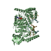
- Assembly
| Deposited unit | 
| ||||||||
|---|---|---|---|---|---|---|---|---|---|
| 1 | 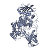
| ||||||||
| 2 | 
| ||||||||
| Unit cell |
|
-Components
| #1: Protein | Mass: 74543.922 Da / Num. of mol.: 2 Source method: isolated from a genetically manipulated source Source: (gene. exp.) Vibrio harveyi (bacteria) / Plasmid: pQE60 / Production host: #2: Water | ChemComp-HOH / |  Mass: 18.015 Da / Num. of mol.: 923 / Source method: isolated from a natural source / Formula: H2O Mass: 18.015 Da / Num. of mol.: 923 / Source method: isolated from a natural source / Formula: H2O |
|---|
-Experimental details
-Experiment
| Experiment | Method: X-RAY DIFFRACTION / Number of used crystals: 1 |
|---|
- Sample preparation
| Crystal | Density Matthews: 3.58 Å3/Da / Density % sol: 65.67 % |
|---|---|
| Crystal grow | Temperature: 293 K / Method: evaporation / pH: 4.5 / Details: 0.1 M sodium acetate pH 4.6, 1.4 M sodium malonate |
-Data collection
| Diffraction | Mean temperature: 100 K | ||||||||||||||||||||||||||||||||||||||||||||||||||||||||||||||||||||||||||||||||||||||||||||||||||||||||||||||||||||||||||||||||||||||||||||||||||||||||||||||||||||||||||||||||||||||||||||||||||||||||||||||||||
|---|---|---|---|---|---|---|---|---|---|---|---|---|---|---|---|---|---|---|---|---|---|---|---|---|---|---|---|---|---|---|---|---|---|---|---|---|---|---|---|---|---|---|---|---|---|---|---|---|---|---|---|---|---|---|---|---|---|---|---|---|---|---|---|---|---|---|---|---|---|---|---|---|---|---|---|---|---|---|---|---|---|---|---|---|---|---|---|---|---|---|---|---|---|---|---|---|---|---|---|---|---|---|---|---|---|---|---|---|---|---|---|---|---|---|---|---|---|---|---|---|---|---|---|---|---|---|---|---|---|---|---|---|---|---|---|---|---|---|---|---|---|---|---|---|---|---|---|---|---|---|---|---|---|---|---|---|---|---|---|---|---|---|---|---|---|---|---|---|---|---|---|---|---|---|---|---|---|---|---|---|---|---|---|---|---|---|---|---|---|---|---|---|---|---|---|---|---|---|---|---|---|---|---|---|---|---|---|---|---|---|---|
| Diffraction source | Source: SYNCHROTRON / Site: SLS  / Beamline: X10SA / Wavelength: 0.9998 Å / Beamline: X10SA / Wavelength: 0.9998 Å | ||||||||||||||||||||||||||||||||||||||||||||||||||||||||||||||||||||||||||||||||||||||||||||||||||||||||||||||||||||||||||||||||||||||||||||||||||||||||||||||||||||||||||||||||||||||||||||||||||||||||||||||||||
| Detector | Type: DECTRIS PILATUS 6M / Detector: PIXEL / Date: Sep 20, 2012 | ||||||||||||||||||||||||||||||||||||||||||||||||||||||||||||||||||||||||||||||||||||||||||||||||||||||||||||||||||||||||||||||||||||||||||||||||||||||||||||||||||||||||||||||||||||||||||||||||||||||||||||||||||
| Radiation | Protocol: SINGLE WAVELENGTH / Monochromatic (M) / Laue (L): M / Scattering type: x-ray | ||||||||||||||||||||||||||||||||||||||||||||||||||||||||||||||||||||||||||||||||||||||||||||||||||||||||||||||||||||||||||||||||||||||||||||||||||||||||||||||||||||||||||||||||||||||||||||||||||||||||||||||||||
| Radiation wavelength | Wavelength: 0.9998 Å / Relative weight: 1 | ||||||||||||||||||||||||||||||||||||||||||||||||||||||||||||||||||||||||||||||||||||||||||||||||||||||||||||||||||||||||||||||||||||||||||||||||||||||||||||||||||||||||||||||||||||||||||||||||||||||||||||||||||
| Reflection | Resolution: 2.37→90.66 Å / Num. obs: 84783 / % possible obs: 99.4 % / Observed criterion σ(I): -3 / Redundancy: 6.811 % / Biso Wilson estimate: 44.968 Å2 / CC1/2: 0.994 / Rmerge(I) obs: 0.146 / Rrim(I) all: 0.159 / Χ2: 1.023 / Net I/σ(I): 9.93 / Num. measured all: 577490 / Scaling rejects: 0 | ||||||||||||||||||||||||||||||||||||||||||||||||||||||||||||||||||||||||||||||||||||||||||||||||||||||||||||||||||||||||||||||||||||||||||||||||||||||||||||||||||||||||||||||||||||||||||||||||||||||||||||||||||
| Reflection shell | Diffraction-ID: 1
|
-Phasing
| Phasing | Method: molecular replacement |
|---|
- Processing
| Software |
| ||||||||||||||||||||||||||||||||||||||||||||||||||||||||||||
|---|---|---|---|---|---|---|---|---|---|---|---|---|---|---|---|---|---|---|---|---|---|---|---|---|---|---|---|---|---|---|---|---|---|---|---|---|---|---|---|---|---|---|---|---|---|---|---|---|---|---|---|---|---|---|---|---|---|---|---|---|---|
| Refinement | Method to determine structure: MOLECULAR REPLACEMENT Starting model: 3rcn Resolution: 2.37→90.66 Å / Cor.coef. Fo:Fc: 0.938 / Cor.coef. Fo:Fc free: 0.905 / SU B: 8.933 / SU ML: 0.204 / Cross valid method: THROUGHOUT / σ(F): 0 / ESU R: 0.293 / ESU R Free: 0.234 / Stereochemistry target values: MAXIMUM LIKELIHOOD Details: HYDROGENS HAVE BEEN ADDED IN THE RIDING POSITIONS U VALUES : REFINED INDIVIDUALLY
| ||||||||||||||||||||||||||||||||||||||||||||||||||||||||||||
| Solvent computation | Ion probe radii: 0.8 Å / Shrinkage radii: 0.8 Å / VDW probe radii: 1.2 Å / Solvent model: BABINET MODEL WITH MASK | ||||||||||||||||||||||||||||||||||||||||||||||||||||||||||||
| Displacement parameters | Biso max: 112.5 Å2 / Biso mean: 50.287 Å2 / Biso min: 23.66 Å2
| ||||||||||||||||||||||||||||||||||||||||||||||||||||||||||||
| Refinement step | Cycle: final / Resolution: 2.37→90.66 Å
| ||||||||||||||||||||||||||||||||||||||||||||||||||||||||||||
| Refine LS restraints |
| ||||||||||||||||||||||||||||||||||||||||||||||||||||||||||||
| LS refinement shell | Resolution: 2.37→2.432 Å / Rfactor Rfree error: 0 / Total num. of bins used: 20
|
